

The Clinical Significance of Schistocytes: A Prospective Evaluation of the International Council for Standardization in Hematology Schistocyte Guidelines
- PMID: 27795225
- PMCID: PMC5451690
- DOI: 10.4274/tjh.2016.0359
The Clinical Significance of Schistocytes: A Prospective Evaluation of the International Council for Standardization in Hematology Schistocyte Guidelines
Abstract
Objective: The presence of ≥1% schistocytes on a peripheral blood smear (PBS) is an important criterion for the diagnosis of thrombotic microangiopathy (TMA). The reporting of schistocytes has been standardized by the International Council for Standardization in Hematology (ICSH). Despite the availability of guidelines, however, the assessment of schistocytes remains subjective. More recently, the automated fragmented red cell (FRC) parameter has been evaluated. However, local studies are not available.
Materials and methods: A prospective study was performed at the Charlotte Maxeke Johannesburg Academic Hospital in order to evaluate the ICSH recommendations for schistocyte measurement in 146 PBSs with schistocytes. Schistocytes were evaluated by microscopy and ADVIA 2120 automated hematology analyzers.
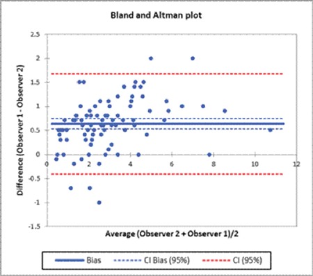
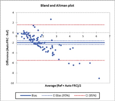
Results: Schistocytes were frequently observed in patients with TMA (n=76), infection (n=20), hematologic malignancy (n=10), renal failure (n=5), and hemoglobinopathy (n=15), and in neonates (n=11). Schistocytes were ≥1% in all PBSs with TMA (n=76) with a mean of 3.44±1.84. Schistocytes of ≥1% were also observed in cases of renal failure and hemoglobinopathy, and in neonates. In these conditions, schistocytes were mainly observed in conjunction with moderate red blood cell changes. The agreement between two morphologists gave a correlation coefficient of 0.63 [confidence interval (CI): 0.52-0.75], while the correlation coefficient between the average of the morphologists and the FRC percentage was -1.97 (CI: -1.60 to -2.34). The ADVIA 2120 underestimated the schistocyte count in patients with TMA.
Conclusion: Observer bias can be decreased by implementing the standardized procedures recommended by the ICSH. However, estimation of schistocytes by the ADVIA 2120 analyzer requires further evaluation as a screening tool. A higher threshold for schistocytes in thrombotic thrombocytopenic purpura is recommended to distinguish this hematological emergency from other conditions associated with ≥1% schistocytes.
Amaç: Periferk kan yaymasında (PKY) ≥%1 şistosit varlığı trombotik mikroanjiopati (TMA) tanısı için önemli bir kriterdir. Şistositlerin raporlanması Hematoloji Standardizasyon Uluslararası Komitesi [International Council for Standardization in Hematology (ICSH)] tarafından standardize edilmiştir. Kılavuzların mevcudiyetine rağmen, şistositlerin değerlendirmesi yine de subjektif kalmaktadır. Son zamanlarda, otomatize fragmente eritrosit (FE) parametresi değerlendirilmektedir. Ne var ki, lokal çalışmalar mevcut değildir. Gereç ve Yöntemler: ICSH önerilerini değerlendirmek için, Charlotte Maxeke Johannesburg Akademik Hastanesi’nde şistosit saptanan 146 PKY’da şistosit ölçümünü değerlendiren prospektif bir çalışma gerçekleştirildi. Şistositler mikroskop ve ADVIA 2120 otomatize hematoloji analizörü ile değerlendirildi. Bulgular: Şistositler, TMA (n=76), enfeksiyon (n=20), hematolojik malignite (n=10), renal yetmezlik (n=5) ve hemoglobinopati (n=15) hastalarında ve yenidoğanlarda (n=11) sıklıkla izlendi. Tüm TMA’lı hastaların (n=76) PKY’lerinde şistositler 3,44±1,84 ortalama ile ≥%1 idi. Şistositler ayrıca renal yetmezlik ve hemoglobinopati olguları ve yenidoğanlarda ≥%1 olarak izlendi. Bu durumlarda, şistositler çoğunlukla ılımlı eritrosit değişiklikleri ile ilişkili olarak gözlendi. İki morfolojist arasındaki anlaşma 0,63 [güven aralığı (GA): 0,52-0,75] korelasyon katsayısı verirken, morfolojistlerin ortalaması ve FE yüzdesi arasındaki korelasyon katsayısı -1.97 (GA: -1,60 - -2,34) idi. ADVIA 2120 ile TMA’lı hastalarda şistosit sayısı daha düşük ölçüldü. Sonuç: ICSH tarafından önerilen standardize prosedürlerin uygulanması ile gözlemci önyargısı azaltılabilir. Ne var ki, tarama aracı olarak ADVIA 2120 analizörü ile şistosit ölçümü daha ileri değerlendirme gerektirmektedir. Trombotik trombositopenik purpurada, şistositlerin ≥%1 olduğu diğer durumlardan bu hematolojik acili ayırt etmek için daha yüksek bir şistosit eşik değeri önerilmektedir.
Conflict of interest statement
Conflict of Interest: The authors of this paper have no conflicts of interest, including specific financial interests, relationships, and/or affiliations relevant to the subject matter or materials included.
Figures
References
-
- Huh HJ, Chung JW, Chae SL. Microscopic schistocyte determination according to International Council for Standardization in Hematology recommendations in various diseases. Int J Lab Hematol. 2013;35:542–547. - PubMed
-
- Lesesve JF, Salignac S, Lecompte T. Laboratory measurement of schistocytes. Int J Lab Hematol. 2007;29:149–151. - PubMed
-
- Zini G, d’Onofrio G, Briggs C, Erber W, Jou JM, Lee SH, McFadden S, Vives-Corrons JL, Yutaka N, Lesesve JF Internatiol Council for Standardization in Haematology (ICSH) ICSH recommendations for identification, diagnostic value, and quantitation of schistocytes. Int J Lab Hematol. 2012;34:107–116. - PubMed
-
- Lesesve JF, El Adssi H, Watine J, Oosterhuis W, Regnier F. Evaluation of ICSH schistocyte measurement guidelines in France. Int J Lab Hematol. 2013;35:601–607. - PubMed
-
- Banno S, Ito Y, Tanaka C, Hori T, Fujimoto K, Suzuki T, Hashimoto T, Ueda R, Mizokami M. Quantification of red blood cell fragmentation by the automated hematology analyzer XE-2100 in patients with living donor liver transplantation. Clin Lab Haematol. 2005;27:292–296. - PubMed
MeSH terms
Substances
LinkOut - more resources
Full Text Sources
Other Literature Sources