. 2016 Dec 19;37(1):e00236-16.
doi: 10.1128/MCB.00236-16. Print 2017 Jan 1.

PHD2 Is a Regulator for Glycolytic Reprogramming in Macrophages

Affiliations

PHD2 Is a Regulator for Glycolytic Reprogramming in Macrophages

Annemarie Guentsch et al. Mol Cell Biol. .

Abstract

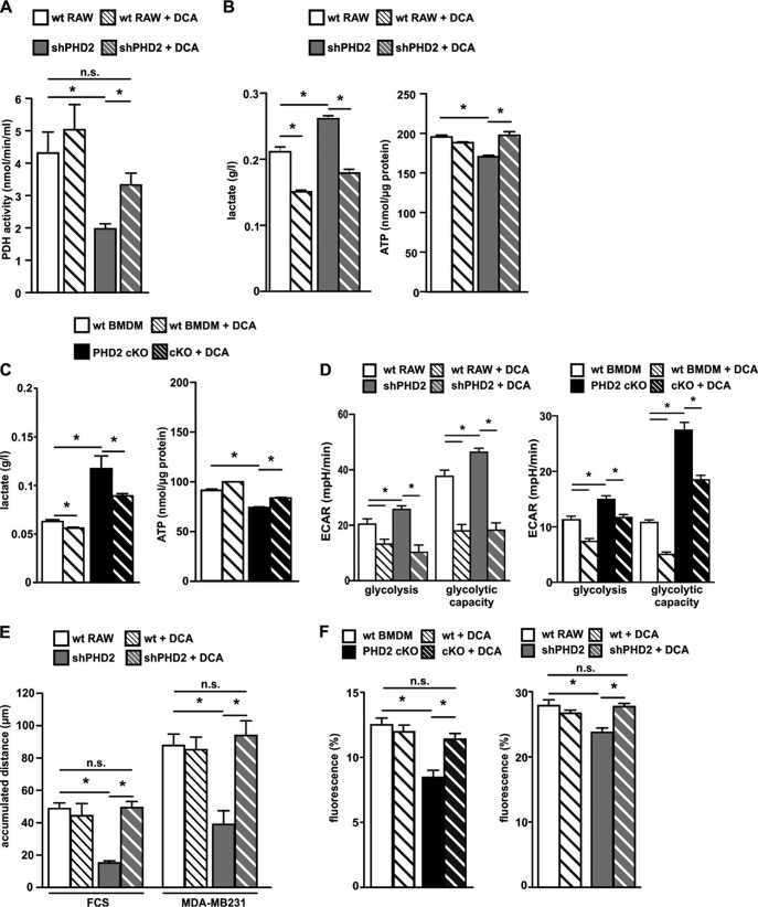
The prolyl-4-hydroxylase domain (PHD) enzymes are regarded as the molecular oxygen sensors. There is an interplay between oxygen availability and cellular metabolism, which in turn has significant effects on the functionality of innate immune cells, such as macrophages. However, if and how PHD enzymes affect macrophage metabolism are enigmatic. We hypothesized that macrophage metabolism and function can be controlled via manipulation of PHD2. We characterized the metabolic phenotypes of PHD2-deficient RAW cells and primary PHD2 knockout bone marrow-derived macrophages (BMDM). Both showed typical features of anaerobic glycolysis, which were paralleled by increased pyruvate dehydrogenase kinase 1 (PDK1) protein levels and a decreased pyruvate dehydrogenase enzyme activity. Metabolic alterations were associated with an impaired cellular functionality. Inhibition of PDK1 or knockout of hypoxia-inducible factor 1α (HIF-1α) reversed the metabolic phenotype and impaired the functionality of the PHD2-deficient RAW cells and BMDM. Taking these results together, we identified a critical role of PHD2 for a reversible glycolytic reprogramming in macrophages with a direct impact on their function. We suggest that PHD2 serves as an adjustable switch to control macrophage behavior.

Keywords: PDK; dioxygenases; hypoxia; macrophages; prolyl-4-hydroxylase domain.


Figures

FIG 1
FIG 1
PHD2 knockdown RAW cells and PHD2 knockout (PHD2 cKO) BMDM display increased PDK1 expression and activity. (A) wt RAW and shPHD2 knockdown cells as well as wt BMDM and PHD2 cKO macrophages were incubated for 24 h at 20% or 1% O2. RNA levels of the indicated genes were analyzed by qRT-PCR. RNA levels in wt RAW cells and wt BMDM were set to 1. Fold changes of the RNA levels for the indicated genes in shPHD2 cells, PHD2 cKO BMDM, or wt cells in hypoxia were determined by comparison to the levels in wt cells in normoxia (n = 3 to 6 independent samples per condition). (B) Annexin V (AV) single-positive cells were analyzed in wt BMDM and PHD2 cKO macrophages, with and without treatment with 1 mM DMOG for 24 h. (C) HIF-1α, HIF-2α, PHD2, and β-actin protein levels in wt RAW and shPHD2 cells as well as wt BMDM and PHD2 cKO macrophages in normoxia (20% O2) or hypoxia (1% O2 for 24 h). (D) Phospho-PDH, total PDH, PDK, and β-actin protein levels in wt RAW and shPHD2 cells as well as wt BMDM and PHD2 cKO macrophages in normoxia (20% O2) or hypoxia (1% O2 for 24 h). (E) PDH activities in normoxia or hypoxia (1% O2 for 24 h) for wt RAW and shPHD2 cells and wt RAW cells treated with 1 mM DMOG for 24 h (n = 6 independent samples per condition). Data are means and SEM. *, P < 0.05.
FIG 2
FIG 2
Macrophages shift their metabolism toward anaerobic glycolysis as a consequence of a reduction of PHD2 expression. (A) wt RAW cells, shPHD2 RAW cells, wt BMDM, and PHD2 cKO cells or wt RAW cells treated with 1 mM DMOG for 24 h were tested for their oxygen consumption rate (OCR) after addition of oligomycin, FCCP, and rotenone plus antimycin A (Rot+AA) (n = 6 [RAW cells] or 10 [BMDM] independent samples per condition). (B) Basal respiration and maximum respiration were analyzed in wt RAW cells, shPHD2 RAW cells, wt BMDM, and PHD2 cKO cells based on the data shown in panel A. The OCR after addition of Rot+AA was subtracted from the OCRs after addition of oligomycin and FCCP to obtain basal respiration and maximum respiration values, respectively. (C and D) The extracellular acidification rate (ECAR) was determined for wt RAW and shPHD2 RAW cells (C) as well as wt BMDM and PHD2 cKO cells (D) or wt RAW cells treated with 1 mM DMOG for 24 h after addition of glucose, oligomycin, and 2-desoxyglucose (2-DG) (n = 7 independent samples per condition). (E and F) Glycolysis and anaerobic glycolytic capacity were analyzed based on the data shown in panels C and D. The ECAR after addition of 2-DG was subtracted from the ECARs after addition of glucose and oligomycin to obtain glycolysis and glycolytic capacity values, respectively. (G) Lactate levels in the supernatants of wt RAW cells, shPHD2 RAW cells, wt BMDM, and PHD2 cKO cells were determined after incubation of the cells under the indicated conditions (n = 4 independent samples per condition). Data are means and SEM. *, P < 0.05. (H) Lactate levels in the supernatants of wt BMDM and cKO cells were determined after incubation of the cells at 20% O2 or 1% O2, with or without addition of glucose to the cell culture medium. Cells were incubated for 24 h in the respective cell culture medium (n = 4 independent samples per condition).
FIG 3
FIG 3
Decreased ATP levels and unaltered polarization in PHD2-deficient macrophages. (A) (Left and middle) wt RAW cells, shPHD2 RAW cells, wt BMDM, and PHD2 cKO cells were incubated at 20% O2 or 1% O2 for 24 h, and subsequently, intracellular ATP levels were determined (n = 6 independent samples per condition). (Right) wt RAW cells were incubated with 1 mM DMOG for the indicated times, and ATP levels were then determined (n = 6 or 7 independent samples per condition). (B and C) RNA levels for M1 and M2 markers in resting wt BMDM and cKO cells (B) or after stimulation with IL-4 (20 nM) or LPS (100 ng/ml) and IFN-γ (20 nM) for 24 h (C). Numbers of copies of the respective RNAs were quantified relative to the number of RNA copies of the housekeeping gene mS12. (D) RNA levels for M1 and M2 markers in resting wt BMDM and PHD2 cKO cells were analyzed after treatment of the cells with 1 mM DMOG for 24 h. Fold changes of the RNA levels for the indicated genes in DMOG-treated wt or cKO BMDM were determined by comparison to the levels in the nontreated cells (n = 3 to 6 independent samples per condition). Data are means and SEM. *, P < 0.05.
FIG 4
FIG 4
A reduction of PHD2 expression in RAW cells or BMDM results in defects in macrophage migration and phagocytosis. (A) wt RAW cells, shPHD2 RAW cells, wt BMDM, and PHD2 cKO cells were tested for their migration capacity in Boyden chambers by use of FCS or conditioned medium from MDA-MB231 cells as a stimulant (n = 4 independent samples). (B) The accumulated migration distances over 6 h for wt RAW cells, shPHD2 RAW cells, wt BMDM, and PHD2 cKO cells were tested in single-cell migration experiments using FCS or conditioned medium from MDA-MB231 cells as a stimulant (n = 59 to 64 cells per condition [RAW cells] or 51 to 71 cells per condition [BMDM]). (C) wt RAW cells were incubated at 20% O2, with or without 1 mM DMOG, or at 1% O2 for 6 h. The accumulated migration distance was tested in single-cell migration experiments using FCS or conditioned medium from MDA-MB231 cells as a stimulant. For cells analyzed at 1% O2, the hypoxic conditions were kept during the single-cell migration experiments, without reoxygenation (n = 50 to 53 cells per condition). (D) wt RAW cells, shPHD2 RAW cells, wt BMDM, and PHD2 cKO cells were incubated at 20% O2, with or without 1 mM DMOG, or at 1% O2 for 20 h in total. Subsequently, the capacity of the cells to phagocytose labeled beads was analyzed. Fluorescent beads were added to the cells for 4 h without reoxygenation. Data are means and SEM (n = 5 independent samples per condition). *, P < 0.05. (E) RNA levels for chemokine receptors in resting wt BMDM and PHD2 cKO cells after incubation under normoxic or hypoxic (1% O2) conditions for 24 h. Fold changes of the RNA levels for the indicated genes in the PHD2 cKO BMDM were determined by comparison to the levels in the wt cells in normoxia (n = 3 independent samples per condition). Data are means and SEM. *, P < 0.05.
FIG 5
FIG 5
Inhibition of PDK1 by dichloroacetate (DCA) reverses the metabolic phenotype and the migration defect in PHD2-deficient macrophages. (A) PDH activity was determined in lysates of wt RAW and shPHD2 RAW cells after incubation of the cells with or without 5 mM DCA for 24 h (n = 6 independent samples per condition). (B and C) Lactate levels in the supernatants and intracellular ATP levels of wt RAW and shPHD2 RAW cells (B) as well as wt BMDM and cKO macrophages (C) after incubation of the cells with or without 5 mM DCA for 24 h. (D) Glycolysis and glycolytic capacity were analyzed in wt RAW cells, shPHD2 RAW cells, wt BMDM, and PHD2 cKO cells after incubating the cells with or without 5 mM DCA for 24 h (n = 6 to 10 independent samples per condition). (E) Accumulated migration distances over 6 h for wt RAW and shPHD2 RAW cells after incubation of the cells with or without 5 mM DCA for 24 h were determined in single-cell migration experiments using FCS or conditioned medium from MDA-MB231 cells as a stimulant (n = at least 20 cells per condition). (F) wt RAW cells, shPHD2 RAW cells, wt BMDM, and PHD2 cKO cells were incubated with or without 5 mM DCA for 24 h. Subsequently, the capacity of the cells to phagocytose labeled beads was analyzed. Data are means and SEM (n = 5 to 8 independent samples per condition). *, P < 0.05; n.s., not significant.
FIG 6
FIG 6
No metabolic phenotype in PHD3-deficient macrophages. (A) wt BMDM and PHD3 cKO macrophages were incubated for 24 h at 20% or 1% O2. RNA levels for the indicated genes were analyzed by qRT-PCR. RNA levels in wt BMDM were set to 1. Fold changes of the RNA levels for the indicated genes in PHD3 cKO BMDM or wt cells in hypoxia were determined by comparison to the levels in the wt cells in normoxia (n = 3 independent samples per condition). (B) ATP (n = 5 independent samples) and lactate (n = 4 independent samples) levels as well as accumulated migration distances were determined for wt BMDM and PHD3 cKO macrophages.
FIG 7
FIG 7
HIF-1α mediates the metabolic alterations in PHD2-deficient macrophages. (A) wt BMDM and dcKO macrophages were incubated for 24 h at 20% or 1% O2. RNA levels for the indicated genes were analyzed by qRT-PCR. RNA levels in wt BMDM were set to 1. Fold changes of the RNA levels for the indicated genes in dcKO BMDM or wt cells in hypoxia were determined by comparison to the levels in the wt cells in normoxia (n = 3 independent samples per condition). Data are means and SEM. *, P < 0.05 compared to wt cells at 20% O2; #, P < 0.05 compared to wt cells at 1% O2. (B) Intracellular ATP levels were determined for wt BMDM, PHD2 cKO cells, and dcKO cells after incubation for 24 h in 20% O2 or 1% O2. (C) OCRs and lactate levels in the supernatants of wt BMDM, PHD2 cKO cells, and dcKO cells. (D) The accumulated migration distances over 6 h for wt BMDM, PHD2 cKO cells, and dcKO cells were determined in single-cell migration experiments using FCS or conditioned medium from MDA-MB231 cells as a stimulant (n = at least 20 cells per condition). Data are means and SEM. *, P < 0.05.

References

    1. Wynn TA, Vannella KM. 2016. Macrophages in tissue repair, regeneration, and fibrosis. Immunity 44:450–462. doi:10.1016/j.immuni.2016.02.015. - DOI - PMC - PubMed
    1. Goda N, Kanai M. 2012. Hypoxia-inducible factors and their roles in energy metabolism. Int J Hematol 95:457–463. doi:10.1007/s12185-012-1069-y. - DOI - PubMed
    1. Bishop T, Ratcliffe PJ. 2015. HIF hydroxylase pathways in cardiovascular physiology and medicine. Circ Res 117:65–79. doi:10.1161/CIRCRESAHA.117.305109. - DOI - PMC - PubMed
    1. Kaelin WG., Jr 2005. Proline hydroxylation and gene expression. Annu Rev Biochem 74:115–128. - PubMed
    1. Schofield CJ, Ratcliffe PJ. 2004. Oxygen sensing by HIF hydroxylases. Nat Rev Mol Cell Biol 5:343–354. - PubMed

Publication types

Substances

LinkOut - more resources