. 2016 Dec 13;199(1):e00613-16.
doi: 10.1128/JB.00613-16. Print 2017 Jan 1.

Genes Required for Bacillus anthracis Secondary Cell Wall Polysaccharide Synthesis

Affiliations

Genes Required for Bacillus anthracis Secondary Cell Wall Polysaccharide Synthesis

So-Young Oh et al. J Bacteriol. .

Abstract

The secondary cell wall polysaccharide (SCWP) is thought to be essential for vegetative growth and surface (S)-layer assembly in Bacillus anthracis; however, the genetic determinants for the assembly of its trisaccharide repeat structure are not known. Here, we report that WpaA (BAS0847) and WpaB (BAS5274) share features with membrane proteins involved in the assembly of O-antigen lipopolysaccharide in Gram-negative bacteria and propose that WpaA and WpaB contribute to the assembly of the SCWP in B. anthracis Vegetative forms of the B. anthracis wpaA mutant displayed increased lengths of cell chains, a cell separation defect that was attributed to mislocalization of the S-layer-associated murein hydrolases BslO, BslS, and BslT. The wpaB mutant was defective in vegetative replication during early logarithmic growth and formed smaller colonies. Deletion of both genes, wpaA and wpaB, did not yield viable bacilli, and when depleted of both wpaA and wpaB, B. anthracis could not maintain cell shape, support vegetative growth, or assemble SCWP. We propose that WpaA and WpaB fulfill overlapping glycosyltransferase functions of either polymerizing repeat units or transferring SCWP polymers to linkage units prior to LCP-mediated anchoring of the polysaccharide to peptidoglycan.

Importance: The secondary cell wall polysaccharide (SCWP) is essential for Bacillus anthracis growth, cell shape, and division. SCWP is comprised of trisaccharide repeats (→4)-β-ManNAc-(1→4)-β-GlcNAc-(1→6)-α-GlcNAc-(1→) with α-Gal and β-Gal substitutions; however, the genetic determinants and enzymes for SCWP synthesis are not known. Here, we identify WpaA and WpaB and report that depletion of these factors affects vegetative growth, cell shape, and S-layer assembly. We hypothesize that WpaA and WpaB are involved in the assembly of SCWP prior to transfer of this polymer onto peptidoglycan.

Keywords: Bacillus anthracis; S-layers; Wzy repeat polymerase; cell wall polysaccharide; envelope assembly.


Figures

FIG 1
FIG 1
Wpa proteins of Bacillus anthracis. (A) Illustration of gene clusters associated with secondary cell wall polysaccharide (SCWP) synthesis in B. anthracis, including the S-layer cluster with csaB (encoding the ketal-pyruvyl transferase of the SCWP), sap and eag (encoding S-layer proteins), patA1-patB1-patA2-patB2 (encoding O-acetyl transferases of the SCWP), and wpaA (bas0847; encoding a PF13425 [O-antigen_lig] family member). bslS, encoding S-layer-associated murein hydrolase S, is located in the vicinity of wpaA. bslT, encoding S-layer-associated murein hydrolase T, is located adjacent to bslO. Finally, wpaB (bas5274) is located in an operon for SCWP synthesis together with tagA1. (B) PFAM-predicted transmembrane topology of PF13425 family members from Streptococcus pneumoniae (O-antigen ligase-like protein; accession number CKE53299) and B. anthracis (WpaA [BAS0847] and WpaB [BAS5274]). (C) Experimentally determined topologies of Wzy and WaaL from P. aeruginosa (34). The numbers at top and bottom indicate positions of amino acid residues in the sequences of the respective proteins. Numbering of transmembrane segments is indicated in the middle as 1 through 12 (WaaL and PF13425) and 1 through 14 (Wzy). PL, periplasmic loop.
FIG 2
FIG 2
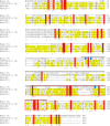
Amino acid sequence alignment of the prototypic O-antigen ligase WaaL of P. aeruginosa (Pa) (accession number CTQ39963.1), the S. pneumoniae (Sp) protein with accession number CKE53299.1, and B. anthracis BAS0847 (YP_027123.1) and BAS5274 (YP_031512.1) using ClustalO (http://www.ebi.ac.uk/Tools/msa/clustalo/). The large periplasmic loop PL5 of WaaL (34) that defines the prototypic PF04932 (Wzy_C) domain is shown with a line above the amino acid sequence. The conserved RX3V motif and catalytic H303 are highlighted in blue in the WaaL sequence. The red box marks the position of the PF13425 (O-antigen_lig) domain. Residues highlighted in yellow are shared by at least two of the three S. pneumoniae and B. anthracis PF13425 proteins. Residues identical between all four proteins are highlighted in red and marked with an asterisk, whereas double and single dots mark residues that are highly and weakly conserved, respectively, in all four sequences.
FIG 3
FIG 3
Mislocalization of murein hydrolases causes the chain length phenotype in B. anthracis ΔwpaA mutants. Spores (1 × 10⁶/ml) derived from various B. anthracis strains were germinated and grown in BHI at 37°C for 4 h prior to fixation for microscopy or cell fractionation and immunoblot analysis. (A) The lengths of 150 chains of vegetative cells were measured and graphed with a box-and-whiskers plot, where the box denotes the 25th and 75th percentiles, the whiskers denote the minimum and maximum measured chain length, and the center line indicates the median value. Data were analyzed by unpaired Student's t tests for significant differences compared to the wild type. *, P < 0.001. (B) Subcellular localization of S-layer and S-layer-associated murein hydrolases in the ΔwpaA mutant. Cultures from germinated spores were fractionated into medium (M), S-layer (S), and cellular (C) fractions. Samples were analyzed by 12% SDS-PAGE and Coomassie staining. (C) Samples obtained in the experiment whose results are shown in panel B were subjected to immunoblot analysis utilizing polyclonal antisera raised against S-layer protein (Sap), S-layer-associated murein hydrolases (BslO, BslS, and BslT), or a membrane lipoprotein (PrsA1). Numbers to the left in panels B and C indicate the migratory positions of molecular mass markers in kilodaltons.
FIG 4
FIG 4
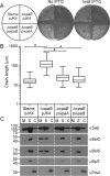
Mislocalization of BslO and BslS, chain length, and growth phenotypes in B. anthracis ΔwpaB mutants. (A) Small-colony phenotype of the ΔwpaB mutant. Spores (1 × 10³) were inoculated on BHI agar, and vegetative forms grown with or without 1 mM IPTG at 37°C for 24 h. (B) Box-and-whisker plot representing measured vegetative-form chain lengths of wild-type B. anthracis and the ΔwpaB mutant. Samples were prepared and analyzed as described in the legend to Fig. 3A. (C) B. anthracis cultures were fractionated into medium (M), S-layer (S), and cellular (C) fractions, which were analyzed by immunoblotting as described in the legend to Fig. 3B. Numbers to the left indicate the migratory positions of molecular mass markers in kilodaltons.
FIG 5
FIG 5
Deposition and distribution of S-layer and S-layer-associated proteins in the envelope of wpaB mutant B. anthracis. Spores derived from wild-type and ΔwpaB mutant B. anthracis strains were germinated and grown in BHI at 37°C for 4 h prior to fixation. Samples were stained with BODIPY-vancomycin (B-vancomycin) or rabbit antiserum raised against purified Sap (αSap) (A) or BslS (αBslS) (B) and viewed via differential interference contrast (DIC) or fluorescence microscopy, and images were acquired and merged. Scale bars denote 5 μm.
FIG 6
FIG 6
B. anthracis wpaA and wpaB are indispensable for vegetative growth. (A) CP-51 phage lysate was prepared from B. anthracis strains carrying spectinomycin-resistance determinants in a ΔbslA(bslA::aad9) or ΔwpaB(wpaB::aad9) background and incubated with wild-type or ΔwpaA mutant bacilli harboring plasmid vector pJK4 or pLM5 (empty vectors), pwpaA, pwpaAts, pwpaB, or pwpaBts. Samples were plated and incubated for 30 h at 30°C, and colonies were enumerated. The mean numbers of colonies and associated standard errors from 3 independent experimental determinations were plotted. (B) Spores (1 × 10³) were inoculated on BHI agar with or without 1 mM IPTG and grown at 30°C or 42°C for 24 h. (C) B. anthracis strains were assessed for growth by inoculating spores (1 × 10⁶/ml) into BHI with 1 mM IPTG at 30°C or into BHI without IPTG at 42°C. Vegetative growth was monitored by recording optical density (A600) over time.
FIG 7
FIG 7
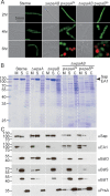
Depletion of wpaA and wpaB affects B. anthracis cell shape, survival, and S-layer assembly. (A) Fluorescence micrographs of B. anthracis cells incubated for 2, 4, or 6 h in BHI without IPTG at 42°C. Bacterial cells were derived from germinated wild-type B. anthracis Sterne or ΔwpaAB(pwpaAts) or ΔwpaAB(pwpaBts) strain spores. At each time point, bacilli were stained with Syto9 (green) and propidium iodide (red) and visualized by fluorescence microscopy to monitor the viability of bacterial populations as a function of the membrane integrity of the cell. Cells with compromised membranes stain red, whereas cells with intact membranes stain green. Merged fluorescence microscopy images are presented. (B) Subcellular fractionation of B. anthracis strains grown for 6 h into medium (M), S-layer (S), and cellular (C) fractions. Proteins were separated on 12% SDS-PAGE and stained with Coomassie blue. (C) Samples obtained in the experiment whose results are shown in panel B were analyzed by immunoblotting with antibodies raised against purified Sap (αSap), EA1 (αEA1), BslO (αBslO), BslS (αBslS), BslT (αBslT), and PrsA1 (αPrsA). Numbers to the left in panels B and C indicate the migratory positions of molecular mass markers in kilodaltons.
FIG 8
FIG 8
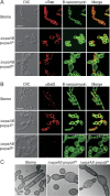
Localization of Sap and BslS in the envelope of wpaAB-depleted B. anthracis strains. Immunofluorescence micrographs stained with BODIPY-vancomycin (B-vancomycin) and anti-Sap antibody (αSap) (A) or anti-BslS antibody (αBslS) (B) and transmission electron micrographs (C) of strains after 6 h of incubation in BHI without IPTG at 42°C following the germination of spores derived from B. anthracis Sterne or the ΔwpaAB(pwpaAts) or ΔwpaAB(pwpaBts) mutant. Scale bars denote 5 μm (A and B) and 1 μm (C).
FIG 9
FIG 9
SCWP synthesis defects of wpaAB-depleted B. anthracis. Size exclusion high-performance liquid chromatography (SEC-HPLC) chromatographs of SCWP isolated from B. anthracis Sterne or the ΔwpaAB(pwpaAts) or ΔwpaAB(pwpaBts) mutant are shown. Spores of the three strains were germinated and incubated in BHI without IPTG at 42°C for 6 h. Murein sacculi were isolated and treated with hydrofluoric acid. Released SCWP was separated by SEC-HPLC, and absorbance was recorded at 206 nm (mAu, milli-absorbance units). The SEC-HPLC data are representative of two independent experiments.

References

    1. Salton MRJ. 1964. The bacterial cell wall. Elsevier, Amsterdam, Netherlands.
    1. Shockman GD, Barrett JF. 1983. Structure, function, and assembly of cell walls of gram-positive bacteria. Annu Rev Microbiol 37:501–527. doi:10.1146/annurev.mi.37.100183.002441. - DOI - PubMed
    1. Kojima N, Araki Y, Ito E. 1983. Structure of linkage region between ribitol teichoic acid and peptidoglycan in cell walls of Staphylococcus aureus H. J Biol Chem 258:9043–9045. - PubMed
    1. D'Elia MA, Millar KE, Beveridge TJ, Brown ED. 2006. Wall teichoic acid polymers are dispensable for cell viability in Bacillus subtilis. J Bacteriol 188:8313–8316. doi:10.1128/JB.01336-06. - DOI - PMC - PubMed
    1. Kawai Y, Marles-Wright J, Cleverley RM, Emmins R, Ishikawa S, Kuwano M, Heinz N, Bui NK, Hoyland CN, Ogasawara N, Lewis RJ, Vollmer W, Daniel RA, Errington J. 2011. A widespread family of bacterial cell wall assembly proteins. EMBO J 30:4931–4941. doi:10.1038/emboj.2011.358. - DOI - PMC - PubMed

Publication types

MeSH terms

Substances

LinkOut - more resources