. 2016 Oct 25;7(5):e01023-16.
doi: 10.1128/mBio.01023-16.

Dual-Reporter Mycobacteriophages (Φ2DRMs) Reveal Preexisting Mycobacterium tuberculosis Persistent Cells in Human Sputum

Affiliations

Dual-Reporter Mycobacteriophages (Φ2DRMs) Reveal Preexisting Mycobacterium tuberculosis Persistent Cells in Human Sputum

Paras Jain et al. mBio. .

Abstract

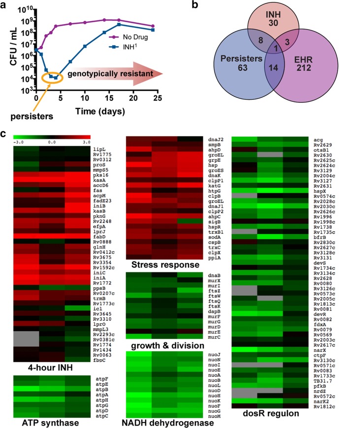
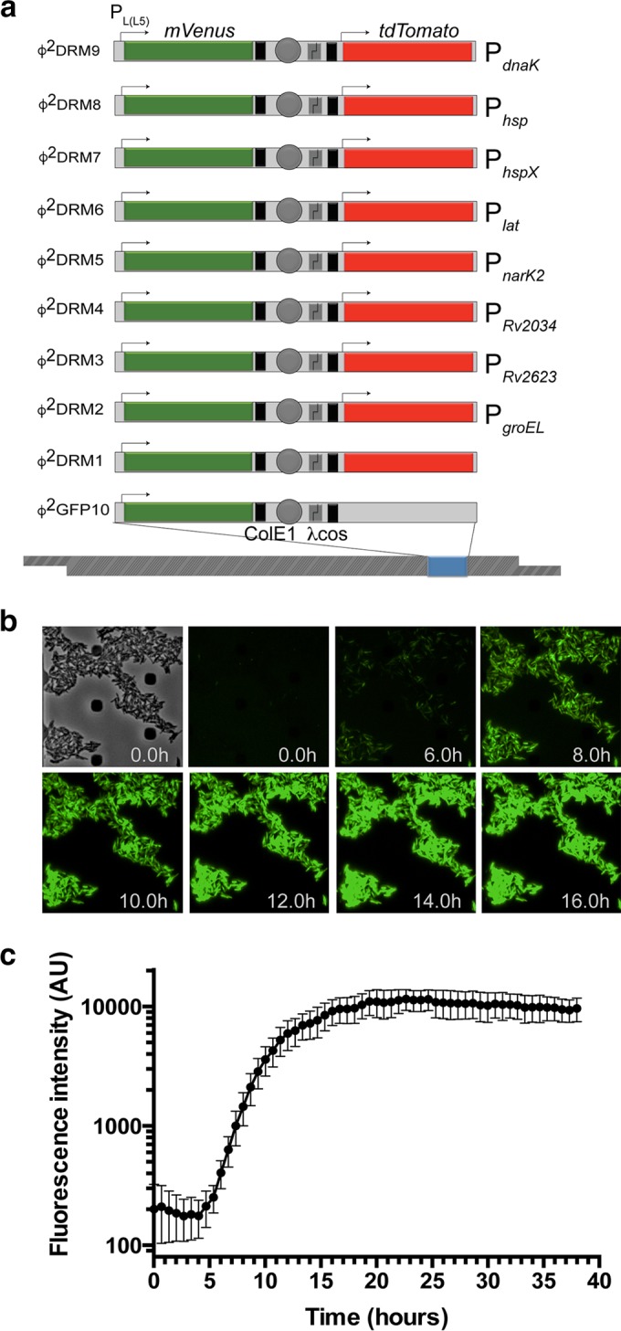
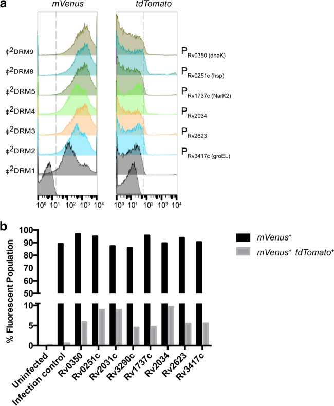
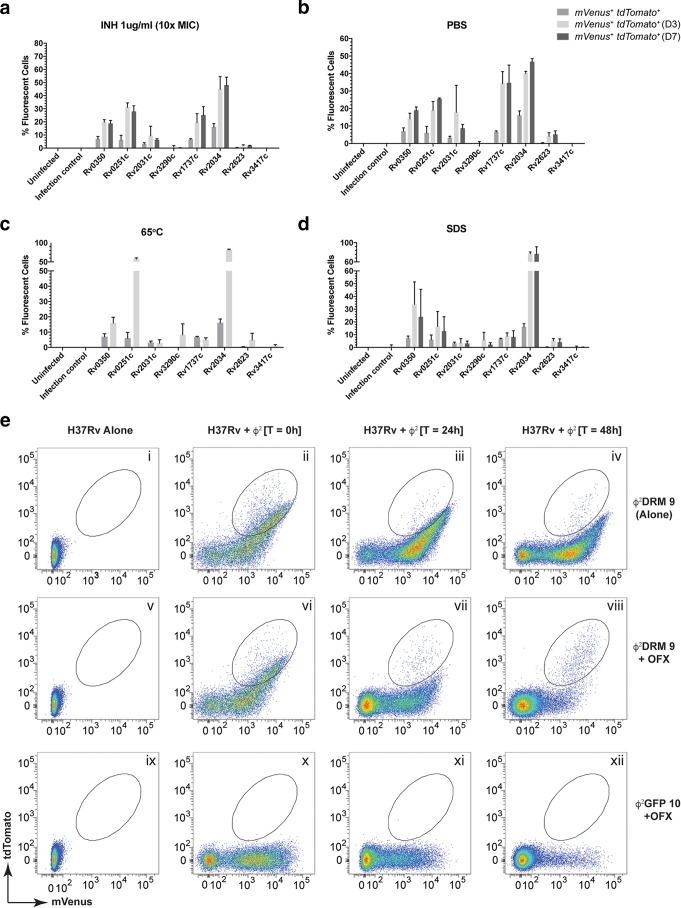
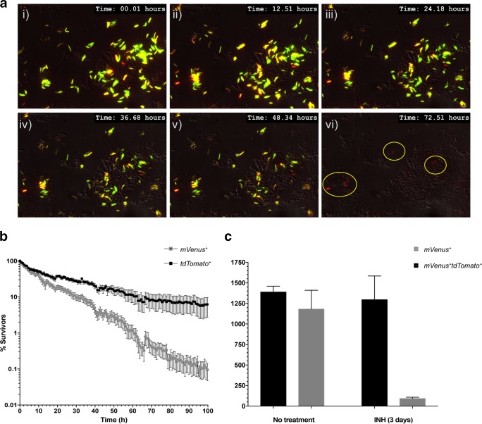
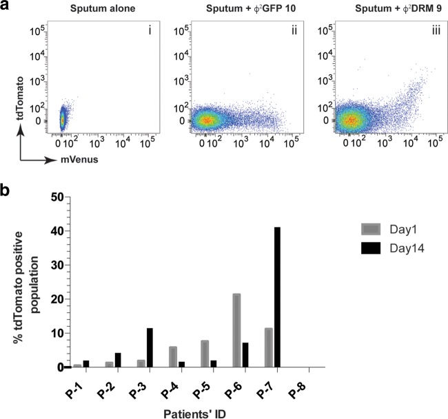
Persisters are the minor subpopulation of bacterial cells that lack alleles conferring resistance to a specific bactericidal antibiotic but can survive otherwise lethal concentrations of that antibiotic. In infections with Mycobacterium tuberculosis, such persisters underlie the need for long-term antibiotic therapy and contribute to treatment failure in tuberculosis cases. Here, we demonstrate the value of dual-reporter mycobacteriophages (Φ2DRMs) for characterizing M. tuberculosis persisters. The addition of isoniazid (INH) to exponentially growing M. tuberculosis cells consistently resulted in a 2- to 3-log decrease in CFU within 4 days, and the remaining ≤1% of cells, which survived despite being INH sensitive, were INH-tolerant persisters with a distinct transcriptional profile. We fused the promoters of several genes upregulated in persisters to the red fluorescent protein tdTomato gene in Φ2GFP10, a mycobacteriophage constitutively expressing green fluorescent protein (GFP), thus generating Φ2DRMs. A population enriched in INH persisters exhibited strong red fluorescence, by microscopy and flow cytometry, using a Φ2DRM with tdTomato controlled from the dnaK promoter. Interestingly, we demonstrated that, prior to INH exposure, a population primed for persistence existed in M. tuberculosis cells from both cultures and human sputa and that this population was highly enriched following INH exposure. We conclude that Φ2DRMs provide a new tool to identify and quantitate M. tuberculosis persister cells.

Importance: Tuberculosis (TB) is again the leading cause of death from a single infectious disease, having surpassed HIV. The recalcitrance of the TB pandemic is largely due to the ability of the pathogen Mycobacterium tuberculosis to enter a persistent state in which it is less susceptible to antibiotics and immune effectors, necessitating lengthy treatment. It has been difficult to study persister cells, as we have lacked tools to isolate these rare cells. In this article, we describe the development of dual-reporter mycobacteriophages that encode a green fluorescent marker of viability and in which the promoters of genes we have identified as induced in the persister state are fused to a gene encoding a red fluorescent protein. We show that these tools can identify heterogeneity in a cell population that correlates with propensity to survive antibiotic treatment and that the proportions of these subpopulations change in M. tuberculosis cells within human sputum during the course of treatment.


Figures

FIG 1
FIG 1
INH-sensitive survivors of INH treatment induce a transcriptional program consistent with a slow-growing stress-resistant state. (a) Treatment of M. tuberculosis with INH kills >99% cells in 4 days, leaving a largely INH-sensitive population from which an INH-resistant population emerges. (b) Survivors of 4 days of INH treatment induce genes consistent with INH sensitivity; the persister stimulon includes a subset of the enduring hypoxic response (EHR). Persister stimulon is defined here narrowly as genes statistically significantly induced >twofold at a false discovery rate (FDR) of <0.4% by SAM. EHR and INH stimulons were similarly defined by Rustad et al. (21) and Fu (18). (c) Heat maps of the persister stimulon in biological triplicates mapped onto genes induced by 4-h INH treatment, stress-responsive genes, genes involved in growth and cell division, the ATP synthase, the type I NADH dehydrogenase, and the dosR regulon. The scale indicates ±3-log2-fold changes relative to expression at day 0.
FIG 2
FIG 2
Design and fluorescence analysis of Φ2DRM constructs. (a) Schematics of the various Φ2DRMs used in the study. The expression of mVenus in Φ2GFP10 and all Φ2DRMs is driven from the constitutive PL(L5) promoter. A unique promoter, listed on the right of each schematic, drives the expression of tdTomato in each Φ2DRM. Transcription terminators, placed upstream from tdTomato and downstream from mVenus to avoid transcriptional read-through, are indicated by black boxes. Arrows indicate the relative positions of promoters. (b) Expression kinetics of mVenus from PL(L5) in M. tuberculosis by time lapse microscopy. Expression of mVenus was detectable after approximately 4 h of phage infection. (c) Quantitative plot of the kinetics of mVenus expression over time. Fluorescence intensity increased progressively over time, plateauing approximately 18 h after the addition of phage. AU, arbitrary units. The error bars represent SD of fluorescent intensity measured at three independent fields.
FIG 3
FIG 3
Fraction of M. tuberculosis cells expressing “persister upregulated genes” in logarithmic-phase cultures. (a) Left, log-phase M. tuberculosis cells infected with representative Φ2DRMs show similar distributions of mVenus expression; right, fractions of the parent populations expressing “persister upregulated genes” quantified by tdTomato expression. The promoter driving tdTomato expression is indicated on the right of each histogram. (b) Plot representing the percentages of mVenus+ and mVenus+ tdTomato+ populations after infection with individual Φ2DRMs. The double-positive cells indicate the fractions of cells expressing “persister upregulated genes” in logarithmic-phase M. tuberculosis. Promoters of the following genes were used to regulate tdTomato expression (the gene name or predicted function is indicated [http://tuberculist.epfl.ch/index.html]): Rv0350, dnaK; Rv0251c, hsp; Rv2031c, hspX, acr; Rv3290c, lat; Rv1737c, narK2; Rv2034, Ars repressor protein gene; Rv2623, universal stress protein family protein gene; Rv3417c, groEL1.
FIG 4
FIG 4
Activation of persister upregulated gene promoters in M. tuberculosis cells under stress. (a to d) The stress conditions used were INH (a), starvation (b), high temperature (c), and SDS (d). Gene promoters controlling tdTomato are indicated on each x axis. The percentage of the population turning on a specific promoter is plotted on each y axis. Error bars represent SEM from 3 independent experiments. For full gene names, see the legend to Fig. 3b. (e) Use of flow cytometry to monitor enrichment of a specific M. tuberculosis population after ofloxacin (OFX) treatment. Logarithmic-phase H37Rv was treated with OFX for 24 and 48 h and infected with either Φ2DRM9 (i to viii) or Φ2GFP10 (ix to xii). Cell-only controls (uninfected H37Rv) are shown in panels i, v, and ix. At time t0, a majority of the infected cells fluoresced green (ii, vi, and x). After 24 h of OFX treatment, the relative percentages of the fluorescent populations are reduced in the treated samples (compare panel vi to vii and x to xi). The same trend continued and the fluorescent populations were further reduced in the treated samples at 48 h (compare panel vi to viii and x to xii). At the end of 48 h, an mVenus+ tdTomato+ subpopulation (high tdTomato, low to medium mVenus) survives the treatment (circled, panel viii) and is the only fluorescent population remaining (compare panel iv to viii). The fluorescent population did not decrease in the untreated control cells (compare panels ii, iii, and iv).
FIG 5
FIG 5
Identification of “likely persister” population in M. tuberculosis by Φ2DRMs. (a) Time lapse microscopy. Φ2DRM9-infected M. tuberculosis cells were treated with INH on a microfluidic device and imaged every 15 min for 3 days. Representative time frames from the time lapse movie at 12-h intervals are shown. At treatment initiation, most cells were mVenus+, with a few mVenus+ tdTomato+ cells. At the end of treatment, all surviving cells (circled) were tdTomato+ with very low mVenus fluorescence. (b) Survival kinetics of M. tuberculosis cells expressing high levels of dnaK promoter activity (black squares) in comparison to the bulk population (grey squares). The plot indicates that cells expressing dnaK were able to resist INH treatment longer than cells not expressing dnaK. Error bars represent SEM from 5 independent experiments. (c) Fifty thousand M. tuberculosis cells infected with Φ2DRM9 were sorted by flow cytometry for mVenus+ and mVenus+ tdTomato+ cells before and after INH treatment. The viable counts in the sorted samples were determined by CFU plating. M. tuberculosis cells expressing tdTomato survive preferentially after INH treatment in comparison to cells that express only mVenus. Error bars represent SEM from 3 independent experiments.
FIG 6
FIG 6
Identification of likely persister populations in sputum samples. (a) Sputum samples obtained from TB patients were infected with Φ2DRM9 and analyzed by flow cytometry. Phage Φ2GFP10 was used as an infection control. (b) Plot representing the fractions of mVenus+ tdTomato+ populations in sputum samples of TB patients at the time of disease presentation and after 2 weeks of TB treatment.

References

    1. Hobby GL, Meyer K, Chaffee E. 1942. Observations on the mechanism of action of penicillin. Exp Biol Med 50:281–285. doi: 10.3181/00379727-50-13773. - DOI
    1. Hobby GL, Meyer K, Chaffee E. 1942. Activity of penicillin in vitro. Exp Biol Med 50:277–280. doi: 10.3181/00379727-50-13772. - DOI
    1. Bigger J. 1944. Treatment of staphylococcal infections with penicillin by intermittent sterilisation. Lancet 244:497–500. doi: 10.1016/S0140-6736(00)74210-3. - DOI
    1. McCune RM Jr, Tompsett R. 1956. Fate of Mycobacterium tuberculosis in mouse tissues as determined by the microbial enumeration technique. I. The persistence of drug-susceptible tubercle bacilli in the tissues despite prolonged antimicrobial therapy. J Exp Med 104:737–762. doi: 10.1084/jem.104.5.737. - DOI - PMC - PubMed
    1. McCune RM, Feldmann FM, Lambert HP, McDermott W. 1966. Microbial persistence. I. The capacity of tubercle bacilli to survive sterilization in mouse tissues. J Exp Med 123:445–468. doi: 10.1084/jem.123.3.445. - DOI - PMC - PubMed

Publication types

MeSH terms

Substances