. 2016 Dec 16;91(1):e01813-16.
doi: 10.1128/JVI.01813-16. Print 2017 Jan 1.

CLEC5A-Mediated Enhancement of the Inflammatory Response in Myeloid Cells Contributes to Influenza Virus Pathogenicity In Vivo

Affiliations

CLEC5A-Mediated Enhancement of the Inflammatory Response in Myeloid Cells Contributes to Influenza Virus Pathogenicity In Vivo

Ooiean Teng et al. J Virol. .

Abstract

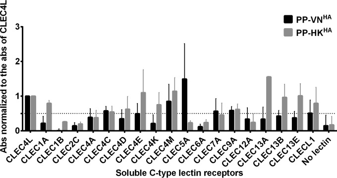
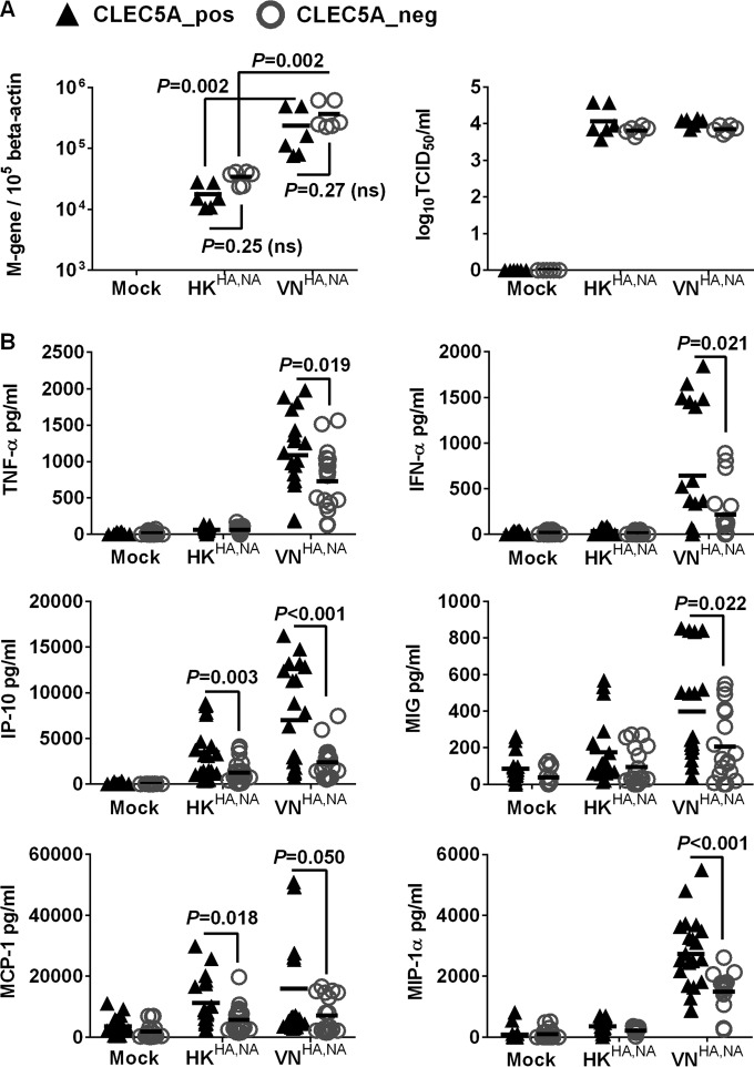
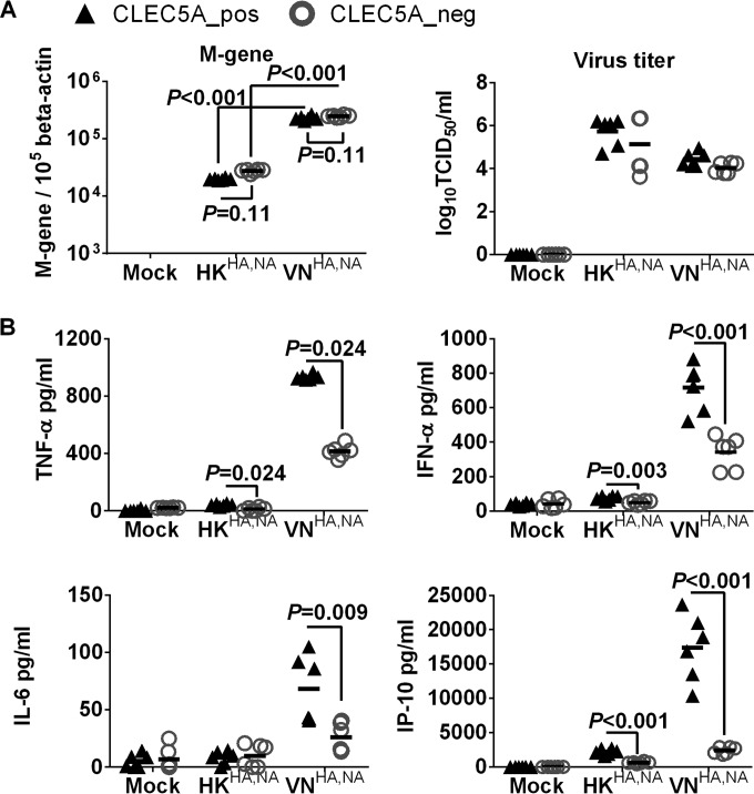
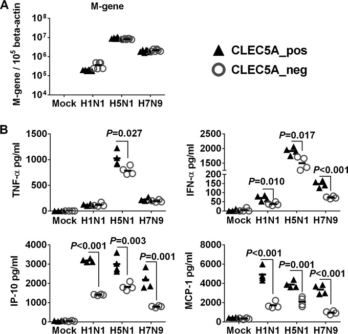
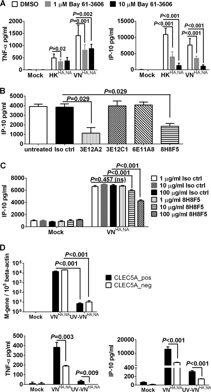
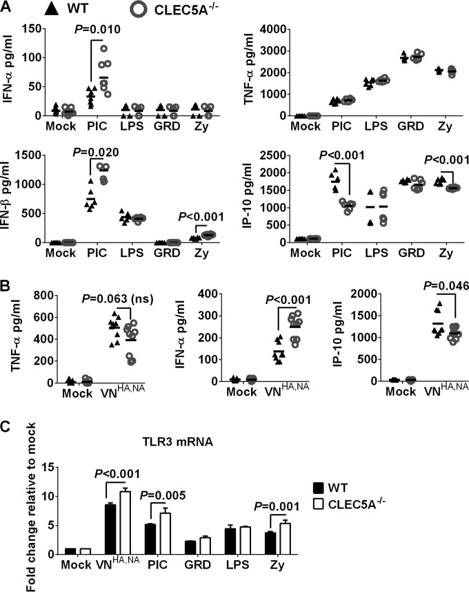
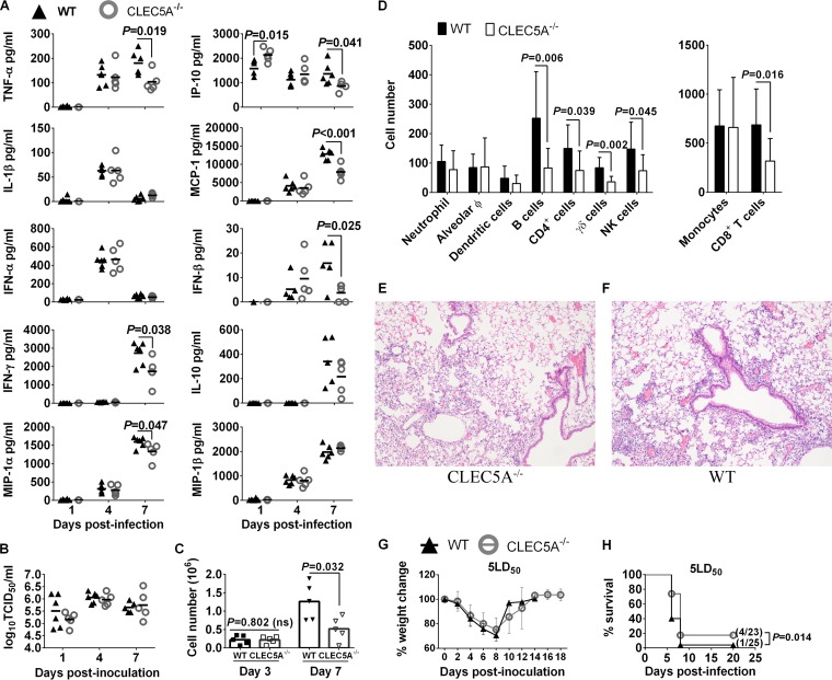
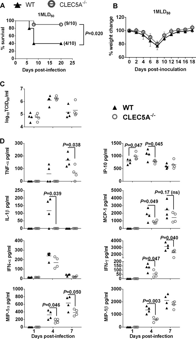
Human infections with influenza viruses exhibit mild to severe clinical outcomes as a result of complex virus-host interactions. Induction of inflammatory mediators via pattern recognition receptors may dictate subsequent host responses for pathogen clearance and tissue damage. We identified that human C-type lectin domain family 5 member A (CLEC5A) interacts with the hemagglutinin protein of influenza viruses expressed on lentiviral pseudoparticles through lectin screening. Silencing CLEC5A gene expression, blocking influenza-CLEC5A interactions with anti-CLEC5A antibodies, or dampening CLEC5A-mediated signaling using a spleen tyrosine kinase inhibitor consistently reduced the levels of proinflammatory cytokines produced by human macrophages without affecting the replication of influenza A viruses of different subtypes. Infection of bone marrow-derived macrophages from CLEC5A-deficient mice showed reduced levels of tumor necrosis factor alpha (TNF-α) and IP-10 but elevated alpha interferon (IFN-α) compared to those of wild-type mice. The heightened type I IFN response in the macrophages of CLEC5A-deficient mice was associated with upregulated TLR3 mRNA after treatment with double-stranded RNA. Upon lethal challenges with a recombinant H5N1 virus, CLEC5A-deficient mice showed reduced levels of proinflammatory cytokines, decreased immune cell infiltration in the lungs, and improved survival compared to the wild-type mice, despite comparable viral loads noted throughout the course of infection. The survival difference was more prominent at a lower dose of inoculum. Our results suggest that CLEC5A-mediated enhancement of the inflammatory response in myeloid cells contributes to influenza pathogenicity in vivo and may be considered a therapeutic target in combination with effective antivirals. Well-orchestrated host responses together with effective viral clearance are critical for optimal clinical outcome after influenza infections.

Importance: Multiple pattern recognition receptors work in synergy to sense viral RNA or proteins synthesized during influenza replication and mediate host responses for viral control. Well-orchestrated host responses may help to maintain the inflammatory response to minimize tissue damage while inducing an effective adaptive immune response for viral clearance. We identified that CLEC5A, a C-type lectin receptor which has previously been reported to mediate flavivirus-induced inflammatory responses, enhanced induction of proinflammatory cytokines and chemokines in myeloid cells after influenza infections. CLEC5A-deficient mice infected with influenza virus showed reduced inflammation in the lungs and improved survival compared to that of the wild-type mice despite comparable viral loads. The survival difference was more prominent at a lower dose of inoculum. Collectively, our results suggest that dampening CLEC5A-mediated inflammatory responses in myeloid cells reduces immunopathogenesis after influenza infections.

Keywords: C-type lectins; CLEC5A; influenza virus; macrophages; spleen tyrosine kinase (Syk).


Figures

FIG 1
FIG 1
Identification of CLEC5A as potential PRR for influenza virus. The relative binding intensity of the pseudoparticles expressing influenza HA derived from H5N1 or H1N1 viruses to a panel of soluble human CLRs is shown. Briefly, 0.5 μg of soluble human CLRs was coated on the plate, followed by incubation with the HA-expressing pseudoparticles. The bound pseudoparticles were detected by anti-H5 or anti-H1 antibodies. The absorbance (Abs) obtained was normalized to the absorbance of CLEC4L. The dotted line represents 50% binding signal compared to that of CLEC4L. The data shown are the means ± standard deviations (SD) from 8 replicates from 4 independent experiments.
FIG 2
FIG 2
CLEC5A mediates enhanced proinflammatory cytokine and chemokine induction in human M-Mϕ after influenza virus infection. M-Mϕ differentiated from the PBMC of 8 independent donors were transfected with CLEC5A gene-specific or nontargeting siRNA, followed by cell sorting to obtain the CLEC5A-positive (CLEC5A_pos) and CLEC5A-negative (CLEC5A_neg) populations. The macrophages were then infected with HKHA,NA or VNHA,NA recombinant viruses at an MOI of 2 for 24 h to determine viral M gene copy numbers in infected M-Mϕ and viral titers in culture supernatants (log10 TCID50/ml in MDCK cells) (A) and proinflammatory cytokines and chemokines in culture supernatant (means ± SD, in pg/ml) (B). P values from Mann-Whitney tests are shown.
FIG 3
FIG 3
CLEC5A mediates enhanced proinflammatory cytokine and chemokine induction in human GM-Mϕ after influenza virus infection. GM-Mϕ differentiated from the PBMC of 2 independent donors were transfected with CLEC5A gene-specific or nontargeting siRNA, followed by cell sorting to obtain the CLEC5A-positive (CLEC5A_pos) and CLEC5A-negative (CLEC5A_neg) populations. The macrophages were then infected with HKHA,NA or VNHA,NA recombinant viruses at an MOI of 2 for 24 h to determine viral M gene copy numbers in infected GM-Mϕ and viral titers in culture supernatant (log10 TCID50/ml in MDCK cells) (A) and proinflammatory cytokines and chemokines in culture supernatants (means ± SD, in pg/ml) (B). P values from Mann-Whitney tests are shown.
FIG 4
FIG 4
CLEC5A-mediated proinflammatory response in human M-Mϕ after infections with influenza viruses of H1N1, H5N1, and H7N9 subtypes. Human M-Mϕ differentiated from PBMC of 2 independent donors were infected with A/Hong Kong/54/98 (H1N1), A/Vietnam/1203/04 (H5N1), or A/Shanghai/2/13 (H7N9) influenza viruses at an MOI of 2 for 24 h to determine viral M gene copy numbers in infected M-Mϕ (A) and proinflammatory cytokines and chemokines in culture supernatants (means ± SD, in pg/ml) (B). P values from Mann-Whitney tests are shown.
FIG 5
FIG 5
Blocking CLEC5A-mediated signaling with Syk inhibitor (Bay 61-3606) and CLEC5A antagonizing antibodies reduced inflammatory response in human M-Mϕ after influenza virus infection. (A) Human M-Mϕ differentiated from the PBMC of 3 independent donors were treated with dimethyl sulfoxide or the Syk inhibitor Bay 61-3606 prior to and throughout the course of infection with HKHA,NA or VNHA,NA virus at an MOI of 2 to determine the TNF-α and IP-10 concentrations (means ± SD, in pg/ml) from culture supernatants at 24 h postinfection. (B) Human M-Mϕ differentiated from the PBMC of 4 independent donors were incubated with 1 μg/ml of murine IgG1 isotype control antibody (Iso ctrl) or clones of anti-human CLEC5A antibodies, which were previously shown to possess antagonizing activity against DV or JEV infections, prior to and throughout the course of infection with VNHA,NA virus at an MOI of 2 to determine the level of IP-10 (means ± SD, in pg/ml) from culture supernatants at 24 h postinfection. (C) Human M-Mϕ differentiated from the PBMC of 4 independent donors were incubated with increasing concentrations of murine IgG1 isotype control antibody or the anti-CLEC5A antibody (clone 8H8F5) prior to and throughout the course of infection with VNHA,NA virus at an MOI of 2 to determine the level of IP-10 (means ± SD, in pg/ml) from culture supernatants at 24 h postinfection. ns, not significant. (D) M-Mϕ differentiated from the PBMC of 2 independent donors were transfected with CLEC5A gene-specific or nontargeting siRNA followed by cell sorting to obtain the CLEC5A-positive (CLEC5A_pos) and CLEC5A-negative (CLEC5A_neg) populations. M-Mϕ were infected with VNHA,NA or UV-inactivated VNHA,NA (UV-VNHA,NA) at an MOI of 2. Viral M gene copy numbers and levels of TNF-α and IP-10 (means ± SD, in pg/ml) in culture supernatant were determined at 24 h postinfection. P values from Mann-Whitney tests are shown.
FIG 6
FIG 6
(A) Inflammatory response in BMM derived from CLEC5A−/− or WT mice after incubation with TLR agonists or after influenza infection. BMM derived from CLEC5A−/− or WT mice were treated with gardiquimod (GRD; 10 μg/ml), lipopolysaccharide (LPS; 100 ng/ml), poly(I·C) (PIC; 50 μg/ml), or zymosan (Zy; 10 μg/ml) for 24 h to determine the level of cytokines and chemokines in the supernatant (means ± SD, in pg/ml, derived from 6 replicates of BMM obtained from 2 mice). (B) BMM derived from CLEC5A−/− or WT mice were infected with VNHA,NA at an MOI of 2 for 24 h to determine the levels of cytokines and chemokines in the supernatant (means ± SD, in pg/ml, derived from 9 replicates of BMM obtained from 3 mice). (C) Copy number of TLR3 mRNA in BMM after activation with TLR agonist or influenza infection at an MOI of 2 for 24 h (means ± SD from 4 replicates of BMM obtained from 2 mice). P values from Mann-Whitney tests are shown.
FIG 7
FIG 7
Lower levels of proinflammatory cytokines and reduced level of immune cell infiltration in the lungs of CLEC5A−/− mice compared to the WT mice after lethal challenge of 5 MLD50 of VNHA,NA virus. WT and CLEC5A−/− mice were infected with 5 MLD50 of VNHA,NA intranasally; mouse lungs were collected at days 1, 4, and 7 postinfection (n = 5 or 6 per group at each time point) to determine the concentrations of cytokines and chemokines (means ± SD, in pg/ml) (A) and virus titers (means ± SD, in log10 TCID50/ml) (B). (C and D) BAL fluid was collected from WT and CLEC5A−/− mice (n = 10 per group at each time point) to monitor the total cell count (means ± SD) detected on days 3 and 7 postinfection (C) and the immunophenotyping result on day 7 postinfection using surface markers (P values from Mann-Whitney tests are shown) (D). Hematoxylin and eosin staining of the CLEC5A−/− (E) and WT (F) mouse lungs at day 7 postinfection with 5 MLD50 of VNHA,NA (n = 2 per group). (G and H) Changes in body weight (G) and survival (H) of WT and CLEC5A−/− mice after challenge with 5 MLD50 (P value from log-rank test is shown).
FIG 8
FIG 8
CLEC5A−/− mice were protected from the sublethal challenge of VNHA,NA virus by reducing the level of proinflammatory cytokine in the lungs. WT and CLEC5A−/− mice were infected with 1 MLD50 of VNHA,NA intranasally. Mouse survival (A) and body weight changes (B) were monitored every other day; P values from log-rank tests are shown. (C and D) Viral titer (means ± SD, in log10 TCID50/ml) (C) and cytokine and chemokine levels (means ± SD, in pg/ml) (D) detected in the mouse lungs at day 1, 4, and 7 postinfection (n = 4 per group at each time point); P values from Mann-Whitney tests are shown.

References

    1. Everitt AR, Clare S, Pertel T, John SP, Wash RS, Smith SE, Chin CR, Feeley EM, Sims JS, Adams DJ, Wise HM, Kane L, Goulding D, Digard P, Anttila V, Baillie JK, Walsh TS, Hume DA, Palotie A, Xue Y, Colonna V, Tyler-Smith C, Dunning J, Gordon SB, GenISIS Investigators, MOSAIC Investigators, Smyth RL, Openshaw PJ, Dougan G, Brass AL, Kellam P. 2012. IFITM3 restricts the morbidity and mortality associated with influenza. Nature 484:519–523. doi:10.1038/nature10921. - DOI - PMC - PubMed
    1. Haller O, Staeheli P, Schwemmle M, Kochs G. 2015. Mx GTPases: dynamin-like antiviral machines of innate immunity. Trends Microbiol 23:154–163. doi:10.1016/j.tim.2014.12.003. - DOI - PubMed
    1. Fukuyama S, Kawaoka Y. 2011. The pathogenesis of influenza virus infections: the contributions of virus and host factors. Curr Opin Immunol 23:481–486. doi:10.1016/j.coi.2011.07.016. - DOI - PMC - PubMed
    1. Kash JC, Taubenberger JK. 2015. The role of viral, host, and secondary bacterial factors in influenza pathogenesis. Am J Pathol 185:1528–1536. doi:10.1016/j.ajpath.2014.08.030. - DOI - PMC - PubMed
    1. Iwasaki A, Pillai PS. 2014. Innate immunity to influenza virus infection. Nat Rev Immunol 14:315–328. doi:10.1038/nri3665. - DOI - PMC - PubMed

MeSH terms