

A Novel Porcine Circovirus Distantly Related to Known Circoviruses Is Associated with Porcine Dermatitis and Nephropathy Syndrome and Reproductive Failure
- PMID: 27795441
- PMCID: PMC5165205
- DOI: 10.1128/JVI.01879-16
A Novel Porcine Circovirus Distantly Related to Known Circoviruses Is Associated with Porcine Dermatitis and Nephropathy Syndrome and Reproductive Failure
Abstract
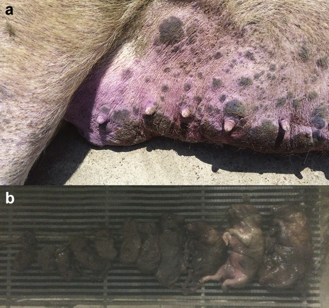
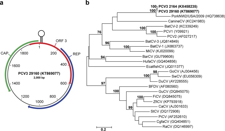
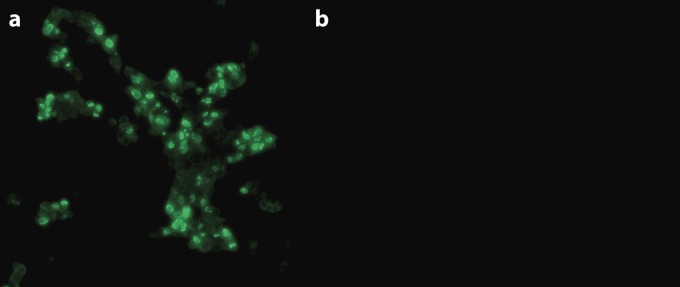
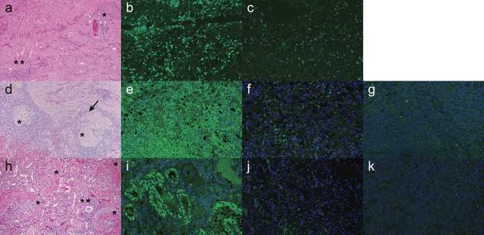
Porcine circovirus-associated disease (PCVAD) is clinically manifested by postweaning multisystemic wasting syndrome (PMWS), respiratory and enteric disease, reproductive failure, and porcine dermatitis and nephropathy syndrome (PDNS). Porcine circovirus 2 (PCV2) is an essential component of PCVAD, although an etiologic role in PDNS is not well established. Here, a novel circovirus, designated porcine circovirus 3 (PCV3), was identified in sows that died acutely with PDNS-like clinical signs. The capsid and replicase proteins of PCV3 are only 37% and 55% identical to PCV2 and bat circoviruses, respectively. Aborted fetuses from sows with PDNS contained high levels of PCV3 (7.57 × 10⁷ genome copies/ml), and no other viruses were detected by PCR and metagenomic sequencing. Immunohistochemistry (IHC) analysis of sow tissue samples identified PCV3 antigen in skin, kidney, lung, and lymph node samples localized in typical PDNS lesions, including necrotizing vasculitis, glomerulonephritis, granulomatous lymphadenitis, and bronchointerstitial pneumonia. Further study of archived PDNS tissue samples that were negative for PCV2 by IHC analysis identified 45 of 48 that were PCV3 positive by quantitative PCR (qPCR), with 60% of a subset also testing positive for PCV3 by IHC analysis. Analysis by qPCR of 271 porcine respiratory disease diagnostic submission samples identified 34 PCV3-positive cases (12.5%), and enzyme-linked immunosorbent assay detection of anti-PCV3 capsid antibodies in serum samples found that 46 (55%) of 83 samples tested were positive. These results suggest that PCV3 commonly circulates within U.S. swine and may play an etiologic role in reproductive failure and PDNS. Because of the high economic impact of PCV2, this novel circovirus warrants further studies to elucidate its significance and role in PCVAD.
Importance: While porcine circovirus 2 (PCV2) was first identified in sporadic cases of postweaning multisystemic wasting syndrome in Canada in the early 1990s, an epidemic of severe systemic disease due to PCV2 spread worldwide in the ensuing decade. Despite being effectively controlled by commercial vaccines, PCV2 remains one of the most economically significant viruses of swine. Here, a novel porcine circovirus (PCV3) that is distantly related to known circoviruses was identified in sows with porcine dermatitis and nephropathy syndrome (PDNS) and reproductive failure. PCV2, which has previously been associated with these clinical presentations, was not identified. High levels of PCV3 nucleic acid were observed in aborted fetuses by quantitative PCR, and PCV3 antigen was localized in histologic lesions typical of PDNS in sows by immunohistochemistry (IHC) analysis. PCV3 was also identified in archival PDNS diagnostic samples that previously tested negative for PCV2 by IHC analysis. The emergence of PCV3 warrants further investigation.
Keywords: abortion; porcine circovirus; porcine dermatitis and nephropathy syndrome.
Copyright © 2016 American Society for Microbiology.
Figures
References
-
- Biagini P, Bendinelli M, Hino S, Kakkola L, Mankertz A, Niel C, Okamoto H, Raidal S, Teo CG, Todd D. 2012. Circoviridae, p 343–349. In King AMQ, Adams MJ, Carstens EB, Lefkowitz EJ (ed), Virus taxonomy: classification and nomenclature of viruses. Ninth report of the International Committee on Taxonomy of Viruses. Elsevier Academic Press, New York, NY.
-
- Li L, Kapoor A, Slikas B, Bamidele OS, Wang C, Shaukat S, Masroor MA, Wilson ML, Ndjango J-BN, Peeters M, Gross-Camp ND, Muller MN, Hahn BH, Wolfe ND, Triki H, Bartkus J, Zaidi SZ, Delwart E. 2010. Multiple diverse circoviruses infect farm animals and are commonly found in human and chimpanzee feces. J Virol 84:1674–1682. doi: 10.1128/JVI.02109-09. - DOI - PMC - PubMed
-
- Garigliany M-M, Börstler J, Jöst H, Badusche M, Desmecht D, Schmidt-Chanasit J, Cadar D. 2015. Characterization of a novel circo-like virus in Aedes vexans mosquitoes from Germany: evidence for a new genus within the family Circoviridae. J Gen Virol 96:915–920. doi: 10.1099/vir.0.000036. - DOI - PubMed
MeSH terms
Substances
LinkOut - more resources
Full Text Sources
Other Literature Sources
Medical
