. 2016:2016:3453059.
doi: 10.1155/2016/3453059. Epub 2016 Oct 4.

Cox-2 Inhibition Protects against Hypoxia/Reoxygenation-Induced Cardiomyocyte Apoptosis via Akt-Dependent Enhancement of iNOS Expression

Affiliations

Cox-2 Inhibition Protects against Hypoxia/Reoxygenation-Induced Cardiomyocyte Apoptosis via Akt-Dependent Enhancement of iNOS Expression

Lei Pang et al. Oxid Med Cell Longev. 2016.

Abstract

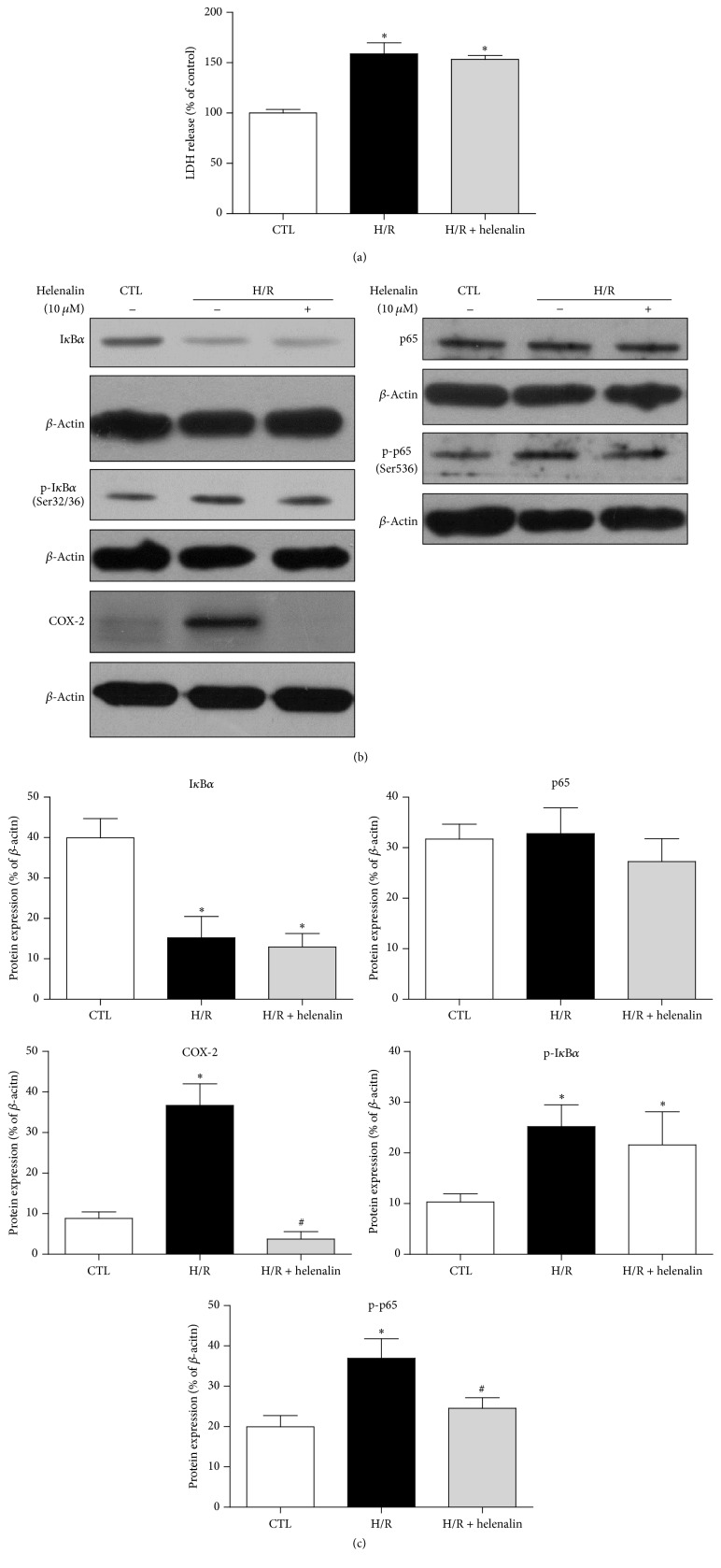
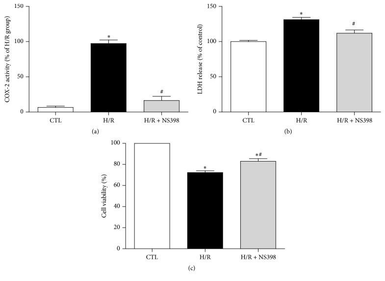
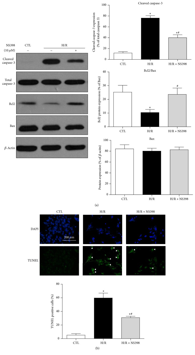
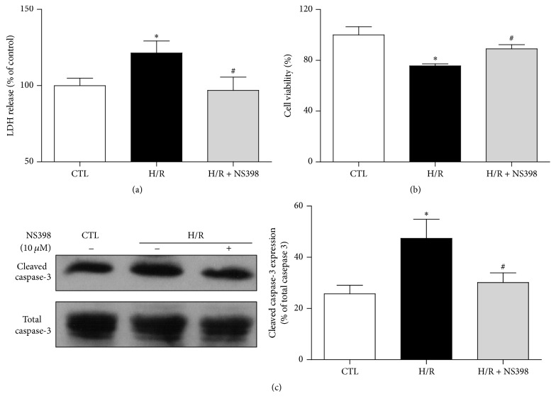
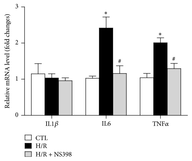
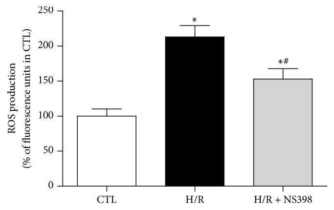
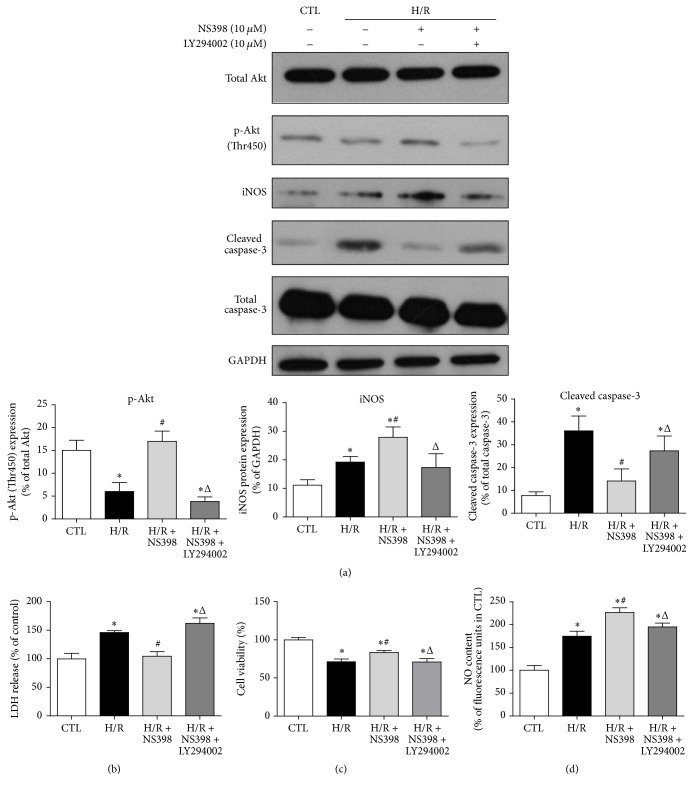
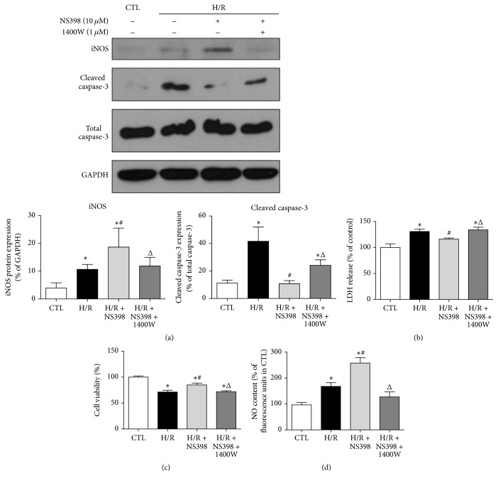
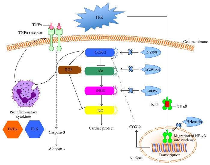
The present study explored the potential causal link between ischemia-driven cyclooxygenase-2 (COX-2) expression and enhanced apoptosis during myocardial ischemia/reperfusion (I/R) by using H9C2 cardiomyocytes and primary rat cardiomyocytes subjected to hypoxia/reoxygenation (H/R). The results showed that H/R resulted in higher COX-2 expression than that of controls, which was prevented by pretreatment with Helenalin (NFκB specific inhibitor). Furthermore, pretreatment with NS398 (COX-2 specific inhibitor) significantly attenuated H/R-induced cell injury [lower lactate dehydrogenase (LDH) leakage and enhanced cell viability] and apoptosis (higher Bcl2 expression and lower level of cleaved caspases-3 and TUNEL-positive cells) in cardiomyocytes. The amelioration of posthypoxic apoptotic cell death was paralleled by significant attenuation of H/R-induced increases in proinflammatory cytokines [interleukin 6 (IL6) and tumor necrosis factor (TNFα)] and reactive oxygen species (ROS) production and by higher protein expression of phosphorylated Akt and inducible nitric oxide synthase (iNOS) and enhanced nitric oxide production. Moreover, the application of LY294002 (Akt-specific inhibitor) or 1400W (iNOS-selective inhibitor) cancelled the cellular protective effects of NS398. Findings from the current study suggest that activation of NFκB during cardiomyocyte H/R induces the expression of COX-2 and that higher COX-2 expression during H/R exacerbates cardiomyocyte H/R injury via mechanisms that involve cross talks among inflammation, ROS, and Akt/iNOS/NO signaling.


Figures

Figure 1
Figure 1
Activation of NFκB mediates the H/R-induced expression of COX-2 in H9C2 cardiomyocytes. (a) The release of LDH (cell injury marker) and (b) representative original Western blots of IκBα, phosphorylated IκBα (S32/36), p65, phosphorylated p65 (S536), and COX-2 in H9C2 cardiomyocytes with or without H/R stimulation (6 h hypoxia followed by 12 h reoxygenation) in the presence or absence of Helenalin (NFκB specific inhibitor, 10 μM, 2 h). (c) Quantification of these proteins after normalization to β-actin. Data are shown as means ± SEM; P < 0.05 CTL versus H/R or H/R + Helenalin, # P < 0.05 H/R versus H/R + Helenalin (one-way ANOVA followed by Tukey's test in (a) and nonparametric Mann–Whitney U test in (c)); n = 4 per group.
Figure 2
Figure 2
Inhibition of COX-2 activity attenuates H/R-induced cell injury. (a) The COX-2 activity, (b) the release of LDH, and (c) cell viability in H9C2 cardiomyocytes with or without H/R treatment in the presence or absence of NS398 (COX-2 specific inhibitor, 10 μM, 1 h). COX-2 activity was expressed against that in H/R group. The LDH leakage and cell viability were expressed against those in CTL group. Data are shown as means ± SEM; P < 0.05 CTL versus H/R or H/R + NS398, # P < 0.05 H/R versus H/R + NS398 (one-way ANOVA followed by Tukey's test); n = 5 per group.
Figure 3
Figure 3
Inhibition of COX-2 activity alleviates H/R-induced cell apoptosis in H9C2 cardiomyocytes. (a) Representative original Western blots of apoptosis related proteins (cleaved caspase-3, total caspase-3, Bcl2, and Bax) in H9C2 cardiomyocytes with or without H/R stimulation in the presence or absence of NS398. Protein presence of cleaved caspase-3 and Bcl2 was normalized to total caspase-3 and Bax, respectively. (b) Representative fluorescence microscopy images of TUNEL staining. White triangle points to positive staining cells (green). DAPI (blue) indicates the nucleus staining. Magnification ×200, scale bars: 100 μm. TUNEL-positive cells% = TUNEL-positive cell numbers/total cell numbers × 100%. Data are shown as means ± SEM; P < 0.05 CTL versus H/R or H/R + NS398, # P < 0.05 H/R versus H/R + NS398 (nonparametric Mann–Whitney U test in (a) and one-way ANOVA followed by Tukey's test in (b)); n = 5 per group.
Figure 4
Figure 4
Inhibition of COX-2 activity attenuates H/R-induced cell injury and apoptosis in primary adult rat cardiomyocytes. (a) The release of LDH and (b) cell viability in primary adult rat cardiomyocytes with or without H/R treatment (45 min hypoxia followed by 2 h reoxygenation) in the presence or absence of NS398. The LDH leakage and cell viability were expressed against those in CTL group. (c) Representative original Western blots of cleaved caspase-3 and total caspase-3 in primary adult rat cardiomyocytes with or without H/R treatment in the presence or absence of NS398. Quantification of cleaved caspase-3 was normalized to total caspase-3. Data are shown as means ± SEM; P < 0.05 CTL versus H/R, # P < 0.05 H/R versus H/R + NS398 (one-way ANOVA followed by Tukey's test in (a), (b) and nonparametric Mann–Whitney U test in (c)); n = 5 per group.
Figure 5
Figure 5
Inhibition of COX-2 activity reduced H/R-induced proinflammatory cytokines expression in H9C2 cardiomyocytes. mRNA expression of proinflammatory genes (IL1β, IL6, and TNFα) in H9C2 cardiomyocytes subjected to H/R stimulation in the presence or absence of NS398. mRNA levels are expressed against those in H9C2 cardiomyocytes with no stimulation. Data are shown as means ± SEM; P < 0.05 CTL versus H/R or H/R + NS398, # P < 0.05 H/R versus H/R + NS398 (one-way ANOVA followed by Tukey's test); n = 6 per group.
Figure 6
Figure 6
Inhibition of COX-2 activity reduced H/R-induced intracellular ROS level in H9C2 cardiomyocytes. ROS production was detected in H9C2 cardiomyocytes with or without H/R stimulation in the presence or absence of NS398 and it is expressed against that in CTL group. Data are shown as means ± SEM; P < 0.05 CTL versus H/R or H/R + NS398, # P < 0.05 H/R versus H/R + NS398 (one-way ANOVA followed by Tukey's test); n = 6 per group.
Figure 7
Figure 7
Inhibition of COX-2 activity protects against H/R-induced cell apoptosis via Akt-dependent enhancement of iNOS expression. (a) Representative original Western blots of total Akt, phosphorylated Akt (T450), iNOS, cleaved caspase-3, and total caspase-3 in H9C2 cardiomyocytes with or without H/R stimulation in the presence or absence of NS398 or LY294002 (Akt-specific inhibitor, 10 μM, 1 h). Phosphorylated Akt, iNOS, and cleaved caspase-3 were normalized to total Akt, GAPDH, and total caspase-3, respectively. (b) The release of LDH, (c) cell viability, and (d) NO content were determined in H9C2 cardiomyocytes with or without H/R stimulation in the presence or absence of NS398 or LY294002 and were expressed against those in CTL group. Data are shown as means ± SEM; P < 0.05 CTL versus H/R or H/R + NS398 or H/R + NS398 + LY294002, # P < 0.05 H/R versus H/R + NS398, and Δ P < 0.05 H/R + NS398 versus H/R + NS398 + LY294002 (nonparametric Mann–Whitney U test in (a) and one-way ANOVA followed by Tukey's test in (b), (c), and (d)); n = 5 per group.
Figure 8
Figure 8
iNOS primarily mediated cardioprotective effects of NS398 in H/R-induced H9C2 cardiomyocytes apoptosis. (a) Representative original Western blots of iNOS, cleaved caspase-3, total caspase-3, and GAPDH in H9C2 cardiomyocytes with or without H/R stimulation in the presence or absence of NS398 or 1400W (iNOS specific inhibitor, 1 μM, 30 min). iNOS and cleaved caspase-3 were normalized to GAPDH and total caspase-3, respectively. (b) The release of LDH, (c) cell viability, and (d) NO content were determined in H9C2 cardiomyocytes with or without H/R stimulation in the presence or absence of NS398 or 1400W and were expressed against those in CTL group. Data are shown as means ± SEM; P < 0.05 CTL versus H/R or H/R + NS398 or H/R + NS398 + 1400W, # P < 0.05 H/R versus H/R + NS398, and Δ P < 0.05 H/R + NS398 versus H/R + NS398 + 1400W (nonparametric Mann–Whitney U test in (a) and one-way ANOVA followed by Tukey's test in (b), (c), and (d)); n = 5 per group.
Figure 9
Figure 9
Schematic of proposed signaling involved in the effect of COX-2 in H/R-induced cardiomyocytes apoptosis. In cardiomyocytes, COX-2 was strongly induced in response to H/R stimulation in NFκB-dependent way. Acute pretreatment with NS398, the COX-2 specific inhibitor, can protect against H/R-induced cell injury and apoptosis through attenuation of proinflammatory cytokines expression, ROS formation, and activation of Akt/iNOS/NO signaling pathway.

References

    1. Moran A. E., Forouzanfar M. H., Roth G. A., et al. The global burden of ischemic heart disease in 1990 and 2010: the Global Burden of Disease 2010 study. Circulation. 2014;129(14):1493–1501. doi: 10.1161/circulationaha.113.004046. - DOI - PMC - PubMed
    1. Hausenloy D. J., Yellon D. M. Myocardial ischemia-reperfusion injury: a neglected therapeutic target. The Journal of Clinical Investigation. 2013;123(1):92–100. doi: 10.1172/jci62874. - DOI - PMC - PubMed
    1. Xia Z., Chen Y., Fan Q., Xue M. Oxidative stress-mediated reperfusion injury: mechanism and therapies. Oxidative Medicine and Cellular Longevity. 2014;2014:2. doi: 10.1155/2014/373081.373081 - DOI - PMC - PubMed
    1. Marnett L. J., Rowlinson S. W., Goodwin D. C., Kalgutkar A. S., Lanzo C. A. Arachidonic acid oxygenation by COX-1 and COX-2. Mechanisms of catalysis and inhibition. The Journal of Biological Chemistry. 1999;274(33):22903–22906. doi: 10.1074/jbc.274.33.22903. - DOI - PubMed
    1. Wong M. S.-K., Vanhoutte P. M. COX-mediated endothelium-dependent contractions: from the past to recent discoveries. Acta Pharmacologica Sinica. 2010;31(9):1095–1102. doi: 10.1038/aps.2010.127. - DOI - PMC - PubMed

MeSH terms

Substances

LinkOut - more resources