

Bioreactor-Based Tumor Tissue Engineering
- PMID: 27795843
- PMCID: PMC5081698
Bioreactor-Based Tumor Tissue Engineering
Abstract
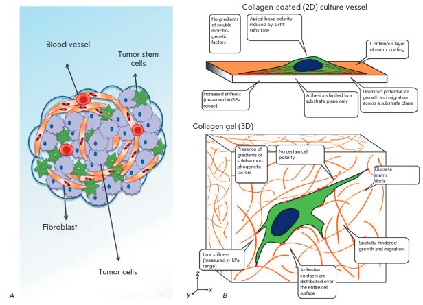
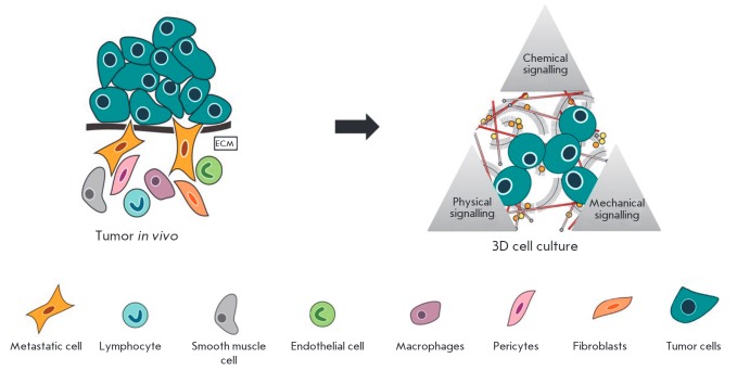
This review focuses on modeling of cancer tumors using tissue engineering technology. Tumor tissue engineering (TTE) is a new method of three-dimensional (3D) simulation of malignant neoplasms. Design and development of complex tissue engineering constructs (TECs) that include cancer cells, cell-bearing scaffolds acting as the extracellular matrix, and other components of the tumor microenvironment is at the core of this approach. Although TECs can be transplanted into laboratory animals, the specific aim of TTE is the most realistic reproduction and long-term maintenance of the simulated tumor properties in vitro for cancer biology research and for the development of new methods of diagnosis and treatment of malignant neoplasms. Successful implementation of this challenging idea depends on bioreactor technology, which will enable optimization of culture conditions and control of tumor TECs development. In this review, we analyze the most popular bioreactor types in TTE and the emerging applications.
Keywords: bioreactors; cancer; models; tissue engineering.
Figures
Similar articles
-
Engineering Vascular Bioreactor Systems to Closely Mimic Physiological Forces In Vitro.Tissue Eng Part B Rev. 2023 Jun;29(3):232-243. doi: 10.1089/ten.TEB.2022.0158. Epub 2022 Dec 8. Tissue Eng Part B Rev. 2023. PMID: 36274223 Review.
-
New generation of bioreactors that advance extracellular matrix modelling and tissue engineering.Biotechnol Lett. 2019 Jan;41(1):1-25. doi: 10.1007/s10529-018-2611-7. Epub 2018 Oct 27. Biotechnol Lett. 2019. PMID: 30368691 Free PMC article. Review.
-
A Perfusion Bioreactor for Longitudinal Monitoring of Bioengineered Liver Constructs.Nanomaterials (Basel). 2021 Jan 21;11(2):275. doi: 10.3390/nano11020275. Nanomaterials (Basel). 2021. PMID: 33494337 Free PMC article.
-
Numerical Simulation of Mass Transfer and Three-Dimensional Fabrication of Tissue-Engineered Cartilages Based on Chitosan/Gelatin Hybrid Hydrogel Scaffold in a Rotating Bioreactor.Appl Biochem Biotechnol. 2017 Jan;181(1):250-266. doi: 10.1007/s12010-016-2210-9. Epub 2016 Aug 15. Appl Biochem Biotechnol. 2017. PMID: 27526111
-
Three-dimensional-construct bioreactor conditioning in human tendon tissue engineering.Tissue Eng Part A. 2011 Oct;17(19-20):2561-72. doi: 10.1089/ten.TEA.2010.0701. Epub 2011 Jul 1. Tissue Eng Part A. 2011. PMID: 21612572
Cited by
-
FABRICA: A Bioreactor Platform for Printing, Perfusing, Observing, & Stimulating 3D Tissues.Sci Rep. 2018 May 15;8(1):7561. doi: 10.1038/s41598-018-25663-7. Sci Rep. 2018. PMID: 29765087 Free PMC article.
-
Key aspects for conception and construction of co-culture models of tumor-stroma interactions.Front Bioeng Biotechnol. 2023 Apr 7;11:1150764. doi: 10.3389/fbioe.2023.1150764. eCollection 2023. Front Bioeng Biotechnol. 2023. PMID: 37091337 Free PMC article. Review.
-
Engineering Breast Cancer Microenvironments and 3D Bioprinting.Front Bioeng Biotechnol. 2018 May 24;6:66. doi: 10.3389/fbioe.2018.00066. eCollection 2018. Front Bioeng Biotechnol. 2018. PMID: 29881724 Free PMC article. Review.
-
Emerging Biomimetic Materials for Studying Tumor and Immune Cell Behavior.Ann Biomed Eng. 2020 Jul;48(7):2064-2077. doi: 10.1007/s10439-019-02384-0. Epub 2019 Oct 15. Ann Biomed Eng. 2020. PMID: 31617045 Free PMC article. Review.
-
The progressive trend of modeling and drug screening systems of breast cancer bone metastasis.J Biol Eng. 2024 Feb 5;18(1):14. doi: 10.1186/s13036-024-00408-5. J Biol Eng. 2024. PMID: 38317174 Free PMC article. Review.
References
-
- Hickman J.A., Graeser R., de Hoogt R., Vidic S., Brito C., Gutekunst M., van der Kuip H., Consortium I.P.. Biotechnol. J. 2014;9(9):1115–1128. - PubMed
-
- Breslin S., O’Driscoll L.. Drug Discov. Today. 2013;18(5-6):240–249. - PubMed
-
- Yamada K.M., Cukierman E.. Cell. 2007;130(4):601–610. - PubMed
-
- Hutmacher D.W., Loessner D., Rizzi S., Kaplan D.L., Mooney D.J., Clements J.A.. Trends Biotechnol. 2010;28(3):125–133. - PubMed
LinkOut - more resources
Full Text Sources