

Cleavage of Human Embryos: Options and Diversity
- PMID: 27795847
- PMCID: PMC5081708
Cleavage of Human Embryos: Options and Diversity
Abstract
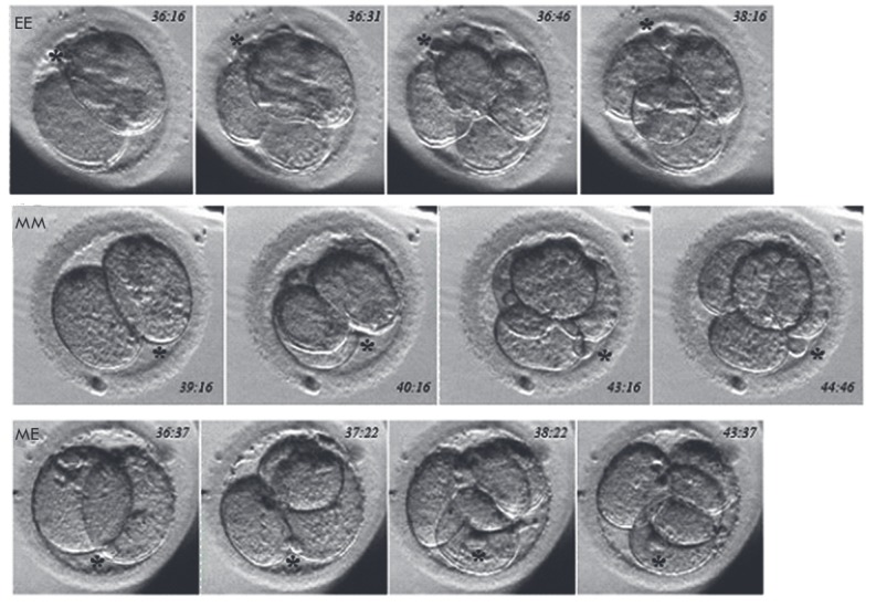
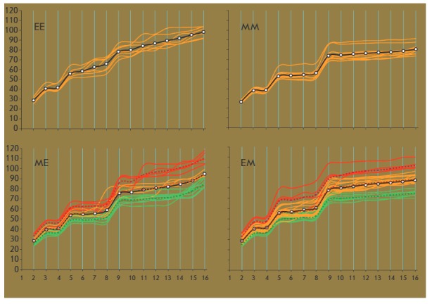
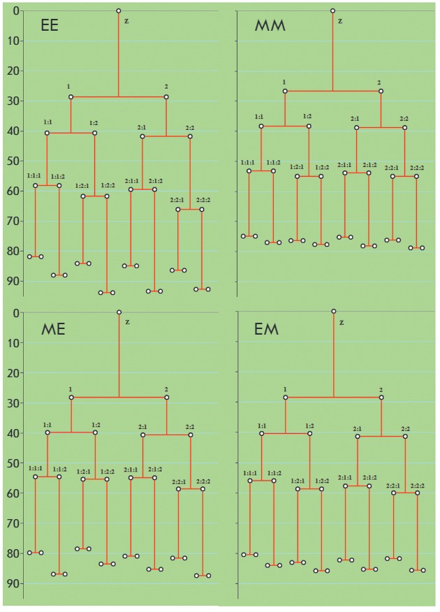
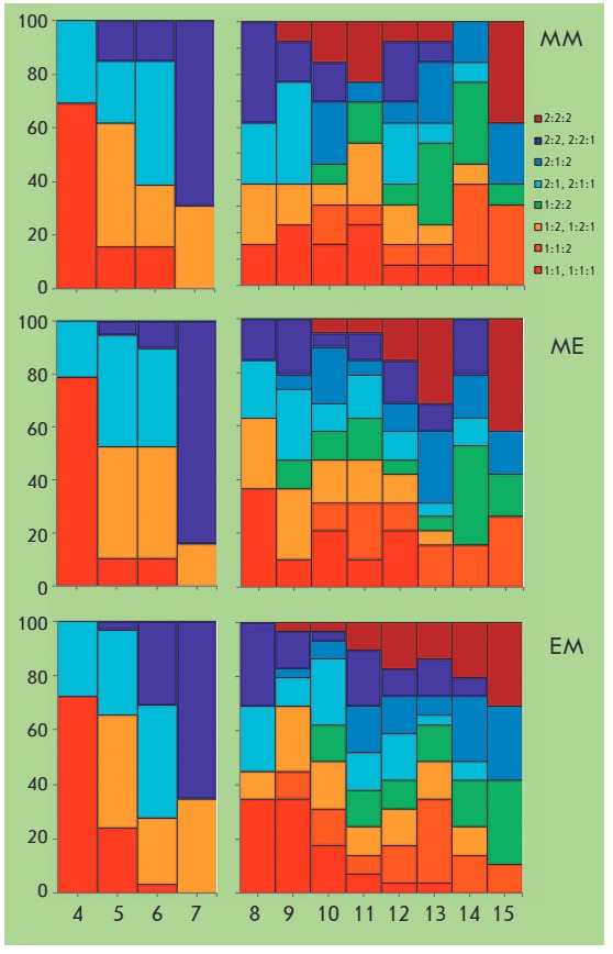
In order to estimate the diversity of embryo cleavage relatives to embryo progress (blastocyst formation), time-lapse imaging data of preimplantation human embryo development were used. This retrospective study is focused on the topographic features and time parameters of the cleavages, with particular emphasis on the lengths of cleavage cycles and the genealogy of blastomeres in 2- to 8-cell human embryos. We have found that all 4-cell human embryos have four developmental variants that are based on the sequence of appearance and orientation of cleavage planes during embryo cleavage from 2 to 4 blastomeres. Each variant of cleavage shows a strong correlation with further developmental dynamics of the embryos (different cleavage cycle characteristics as well as lengths of blastomere cycles). An analysis of the sequence of human blastomere divisions allowed us to postulate that the effects of zygotic determinants are eliminated as a result of cleavage, and that, thereafter, blastomeres acquire the ability of own syntheses, regulation, polarization, formation of functional contacts, and, finally, of specific differentiation. This data on the early development of human embryos obtained using noninvasive methods complements and extend our understanding of the embryogenesis of eutherian mammals and may be applied in the practice of reproductive technologies.
Keywords: blastomere genealogy; cleavage; human embryos; time-lapse analysis.
Figures
Similar articles
-
Spatial arrangement of individual 4-cell stage blastomeres and the order in which they are generated correlate with blastocyst pattern in the mouse embryo.Mech Dev. 2005 Apr;122(4):487-500. doi: 10.1016/j.mod.2004.11.014. Epub 2004 Dec 18. Mech Dev. 2005. PMID: 15804563
-
Blastomere cleavage plane orientation and the tetrahedral formation are associated with increased probability of a good-quality blastocyst for cryopreservation or transfer: a time-lapse study.Fertil Steril. 2019 Jun;111(6):1159-1168.e1. doi: 10.1016/j.fertnstert.2019.02.019. Epub 2019 Apr 12. Fertil Steril. 2019. PMID: 30982605
-
Optimal timing for blastomere biopsy of 8-cell embryos for preimplantation genetic diagnosis.Hum Reprod. 2018 Jan 1;33(1):32-38. doi: 10.1093/humrep/dex343. Hum Reprod. 2018. PMID: 29165686
-
Retrospective analysis: reproducibility of interblastomere differences of mRNA expression in 2-cell stage mouse embryos is remarkably poor due to combinatorial mechanisms of blastomere diversification.Mol Hum Reprod. 2018 Jul 1;24(7):388-400. doi: 10.1093/molehr/gay021. Mol Hum Reprod. 2018. PMID: 29746690
-
Survival and developmental potential of stored human early cleavage stage embryos.Eur J Obstet Gynecol Reprod Biol. 2004 Jul 1;115 Suppl 1:S8-11. doi: 10.1016/j.ejogrb.2004.01.009. Eur J Obstet Gynecol Reprod Biol. 2004. PMID: 15196708 Review.
References
-
- K. Elder., J. Cohen. Human preimplantation embryo selection. Reproductive medicine and assisted reproductive techniques. // Eds. K. Elder, J. Cohen. Informa Helthcare, 2007. 379 p. 2007. Human preimplantation embryo selection. Reproductive medicine and assisted reproductive techniques.
-
- Van den Bergh M., Ebner T., Elder K. Atlas of oocytes, zygotes and embryos in reproductive medicine. NY: Cambridge University Press, 2012. 237 p. 2012.
-
- Pribenszky C., Losonczi E., Molnar M., Lang Z., Matyas S., Rajczy K., Molnar K., Kovacs P., Nagy P., Conceicao J.. Reprod. BioMed. Online. 2010;20:371–379. - PubMed
-
- Kirkegaard K., Agerholm I.E., Ingerslev H.J.. Hum. Reprod. 2012;27(5):1277–1285. - PubMed
-
- Herrero J., Meseguer M.. Fertil. Steril. 2013;99:1030–1034. - PubMed
LinkOut - more resources
Full Text Sources