. 2016 Jun 13;2(6):963-976.
doi: 10.1021/acsbiomaterials.6b00052. Epub 2016 May 9.

Photothermal therapy of glioblastoma multiforme using multiwalled carbon nanotubes optimized for diffusion in extracellular space

Affiliations

Photothermal therapy of glioblastoma multiforme using multiwalled carbon nanotubes optimized for diffusion in extracellular space

Brittany N Eldridge et al. ACS Biomater Sci Eng. .

Abstract

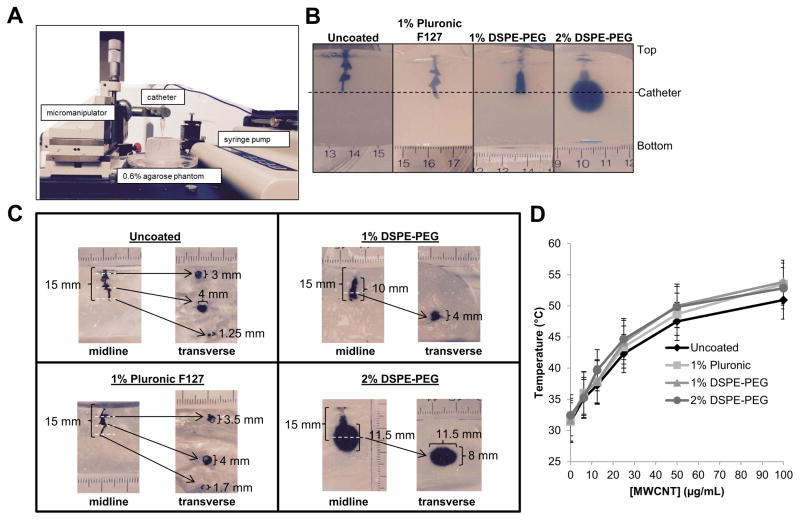
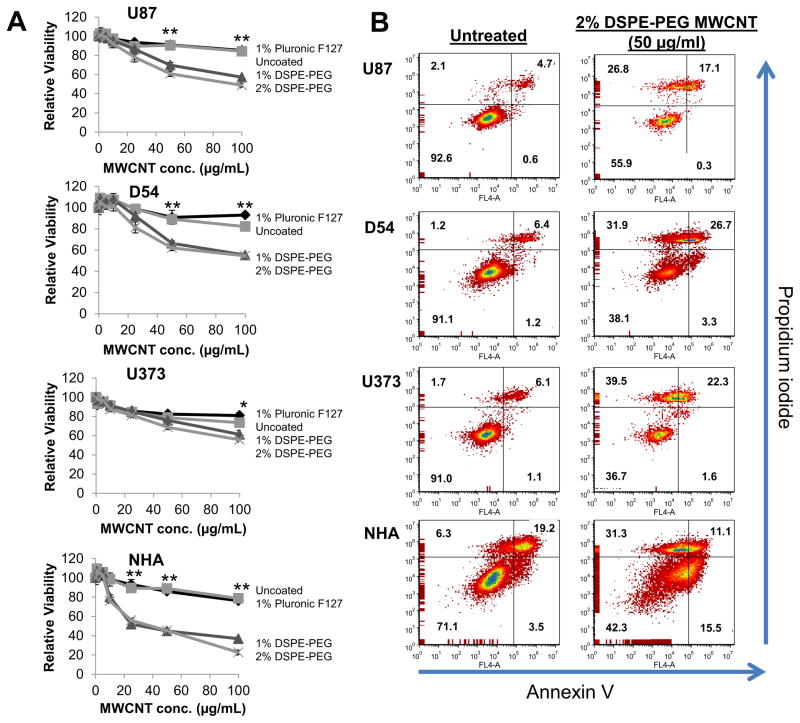
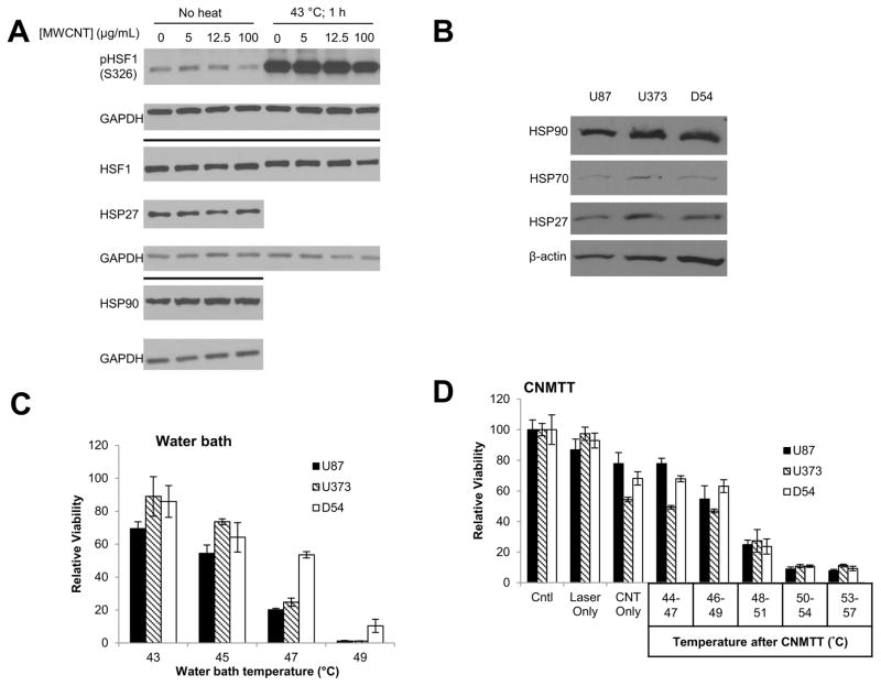
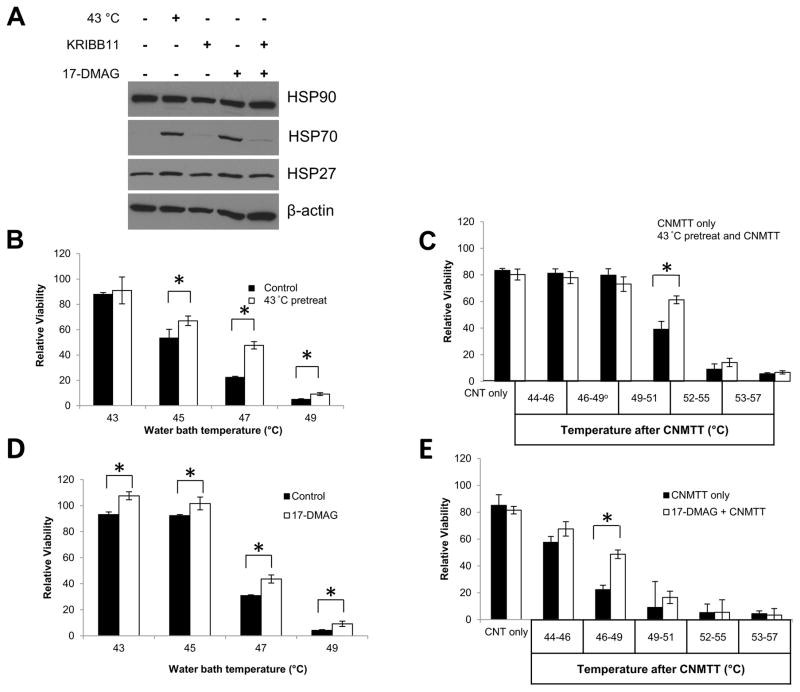
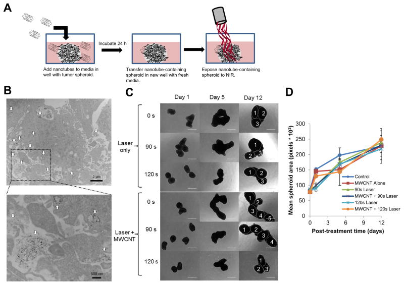
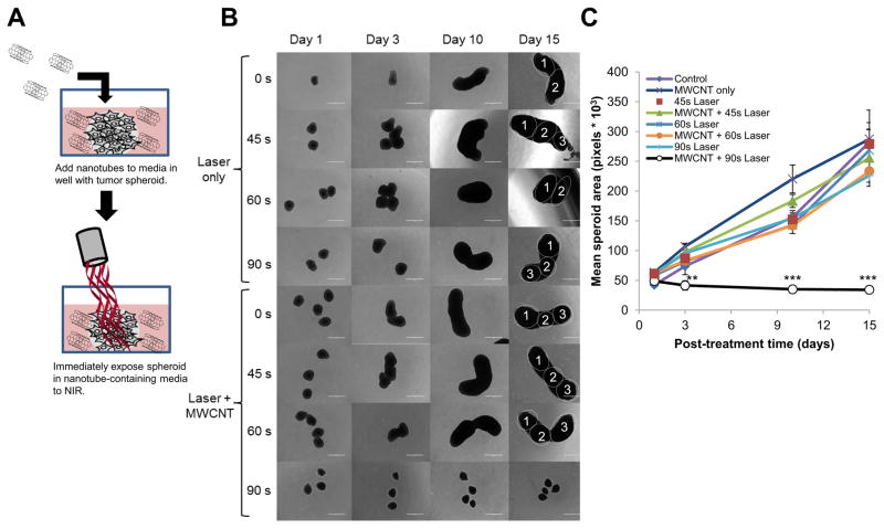
Glioblastoma multiforme (GBM) is the most common and most lethal primary brain tumor with a 5 year overall survival rate of approximately 5%. Currently, no therapy is curative and all have significant side effects. Focal thermal ablative therapies are being investigated as a new therapeutic approach. Such therapies can be enhanced using nanotechnology. Carbon nanotube mediated thermal therapy (CNMTT) uses lasers that emit near infrared radiation to excite carbon nanotubes (CNTs) localized to the tumor to generate heat needed for thermal ablation. Clinical translation of CNMTT for GBM will require development of effective strategies to deliver CNTs to tumors, clear structure-activity and structure-toxicity evaluation, and an understanding of the effects of inherent and acquired thermotolerance on the efficacy of treatment. In our studies, we show that a dense coating of phospholipid-poly(ethylene glycol) on multiwalled CNTs (MWCNTS) allows for better diffusion through brain phantoms, while maintaining the ability to achieve ablative temperatures after laser exposure. Phospholipid-poly(ethylene glycol) coated MWCNTs do not induce a heat shock response (HSR) in GBM cell lines. Activation of the HSR in GBM cells via exposure to sub-ablative temperatures or short term treatment with an inhibitor of heat shock protein 90 (17-(dimethylaminoethylamino)-17-demethoxygeldanamycin (17-DMAG)), induces a protective heat shock response that results in thermotolerance and protects against CNMTT. Finally, we evaluate the potential for CNMTT to treat GBM multicellular spheroids. These data provide pre-clinical insight into key parameters needed for translation of CNMTT including nanoparticle delivery, cytotoxicity, and efficacy for treatment of thermotolerant GBM.

Keywords: Cancer; ablation; brain tumor; convection; heat shock; hyperthermia; nanotechnology.


Figures

Figure 1
Figure 1. Assessment of structure-activity relationships for MWCNTs modified by acid oxidation and coating with various surfactants
A) Acid oxidized MWCNTs dispersed in surfactants were infused at a rate of 0.2 μL/min via a catheter inserted 15 mm below the surface of a 0.6% agarose brain-mimicking hydrogels using a micromanipulator to place the catheter and an infusion pump to regulate flow as shown. B) Representative images illustrating the effect of the coatings on the distribution of the MWCNTs in the hydrogels after infusion of MWCNT dispersions are shown as follows: 1) Uncoated; 2) 1% Pluronic; 3) 1% DSPE-PEG; 4) 2% DSPE-PEG. The depth at which the catheter was placed is indicated by the dashed line. C) Hydrogels infused with MWCNTs were bisected alone the midline and photographed as shown on the left in each panel. The two halves were place back together and transverse sections were made at the points indicated by the white dashed lines. An image of each transverse section is shown on the right of each panel. D) Increasing concentrations of MWCNTs dispersed in surfactants were exposed to a 970 nm diode laser at 3 W/cm2 for 30 seconds. Temperatures of the MWCNT suspensions were determined via thermocouple immediately after laser exposure.
Figure 2
Figure 2. Evaluation of the cytotoxicity induced by MWCNTs modified by acid oxidation and coating with various surfactants
A) U87, U373, and D54 GBM cells or NHA cells were treated for 24 h with increasing doses of acid oxidized MWCNTs dispersed in the indicated surfactants. After 24 h, cells were lysed, pelleted and the supernatant was analyzed for ATP content as a measure of cell viability using the CellTiter-Glo assay. Samples were prepared and measured in sextuplicate and are displayed as the mean ± standard deviation of each measurement. Significant differences in viability between cells treated with 1% or 2% DSPE-PEG coated MWCNTs and uncoated or Pluronic F-127 coated MWCNTs (determined by ANOVA followed by Student’s T-Test when appropriate) are indicated by (*; p<0.05) or (**; p<0.01). B) U87, U373, and D54 GBM cells or NHA cells were treated for 24 h with 2% DSPE-PEG MWCNTs (50 μg/ml). Cells were co-stained with propidium iodide and Annexin V and then evaluated by flow cytometry. the percentages of cells characterized as viable (lower-left quadrant), early apoptotic (lower-right quadrant), late-apoptotic (upper-right quadrant), and necrotic (upper-left quadrant) are shown within each quadrant. At least 10,000 cells were counted for each measurement and the experiments were repeated 2–3 times for each cell line with similar results.
Figure 3
Figure 3. Quantification of heat shock protein expression and response of GBM cells to thermal therapies
A) Adherent U87 cells were treated with increasing concentrations of 2% DSPE-PEG MWCNTs overnight. To determine if the nanotubes influenced activation of the heat shock response, half of the cells were subsequently exposed to 43 ºC for 1 h. Whole cell lysates were collected and probed for proteins involved in the heat shock response via western blot analysis. GAPDH was used as a loading control. B) Basal expression of heat shock proteins in U87, U373, and D54 GBM cells was assessed by western blot analysis and normalized to β-actin. C) GBM cells were heated to increasing temperatures (43–49 ºC) in a circulating water bath to mimic conventional heating methods. Cells were allowed to recover for two days and cell viability was determined by MTT assay. D) GBM cells were heated to increasing temperatures (44–57 ºC) using CNMTT. Cells were allowed to recover for two days and cell viability was determined by MTT assay. For (C) and (D), data are expressed as the mean of triplicate samples and are normalized to the unheated control for each cell line. Significant decreases in viability with each increase in temperature (p<0.05) were detected for all cell lines. The results shown are representative of at least 3 independent experiments.
Figure 4
Figure 4. Determination of the effect of sublethal heat or chemically induced heat shock protein expression on thermotolerance of GBM cells
To induce the HSR, U87 cells were placed in a 43 ºC incubator for 1 hour and recovered at 37 ºC for 4 hours, or treated with a combination of 1 μM 17-DMAG and 10 μM KRIBB11 for 5 hours. KRIBB11 treatment occurred 30 minutes prior to 17-DMAG treatment to allow sufficient time for the inhibitor to take effect. A) Whole cell lysates were collected from all treatment groups and probed for HSP90, HSP70, or HSP27 via western blot analysis. β-actin was used as a loading control. The HSR was induced in U87 cells by placement in a 43 ºC incubator for 1 h and recovery at 37 ºC for 4 h as above. Cells were subsequently heated to increasing temperatures in B) a circulating water bath to mimic conventional heating methods or C) by CNMTT. The HSR was induced in U87 cells by treatment with 1 μM of 17-DMAG for 5 h as above. Cells were subsequently heated to increasing temperatures in D) a circulating water bath E) or by CNMTT. Cells were allowed to recover for two days after each treatment and cell viability was determined by MTT assay. Data are expressed as the mean of triplicate samples normalized to untreated (no heat and no 17-DMAG) controls. Significant differences (p<0.05) between treatment groups determined by Student’s T-Test are indicated by (*). The results shown are representative of 2–3 independent experiments per condition tested.
Figure 5
Figure 5. Imaging the diffusion of 2% DSPE-PEG MWCNTs into three dimensional GBM tumor spheroids and treatment of nanotube containing spheroids by CNMTT
Briefly, U87 cells were grown as three dimensional tumor spheroids and incubated overnight with 2% DSPE-PEG MWCNTs. The spheroids were washed, and then transferred to new wells for use in electron microscopy or photothermal treatment studies. A) Schematic illustrating the experimental design. U87 spheroids incubated overnight with 2% DSPE-PEG MWCNTs were washed, fixed, embedded, sectioned, overlaid onto copper-coated formvar grids and imaged by TEM. In the electron micrographs, B) MWCNTs (indicated by the white arrows) can be seen penetrating throughout the spheroids (upper panel). A higher powered image (lower panel) of the highlighted area indicates that the MWCNTs are both in intracellular compartments and in the extracellular space of spheroids. Groups of 4–6 spheroids alone or spheroids incubated overnight with 2% DSPE-PEG MWCNTs then washed to remove excess nanoparticles were exposed to laser emitted NIR energy (3 W/cm2) for 0–120 s. Spheroid growth over time was monitored and C) representative photomicrographs are shown. Individual spheroids fused into a single cluster are identified in the day 15 images. D) Mean surface area per spheroid was quantified in pixels using Image J software. For area measurements, images of spheroids in addition to those shown in B) were used. No significant differences in spheroid grow were detected between treatment groups.
Figure 6
Figure 6. Treatment of GBM spheroids by CNMTT in the presence of extracellular 2% DSPE-PEG MWCNTs
Briefly, U87 cells were grown as three dimensional tumor spheroids. Groups of 4–6 spheroids were transferred to new wells containing 2% DSPE-PEG MWCNTs (20 μg/ml) in the media, and were exposed to laser emitted NIR energy (3 W/cm2) for 0–90 s. After treatment, the spheroids were washed, and then transferred to new wells with growth media only. A) Schematic illustrating the experimental design. Spheroid growth over time was monitored and B) representative photomicrographs are shown. Individual spheroids fused into a single cluster are identified in the day 15 images. C) Mean surface area per spheroid was quantified in pixels using Image J software. For area measurements, images of spheroids in addition to those shown in B) were used. Significant differences in spheroid grow were detected between spheroids treated with MWCNTs and laser for 90 s as compared to all other treatment groups (determined by ANOVA followed by Student’s T-Test when appropriate) are indicated by (**; p<0.01) or (***; p<0.001).

References

    1. Omuro A, DeAngelis LM. Glioblastoma and other malignant gliomas: a clinical review. JAMA. 2013;310(17):1842–50. - PubMed
    1. Lima FR, Kahn SA, Soletti RC, Biasoli D, Alves T, da Fonseca AC, Garcia C, Romao L, Brito J, Holanda-Afonso R, Faria J, Borges H, Moura-Neto V. Glioblastoma: Therapeutic challenges, what lies ahead. Biochim Biophys Acta. 2012;1826(12):338–49. - PubMed
    1. Sloan AE, Ahluwalia MS, Valerio-Pascua J, Manjila S, Torchia MG, Jones SE, Sunshine JL, Phillips M, Griswold MA, Clampitt M, Brewer C, Jochum J, McGraw MV, Diorio D, Ditz G, Barnett GH. Results of the NeuroBlate System first-in-humans Phase I clinical trial for recurrent glioblastoma: clinical article. J Neurosurg. 2013;118(6):1202–19. - PubMed
    1. Mohammadi AM, Hawasli AH, Rodriguez A, Schroeder JL, Laxton AW, Elson P, Tatter SB, Barnett GH, Leuthardt EC. The role of laser interstitial thermal therapy in enhancing progression-free survival of difficult-to-access high-grade gliomas: a multicenter study. Cancer Med. 2014;3(4):971–9. - PMC - PubMed
    1. Carpentier A, Chauvet D, Reina V, Beccaria K, Leclerq D, McNichols RJ, Gowda A, Cornu P, Delattre JY. MR-guided laser-induced thermal therapy (LITT) for recurrent glioblastomas. Lasers Surg Med. 2012;44(5):361–8. - PubMed

LinkOut - more resources