Review
. 2015 Nov:1:499-518.
doi: 10.1146/annurev-vision-082114-035742. Epub 2015 Oct 14.

Control and Functions of Fixational Eye Movements

Affiliations
Review

Control and Functions of Fixational Eye Movements

Michele Rucci et al. Annu Rev Vis Sci. 2015 Nov.

Abstract

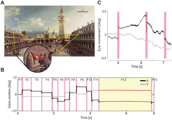
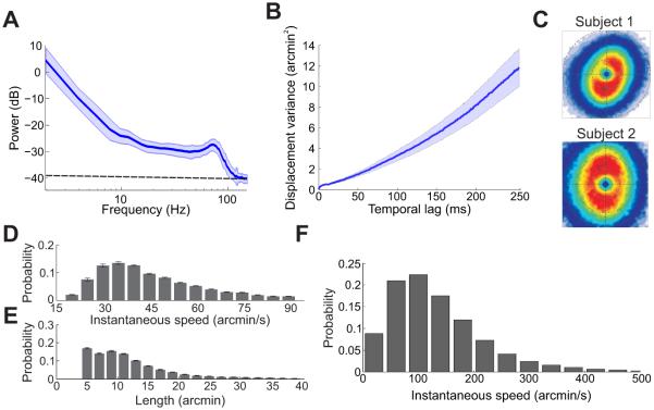
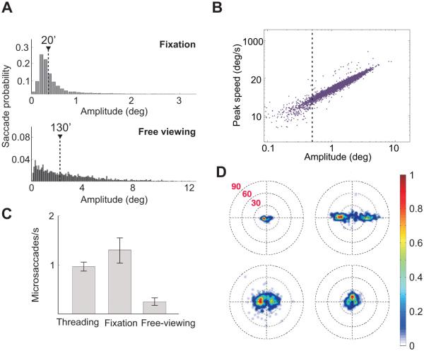
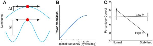
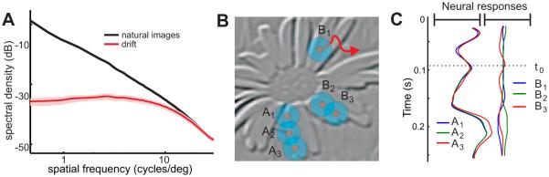
Humans and other species explore a visual scene by rapidly shifting their gaze 2-3 times every second. Although the eyes may appear immobile in the brief intervals in between saccades, microscopic (fixational) eye movements are always present, even when attending to a single point. These movements occur during the very periods in which visual information is acquired and processed and their functions have long been debated. Recent technical advances in controlling retinal stimulation during normal oculomotor activity have shed new light on the visual contributions of fixational eye movements and their degree of control. The emerging body of evidence, reviewed in this article, indicates that fixational eye movements are important components of the strategy by which the visual system processes fine spatial details, enabling both precise positioning of the stimulus on the retina and encoding of spatial information into the joint space-time domain.

Keywords: Ocular drift; ganglion cell; microsaccade; neural encoding; retina; saccade; visual acuity.


Figures

Figure 1
Figure 1
Normal eye movements. (A) As an observer explores a scene, saccades (yellow segments) separate brief periods of “fixation” in which visual information is acquired (red). As shown in the enlargement, small eye movements continually occur during fixation. (B) Horizontal and vertical traces of the oculomotor sequence in A. Magenta bars indicate saccades. (C) Traces of the fixational eye movements present during one fixation (circle in A and yellow region in B). The eye wanders with a seemingly random trajectory (ocular drift) occasionally interrupted by microscopic saccades (microsaccades; magenta).
Figure 2
Figure 2
General characteristics of ocular drifts and tremor. (A) Power spectrum of the oculomotor activity recorded in the periods between saccades/microsaccades. The dashed line represents the level of the eyetracker noise. (B) Variance of the displacement in the line of sight as a function of time. The increment is approximately linear, as distinctive of Brownian motion. (C) Probability distributions of instantaneous drift velocity for two observers during sustained fixation on a marker (Cherici et al. 2012). (D-E) Average distributions of ocular drift speed and path length during free viewing of natural scenes (Kuang et al. 2012). (F) Drift speed distribution for one observer during normal head-free viewing (Aytekin, Victor & Rucci 2014).
Figure 3
Figure 3
General characteristics of microsaccades. (A) Saccade amplitude distribution during sustained fixation and free viewing. Data represent average distributions across six observers. Triangles mark the medians of the distributions. (B) Saccade peak speed of as a function of saccade amplitude during free viewing. The dashed line marks saccades smaller than half a degree. (C) Microsaccade rates in three different tasks: needle threading, sustained fixation on a single dot, and free viewing of a natural scene (Ko, Poletti & Rucci 2010). (D) Individual variability in microsaccades during sustained fixation. Each panel shows the two-dimensional probability distribution of microsaccade displacements for an individual observer (Cherici et al. 2012).
Figure 4
Figure 4
Enhancement of high spatial frequencies resulting from fixational drift. (A) Exposure of retinal receptors (circles) to low (top) and high (bottom) spatial frequency gratings during fixational instability (arrows). The grating at higher frequency gives a larger change in luminance (ΔI). (B) Mean amplification resulting from ocular drift as a function of spatial frequency. Data represent averages across N =5 observers (Mostofi, Boi & Rucci 2014). (C) Results of experiments in which subjects judged the orientation of a noisy grating (±45° either at low or high spatial frequency) in the presence (normal) and absence (stabilized) of normal fixational eye movements. In the stabilized condition, the position of the stimulus was continually updated according to the subject’s eye movements so to eliminate retinal image motion. Removal of fixational modulations via retinal stabilization selectively impaired high spatial frequency vision (Rucci et al. 2007).
Figure 5
Figure 5
Interaction with natural images and possible neural consequences of ocular drift. (A) Comparison between the power of a set of natural images and the temporal power (the sum over all nonzero temporal frequencies) in the modulations caused by ocular drift. Normal drift equalizes power over a broad spatial frequency range (Kuang et al. 2012). (B) Activity in a simulated array of retinal ganglion cells. Each pixel represents the mean instantaneous firing rate at time to of a simulated neuron with receptive field (circles) centered on the pixel. (C) Time-course of the responses of the six neurons shown in B. Note the enhancement of edges in the synchronous modulations.
Figure 6
Figure 6
Microsaccades in a high-acuity task. (A) Threading a needle in a virtual environment. Subjects moved a horizontal bar (the thread) toward the small gap in a vertical bar (the needle). The panel on the right and its enlargement show the spatial distribution of fixations in a trial. Fixations were primarily allocated to the thread (blue circles) and the eye of the needle (green circles). The red crosses mark the thread trajectory. (B) Mean instantaneous frequency and amplitude of microsaccades. The two horizontal lines represent mean rates during sustained fixation (dashed line) and free viewing (dotted line). (C) Probabilities of various types of microsaccades, classified according to their starting and landing points. Microsaccades almost always brought the line of sight on the thread and needle, rarely on the background. (D) Conditional probabilities of realigning the thread following different types of microsaccades. Adjustments were more likely to occur immediately after microsaccades across different objects. (E) Microsaccade landing probability in successful and unsuccessful trials. Microsaccades were more precise in the trials in which the thread was correctly aligned with the needle. All data refer to saccades smaller than 20′. Modified from Ko, Poletti & Rucci (2010).
Figure 7
Figure 7
Consequences of microsaccades for foveal vision. (A) Subjects reported whether two sequentially-presented noisy gratings were parallel or orthogonal. Gratings (11 cycles/deg) were tilted by ±45° and appeared within two rectangular noise bars centered at the desired eccentricity d, first in the left and then in the right bar. (B) Stimuli were either displayed at fixed positions on the screen (Normal) and normally moved on the retina because of fixational eye movements, or at fixed locations on the retina (Stabilized) and moved on the display under computer control to compensate for the subject’s eye movements. Performance decreased sharply with eccentricity under retinal stabilization. Discrimination was also impaired in the trials in which microsaccades did occur but were less precise ((★) p <0.05; (★★) p <0.005; two-tailed paired t-test). (C) Proportions of microsaccades landing on different regions of the display for stimuli at 15′ eccentricity. Most microsaccades moved the line of sight on the bar containing the stimulus. (D) Eye movements in two example trials. Red and blue segments represent microsaccades and drifts, respectively. Subjects were asked to maintain fixation at the center of the display (cross in A). Modified from Poletti, Listorti & Rucci (2013).

References

    1. Adler FH, Fliegelman M. Influence of fixation on the visual acuity. Arch. Ophthalmol. 1934;12:475483.
    1. Adler FH, Meyer GP. The mechanism of the fovea. Trans. Am. Ophthalmol. Soc. 1935;33:266–280. - PMC - PubMed
    1. Ahissar E, Arieli A. Figuring space by time. Neuron. 2001;32:185–201. - PubMed
    1. Ahissar E, Arieli A. Seeing via miniature eye movements: A dynamic hypothesis for vision. Front. Comp. Neurosci. 2012;6:1–27. - PMC - PubMed
    1. Arend LE. Spatial differential and integral operations in human vision: Implications of stabilized retinal image fading. Psychol. Rev. 1973;80:374–395. - PubMed

Publication types

LinkOut - more resources