. 2016 Dec 1;126(12):4527-4536.
doi: 10.1172/JCI87343. Epub 2016 Oct 31.

Endothelial cation channel PIEZO1 controls blood pressure by mediating flow-induced ATP release

Endothelial cation channel PIEZO1 controls blood pressure by mediating flow-induced ATP release

ShengPeng Wang et al. J Clin Invest. .

Abstract

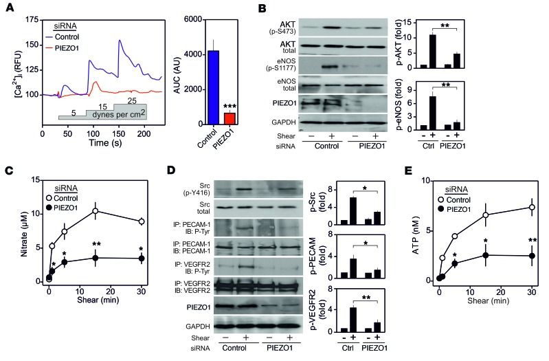
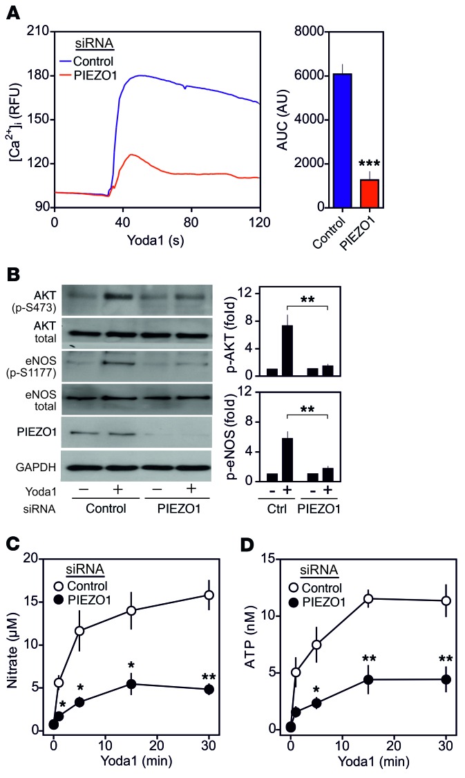
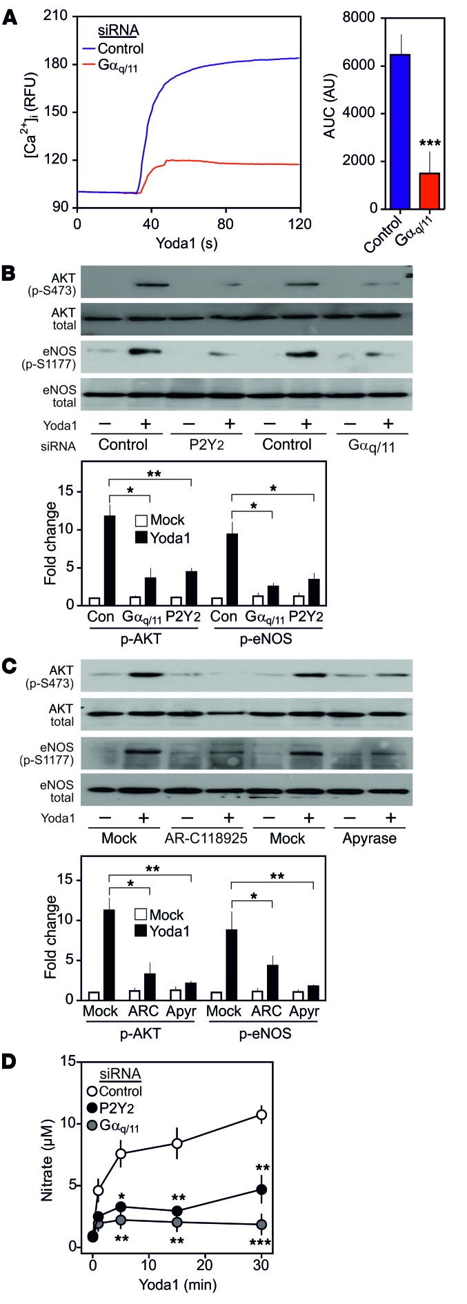
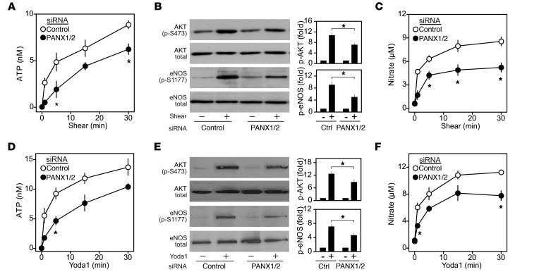
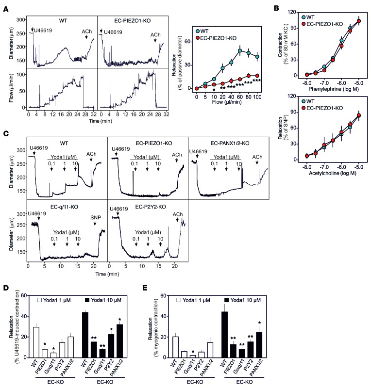
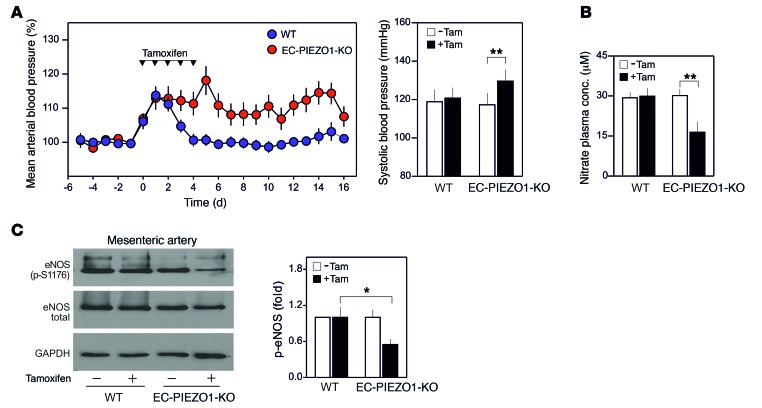
Arterial blood pressure is controlled by vasodilatory factors such as nitric oxide (NO) that are released from the endothelium under the influence of fluid shear stress exerted by flowing blood. Flow-induced endothelial release of ATP and subsequent activation of Gq/G11-coupled purinergic P2Y2 receptors have been shown to mediate fluid shear stress-induced stimulation of NO formation. However, the mechanism by which fluid shear stress initiates these processes is unclear. Here, we have shown that the endothelial mechanosensitive cation channel PIEZO1 is required for flow-induced ATP release and subsequent P2Y2/Gq/G11-mediated activation of downstream signaling that results in phosphorylation and activation of AKT and endothelial NOS. We also demonstrated that PIEZO1-dependent ATP release is mediated in part by pannexin channels. The PIEZO1 activator Yoda1 mimicked the effect of fluid shear stress on endothelial cells and induced vasorelaxation in a PIEZO1-dependent manner. Furthermore, mice with induced endothelium-specific PIEZO1 deficiency lost the ability to induce NO formation and vasodilation in response to flow and consequently developed hypertension. Together, our data demonstrate that PIEZO1 is required for the regulation of NO formation, vascular tone, and blood pressure.


Conflict of interest statement

The authors have declared that no conflict of interest exists.

Figures

Figure 1
Figure 1. PIEZO1 mediates endothelial response to fluid shear stress in vitro.
HUAECs were transfected with scrambled (control) siRNA or siRNA directed against PIEZO1 as described in Methods. (A) Fluo-4–loaded HUAECs (n = 22, control; n = 29, PIEZO1 knockdown; 3 independent experiments) were exposed to the indicated shear forces, and [Ca2+]i was determined as fluorescence intensity (RFU, relative fluorescence units). Bar diagrams show areas under the curve (AUC). Shown are means ± SEM; ***P ≤ 0.001 (2-tailed Student’s t test). (BE) HUAECs (n = 3) were exposed to fluid shear (15 dyn/cm2) for 5 minutes in B and D or for the indicated time periods. For determination of Src activation, 15 seconds of shear was applied. AKT, eNOS, and Src activation (B and D) was determined by Western blotting for phosphorylated AKT, eNOS, and Src kinases and total AKT, eNOS, and Src. (C) Nitrate concentration in the cell medium. PECAM-1 and VEGFR2 activation (D) was determined by immunoprecipitation and Western blotting for tyrosine phosphorylated PECAM-1 and VEGFR2. Knockdown of PIEZO1 was verified by anti-PIEZO1 immunoblotting. Bar diagrams show the densitometric evaluation. (E) Concentration of ATP in the supernatant of HUAECs. Shown are means ± SEM; *P ≤ 0.05; **P ≤ 0.01 (2-way ANOVA and Bonferroni’s post hoc test).
Figure 2
Figure 2. Yoda1 induces endothelial responses similar to fluid shear stress via PIEZO1.
Cells were transfected with scrambled (control) siRNA or siRNA directed against PIEZO1. (A) Fluo-4–loaded HUAECs (n = 24, control; n = 22, PIEZO1 knockdown; 3 independent experiments) were exposed to 1 μM Yoda1, and [Ca2+]i was determined as fluorescence intensity (RFU, relative fluorescence units). Bar diagrams show AUC. Shown are means ± SEM; ***P ≤ 0.001 (2-tailed Student’s t test). (BD) HUAECs were exposed to 1 μM Yoda1 for the indicated times (5 minutes in B). AKT and eNOS activation was determined by Western blotting for phosphorylated AKT and eNOS as well as total AKT and eNOS (n = 3). Knockdown of PIEZO1 was verified by anti-PIEZO1 immunoblotting. Bar diagrams show the densitometric evaluation. (C and D) Nitrate concentration (C, n = 3) and concentration of ATP (D, n = 6) in the cell medium. Shown are the mean ± SEM. *P ≤ 0.05 and **P ≤ 0.01 (2-way ANOVA and Bonferroni’s post-hoc test).
Figure 3
Figure 3. Yoda1-induced, PIEZO1-mediated endothelial effects involve P2Y2 and Gq/G11.
(AD) HUAECs were transfected with scrambled siRNA (control; con) or siRNA directed against P2Y2 or Gαq/Gα11 (A, B, and D) or were preincubated without or with 30 μM of the P2Y2 antagonist AR-C118925 (ARC) or 2 U/ml apyrase (apyr) for 30 minutes (C). (A) Fluo-4–loaded HUAECs (n = 14, control; n = 20, Gαq/11 knockdown; 3 independent experiments) were exposed to 1 μM Yoda1, and [Ca2+]i was determined as fluorescence intensity (RFU, relative fluorescence units). Bar diagrams show AUC. Shown are means ± SEM; ***P ≤ 0.001 (2-tailed Student’s t test). (B and C) Cells were exposed to 1 μM Yoda1 for 5 minutes, and AKT and eNOS activation was determined as described. Bar diagrams show the densitometric evaluation (n = 3). (D) Nitrate concentration in the cell medium (n = 4). Shown are the mean ± SEM. *P ≤ 0.05, **P ≤ 0.01, and ***P ≤ 0.001 (2-way ANOVA and Bonferroni’s post-hoc test).
Figure 4
Figure 4. Role of pannexins in flow- and Yoda1-induced endothelial effects.
(AF) HUAECs were transfected with scrambled (control) siRNA or siRNAs directed against pannexin 1 and pannexin 2 (PANX1/2) and were exposed to fluid shear (15 dyn/cm2) (AC) or 1 μM Yoda1 (DF) for the indicated time periods (5 minutes in B and E). The concentration of ATP (A and D, n = 3) or nitrate (C and F, n = 4) in the cell medium was measured. AKT and eNOS activation (B and E, n = 3) was determined by Western blotting for phosphorylated AKT and eNOS as well as total AKT and eNOS. Bar diagrams show the densitometric evaluation. Shown are the mean ± SEM. *P ≤ 0.05 (2-way ANOVA and Bonferroni’s post-hoc test).
Figure 5
Figure 5. Endothelial PIEZO1 is required for flow-induced vasodilation.
(A) Effect of a stepwise increase in perfusion flow on the diameter of mesenteric arteries from tamoxifen-treated WT mice or EC-PIEZO1-KO mice precontracted with 100 nM of the thromboxane A2 analog U46619. After flow was stopped, 10 μM acetylcholine (ACh) was added. Right panel: Flow-induced vasorelaxation as percentage of the passive vessel diameter (n = 6, WT; n = 7, EC-PIEZO1-KO). (B) Effect of acetylcholine and phenylephrine on the tension of mesenteric artery stripes from WT (n = 5) and EC-PIEZO1-KO animals (n = 5). (CE) Effect of Yoda1 at the indicated concentration on the diameter of mesenteric arteries precontracted by U46619 (C and D) or by induction of myogenic tone (E) from WT (n = 4), EC-PIEZO1-KO (n = 4), EC-q/11-KO (n = 3), EC-P2Y2-KO (n = 3), or EC-PANX1/2-KO (n = 3) mice. Vessels were prepared 7–10 days after tamoxifen-dependent induction. Shown are means ± SEM. *P ≤ 0.05, **P ≤ 0.01, and ***P ≤ 0.001 (2-way ANOVA and Bonferroni’s post-hoc test).
Figure 6
Figure 6. Endothelial PIEZO1 controls blood pressure.
(A) Blood pressure in WT (n = 12) and EC-PIEZO1-KO mice (n = 14) before, during, and after induction by tamoxifen. Average blood pressure 5 days before induction was set at 100%. The bar diagram shows systolic arterial blood pressure 4 days before tamoxifen treatment and in the second week after induction. (B) Plasma nitrate levels in WT (n = 8) and EC-PIEZO1-KO mice (n = 8) before and 5 days after induction. (C) Phosphorylation of eNOS at S1176 in lysates of mesenteric arteries prepared 3 days after induction from WT and EC-PIEZO1-KO mice. Bar diagram shows a densitometric evaluation (n = 4). Shown are the mean ± SEM. *P ≤ 0.05 and **P ≤ 0.01 (2-tailed Student’s t test, B and C, and 2-way ANOVA and Bonferroni’s post-hoc test, A).

Comment in

References

    1. Hahn C, Schwartz MA. Mechanotransduction in vascular physiology and atherogenesis. Nat Rev Mol Cell Biol. 2009;10(1):53–62. doi: 10.1038/nrm2596. - DOI - PMC - PubMed
    1. Tarbell JM, Simon SI, Curry FR. Mechanosensing at the vascular interface. Annu Rev Biomed Eng. 2014;16:505–532. doi: 10.1146/annurev-bioeng-071813-104908. - DOI - PMC - PubMed
    1. Davies PF. Flow-mediated endothelial mechanotransduction. Physiol Rev. 1995;75(3):519–560. - PMC - PubMed
    1. Félétou M, Köhler R, Vanhoutte PM. Endothelium-derived vasoactive factors and hypertension: possible roles in pathogenesis and as treatment targets. Curr Hypertens Rep. 2010;12(4):267–275. doi: 10.1007/s11906-010-0118-2. - DOI - PMC - PubMed
    1. Busse R, Fleming I. Regulation of endothelium-derived vasoactive autacoid production by hemodynamic forces. Trends Pharmacol Sci. 2003;24(1):24–29. doi: 10.1016/S0165-6147(02)00005-6. - DOI - PubMed

Publication types

MeSH terms