

Review
doi: 10.1080/19491034.2016.1252893.
Epub 2016 Oct 31.
Shared mechanisms in physiological and pathological nucleoplasmic reticulum formation
Affiliations
- PMID: 27797635
- PMCID: PMC5287099
- DOI: 10.1080/19491034.2016.1252893
Item in Clipboard
Review
Shared mechanisms in physiological and pathological nucleoplasmic reticulum formation
Nucleus.
.
Abstract
The mammalian nuclear envelope (NE) can develop complex dynamic membrane-bounded invaginations in response to both physiological and pathological stimuli. Since the formation of these nucleoplasmic reticulum (NR) structures can occur during interphase, without mitotic NE breakdown and reassembly, some other mechanism must drive their development. Here we consider models for deformation of the interphase NE, together with the evidence for their potential roles in NR formation.
Keywords: NE; NR; chromosome territories; cytoskeleton; gene expression; membrane curvature; nuclear envelope; nucleoplasmic reticulum.
Figures
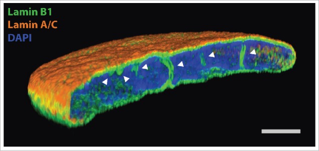
Super resolution light microscopy on normal human dermal fibroblasts, labeled with anti-lamin B1 antibody (green), anti-lamin A/C antibody (orange) and DAPI (blue). White arrowheads point to intranuclear NR tubules. Scale bar, 2 µm.
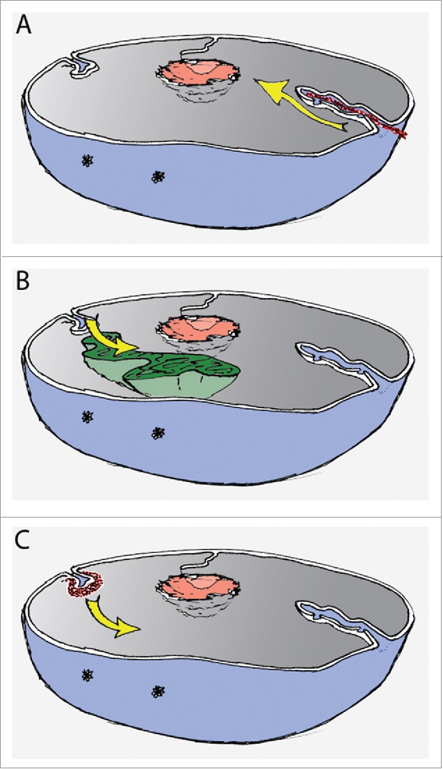
Schematic representation of nucleus with possible mechanisms driving NR formation. (A) Pushing the NE by cytoskeleton (red) as visualised by yellow arrow; (B) Pulling the NE by chromatin movement (green), movement indicated by yellow arrow; (C) Focal and de novo assembly and growth (yellow arrow) of NR invaginations (red) by dedicated machinery.
References
-
- Prunuske AJ, Ullman KS. The nuclear envelope: form and reformation. Curr Opin Cell Biol 2006; 18:108-16; PMID:16364623; http://dx.doi.org/ 10.1016/j.ceb.2005.12.004 - DOI - PMC - PubMed
-
- Kamei H. Relationship of nuclear invaginations to perinuclear rings composed of intermediate filaments in MIA PaCa-2 and some other cells. Cell Structure Function 1994; 19:123-32; PMID:7954871; http://dx.doi.org/ 10.1247/csf.19.123 - DOI - PubMed
-
- Malhas A, Goulbourne C, Vaux DJ. The nucleoplasmic reticulum: form and function. Trends Cell Biol 2011; 21:362-73; PMID:21514163; http://dx.doi.org/ 10.1016/j.tcb.2011.03.008 - DOI - PubMed
-
- Echevarria W, Leite MF, Guerra MT, Zipfel WR, Nathanson MH. Regulation of calcium signals in the nucleus by a nucleoplasmic reticulum. Nat Cell Biol 2003; 5:440-6; PMID:12717445; http://dx.doi.org/ 10.1038/ncb980 - DOI - PMC - PubMed
-
- Fricker M, Hollinshead M, White N, Vaux D. Interphase nuclei of many mammalian cell types contain deep, dynamic, tubular membrane-bound invaginations of the nuclear envelope. J Cell Biol 1997; 136:531-44; PMID:9024685; http://dx.doi.org/ 10.1083/jcb.136.3.531 - DOI - PMC - PubMed
Publication types
MeSH terms
Substances
Grants and funding
LinkOut - more resources
Full Text Sources
Other Literature Sources