. 2018 Feb;67(2):320-332.
doi: 10.1136/gutjnl-2016-311585. Epub 2016 Oct 21.

IL-6 and PD-L1 antibody blockade combination therapy reduces tumour progression in murine models of pancreatic cancer

Affiliations

IL-6 and PD-L1 antibody blockade combination therapy reduces tumour progression in murine models of pancreatic cancer

Thomas A Mace et al. Gut. 2018 Feb.

Abstract

Objective: Limited efficacy of immune checkpoint inhibitors in pancreatic ductal adenocarcinoma (PDAC) has prompted investigation into combination therapy. We hypothesised that interleukin 6 (IL-6) blockade would modulate immunological features of PDAC and enhance the efficacy of anti-programmed death-1-ligand 1 (PD-L1) checkpoint inhibitor therapy.

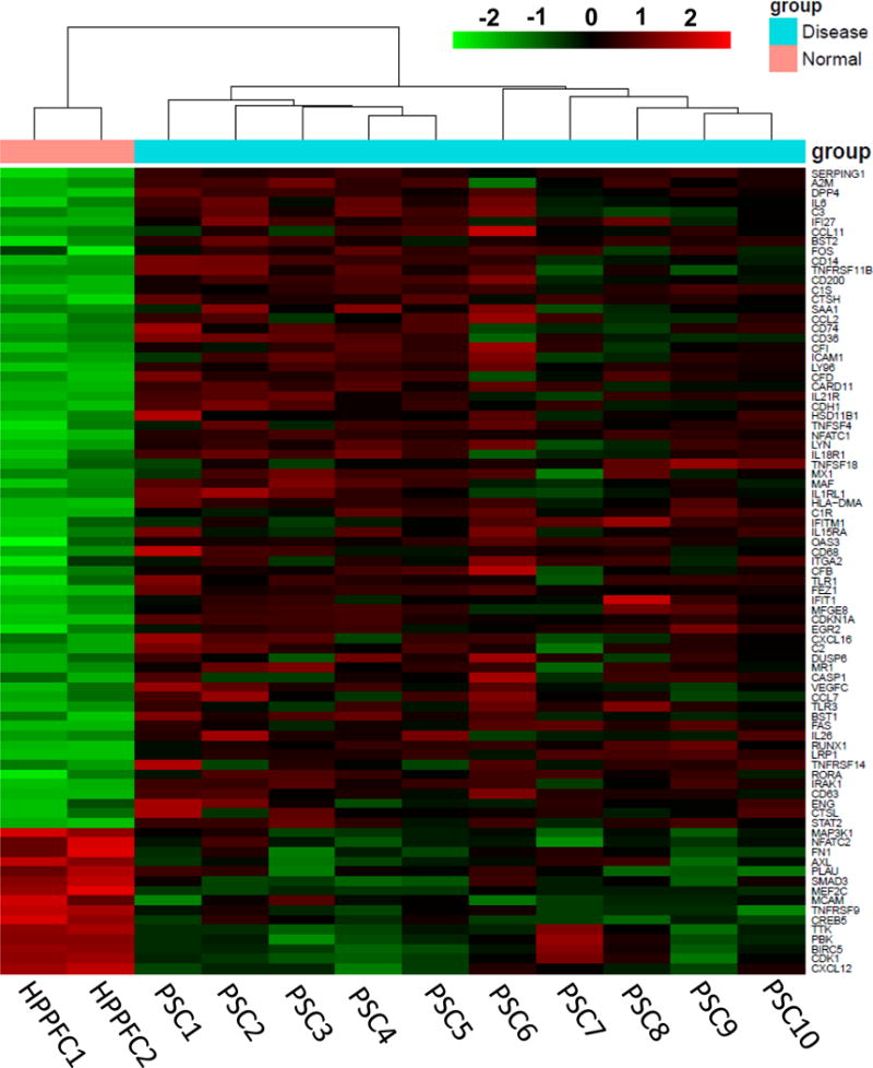
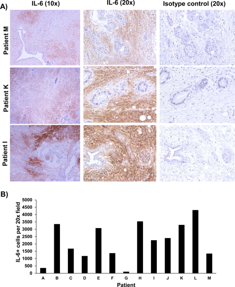
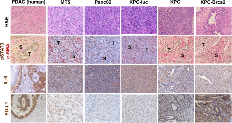
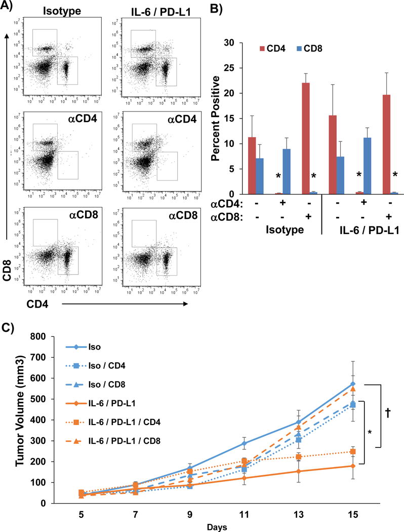
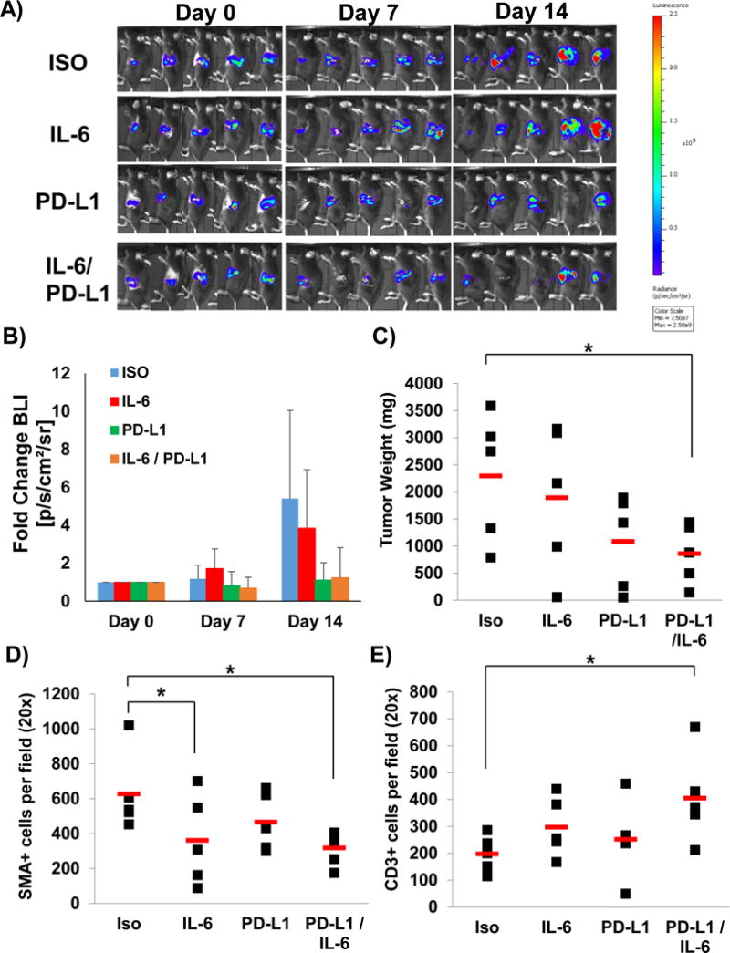
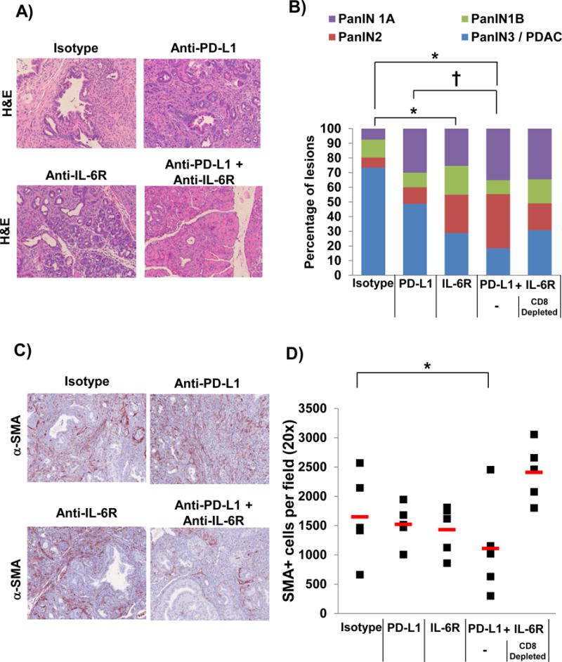
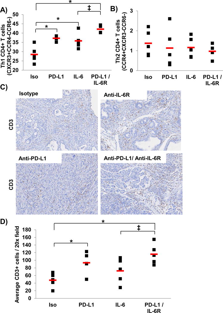
Design: Transcription profiles and IL-6 secretion from primary patient-derived pancreatic stellate cells (PSCs) were analyzed via Nanostring and immunohistochemistry, respectively. In vivo efficacy and mechanistic studies were conducted with antibodies (Abs) targeting IL-6, PD-L1, CD4 or CD8 in subcutaneous or orthotopic models using Panc02, MT5 or KPC-luc cell lines; and the aggressive, genetically engineered PDAC model (KrasLSL-G12D, Trp53LSL-R270H, Pdx1-cre, Brca2F/F (KPC-Brca2 mice)). Systemic and local changes in immunophenotype were measured by flow cytometry or immunohistochemical analysis.

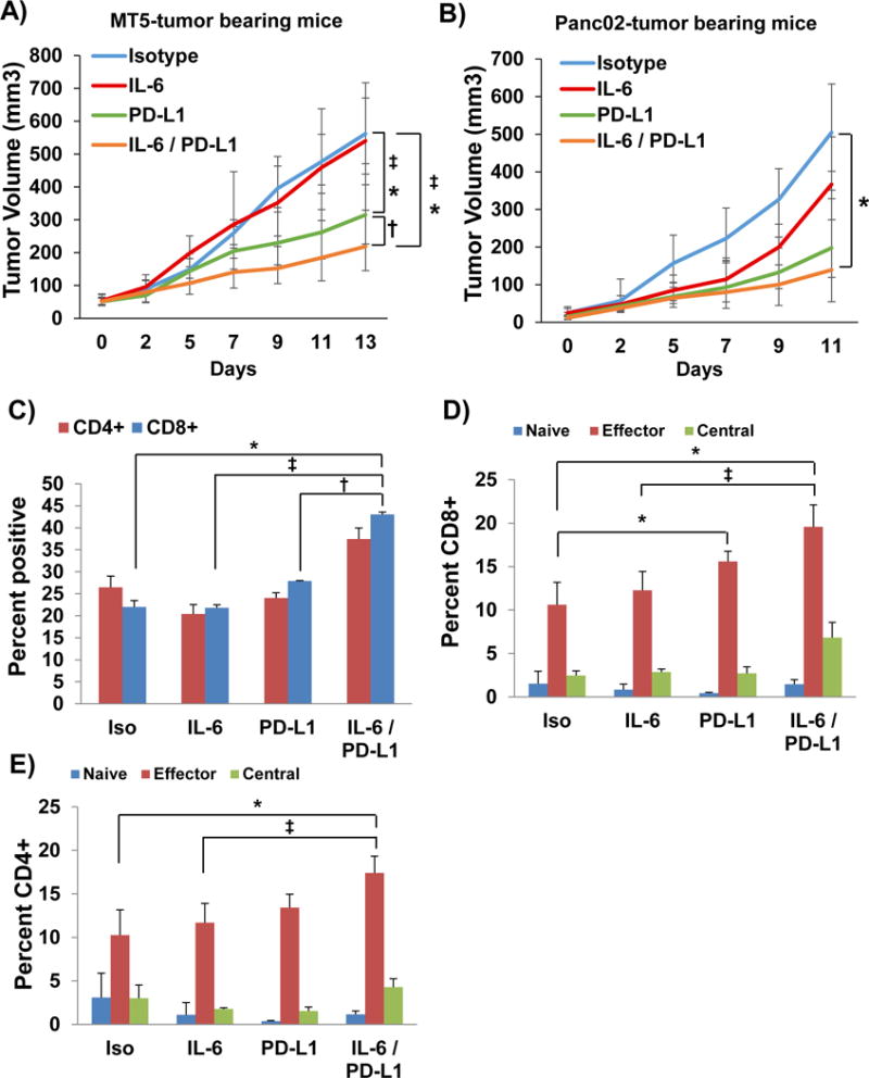
Results: PSCs (n=12) demonstrated prominent IL-6 expression, which was localised to stroma of tumours. Combined IL-6 and PD-L1 blockade elicited efficacy in mice bearing subcutaneous MT5 (p<0.02) and Panc02 tumours (p=0.046), which was accompanied by increased intratumoural effector T lymphocytes (CD62L-CD44-). CD8-depleting but not CD4-depleting Abs abrogated the efficacy of combined IL-6 and PD-L1 blockade in mice bearing Panc02 tumours (p=0.0016). This treatment combination also elicited significant antitumour activity in mice bearing orthotopic KPC-luc tumours and limited tumour progression in KPC-Brca2 mice (p<0.001). Histological analysis revealed increased T-cell infiltration and reduced α-smooth muscle actin cells in tumours from multiple models. Finally, IL-6 and PD-L1 blockade increased overall survival in KPC-Brca2 mice compared with isotype controls (p=0.0012).

Conclusions: These preclinical results indicate that targeted inhibition of IL-6 may enhance the efficacy of anti-PD-L1 in PDAC.

Keywords: IMMUNOTHERAPY; INTERLEUKINS; PANCREATIC CANCER.


Conflict of interest statement

Competing interests: None declared.

Figures

Figure 1
Figure 1. Profile of mRNA transcripts from patient-derived PSC
RNA was isolated from 10 patient-derived pancreatic cancer stellate cells and analyzed utilizing the Nanostring nCounter PanCan Immune Profiling Panel. Data are expressed as the fold change in expression as compared to a normal human pancreatic fibroblast cell line and relative to several housekeeping genes. Genes are ranked from highest fold-change to lowest. Heat map with hierarchical clustering for genes with at least 2 fold change up or down with a p<0.01 cutoff. Significantly higher expression is shown in red and lower expression in green.
Figure 2
Figure 2. IL-6 expression is enriched in the stromal compartments of human PDAC tissue
Human PDAC tumors (among n=13 patients) stained for IL-6 expression by immunohistochemistry. A) IL-6 (Brown Chromogen) IHC staining from three representative patient samples is displayed at 10× and 20× magnification. Tissue sections from each patient were stained with an isotype control Ab to account for background. B) Number of IL-6 positive cells per field was quantified.
Figure 3
Figure 3. PDAC GEMM recapitulate IL-6 pathway activation and PD-L1 expression observed in human disease
IHC analysis of representative H&E, pSTAT3 (brown)/α–SMA (pink), IL-6 (brown), and PD-L1 (brown) staining on tissue sections from human pancreatic cancer, orthotopic (KPC-luc), subcutaneous (MT5, Panc02), or pancreata from the following GEMM: KPC (KrasLSL-G12D, Trp53LSL-R270H, Pdx1-cre), and KPC-Brca2 (KrasLSL-G12D, Trp53LSL-R270H, Pdx1.cre, Brca2F/F). All images are presented at 40× magnification.
Figure 4
Figure 4. IL-6 and PD-L1 antibody blockade combination therapy decreases PDAC tumor progression and increases the percentage of intratumoral effector T cells
A) MT5 or B) Panc02 murine pancreatic tumor cells were subcutaneously injected into C57BL/6 mice with treatment beginning when tumors reached 50–100mm3. Mice were treated with 200μg (intraperitoneal injection 3 times/week) with isotype control, anti-IL-6 and/or anti-PD-L1 antibodies (n=5–6 mice/group) until mice met pre-specified IACUC-approved early removal criteria. Geometric means ± SD; *p<0.01 compared to Isotype; p<0.03 compared to PD-L1; p<0.05 compared to IL-6. Panc02 tumors were dissociated using Collagenase II and the Miltenyi Biotec gentleMACS dissociator to obtain a single cell suspension. Cells were stained and analyzed by flow cytometry for C) CD4+, CD8+ T cells and activation markers for different D) CD4+ and E) CD8+ T cell subsets (n=3/group). Naïve (CD62L+CD44), Effector Memory (CD62LCD44+), and Central Memory (CD62L+CD44+). Means ± SD; *p<0.002 compared to Isotype; p<0.01 compared to PD-L1; p<0.02 compared to IL-6.
Figure 5
Figure 5. The anti-tumor response to combined IL-6 and PD-L1 blockade is CD8+ T cell dependent
Panc02 murine pancreatic tumor cells were subcutaneously injected into C57BL/6 mice with treatment beginning when tumors reached 50–100mm3. Mice were also depleted of either CD4+ or CD8+ T cells by injecting depletion antibodies on days 5, 6, 8, 11, and 14. A) Representative flow cytometry and B) quantification of CD4 and CD8 staining of splenocytes to confirm depletion. Means ± SD; *p<0.001. C) On day 7, mice were treated with 200 μg (intraperitoneal injection 3 times/week) of isotype controls or anti-IL-6 and anti-PD-L1 antibodies combined (n=5 mice/group) until mice met pre-specified IACUC-approved early removal criteria. Means ± SD; *p<0.001 compared to isotype; p<0.002 CD8 depletion vs CD4 depletion.
Figure 6
Figure 6. IL-6 and PD-L1 antibody combination blockade reduces tumor growth in an orthotopic model of pancreatic cancer
C57BL/6 mice were orthotopically injected with luciferase expressing KPC cells (KPC-luc) in matrigel and A) tumor growth assessed and B) quantified by bioluminescent imaging weekly. C) Tumor weights at completion of study. Means ± SD; *p=0.05530 compared to isotype. Pancreatic tissue was stained for D) α–SMA+ stromal cells (Means ± SD; *p=0.0563 compared to Isotype) and E) CD3+ T cells (Means ± SD; *p=0.0553 compared to Isotype) by IHC quantified at 20× magnification.
Figure 7
Figure 7. IL-6 and PD-L1 antibody blockade combination therapy decreases PDAC tumor progression and α-SMA+ cells in the pancreata from KPC-Brca2 mice
KPC-Brca2 mice were treated at 5–6 weeks of age with 200 μg (intraperitoneal injection 3 times/week) of isotype control, anti-IL-6R and/or anti-PD-L1 antibodies for 2 weeks (n=5 mice/group). A cohort of mice treated with anti-IL-6R and PD-L1 antibodies were also depleted for CD8+ T cells. A) Representative H&E staining of pancreata at 20× magnification and B) quantification of the pathology. Means ± SD; *p<0.005 compared to Isotype; p=0.01) compared to PD-L1; C) Pancreatic tissue was stained for α–SMA+ stromal cells (red) by IHC and D) quantified at 20× magnification. Means ± SD; *p=0.0545 compared to Isotype.
Figure 8
Figure 8. Higher percentage of circulating T cells with Th1 phenotypic properties and intratumoral CD3+ cells in mice treated with IL-6 and PD-L1 antibody blockade
KPC-Brca2 mice were treated at 5–6 weeks of age with 200 μg (intraperitoneal injection 3 times/week) with isotype control, anti-IL-6R and/or anti-PD-L1 antibodies for 2 weeks (n=5 mice/group). Splenocytes were stained and analyzed by flow cytometry for A) T cells with Th1 phenotypic properties (CD4+CXCR4+CCR4CCR6) or B) T cells with Th2 phenotypic properties (CD4+CCR4+CXCR4CCR6). Means ± SD; *p<0.002 compared to Isotype; p=0.028 compared to IL-6. Pancreatic tissue was stained for C) CD3+ T cells (brown) by IHC and D) quantified at 20× magnification. Means ± SD; *p<0.05 compared to Isotype; p=0.052 compared to IL-6.
Figure 9
Figure 9. IL-6 and PD-L1 blockade increases overall survival of KPC-Brca2 mice
KPC-Brca2 beginning at 5 weeks of age mice were treated with isotype control antibodies or antibodies targeting IL-6 and PD-L1 (200μg/each) until mice were moribund and met pre-specified IACUC-approved early removal criteria. Kaplan-Meier survival curves with log-rank test for significance between isotype control and IL-6/PD-L1 antibodies (p=0.0012).

References

    1. Rahib L, Smith BD, Aizenberg R, et al. Projecting cancer incidence and deaths to 2030: the unexpected burden of thyroid, liver, and pancreas cancers in the United States. Cancer Res. 2014;74:2913–21. - PubMed
    1. Vincent A, Herman J, Schulick R, et al. Pancreatic cancer. Lancet. 2011;378:607–20. - PMC - PubMed
    1. Al-Hajeili M, Azmi AS, Choi M. Nab-paclitaxel: potential for the treatment of advanced pancreatic cancer. Onco Targets Ther. 2014;7:187–92. - PMC - PubMed
    1. Zhang Y, Yan W, Collins A, et al. Interleukin-6 is required for pancreatic cancer progression by promoting MAPK signaling activation and oxidative stress resistance. Cancer Res. 2013;73:6359–74. - PMC - PubMed
    1. Lesina M, Kurkowski MU, Ludes K, et al. Stat3/Socs3 activation by IL-6 transsignaling promotes progression of pancreatic intraepithelial neoplasia and development of pancreatic cancer. Cancer Cell. 2011;19:456–69. - PubMed

Publication types

MeSH terms