. 2016 Dec 1;25(23):5069-5082.
doi: 10.1093/hmg/ddw327.

Modelling C9orf72 dipeptide repeat proteins of a physiologically relevant size

Affiliations

Modelling C9orf72 dipeptide repeat proteins of a physiologically relevant size

Janis Bennion Callister et al. Hum Mol Genet. .

Abstract

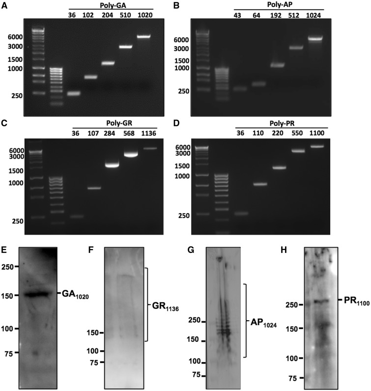
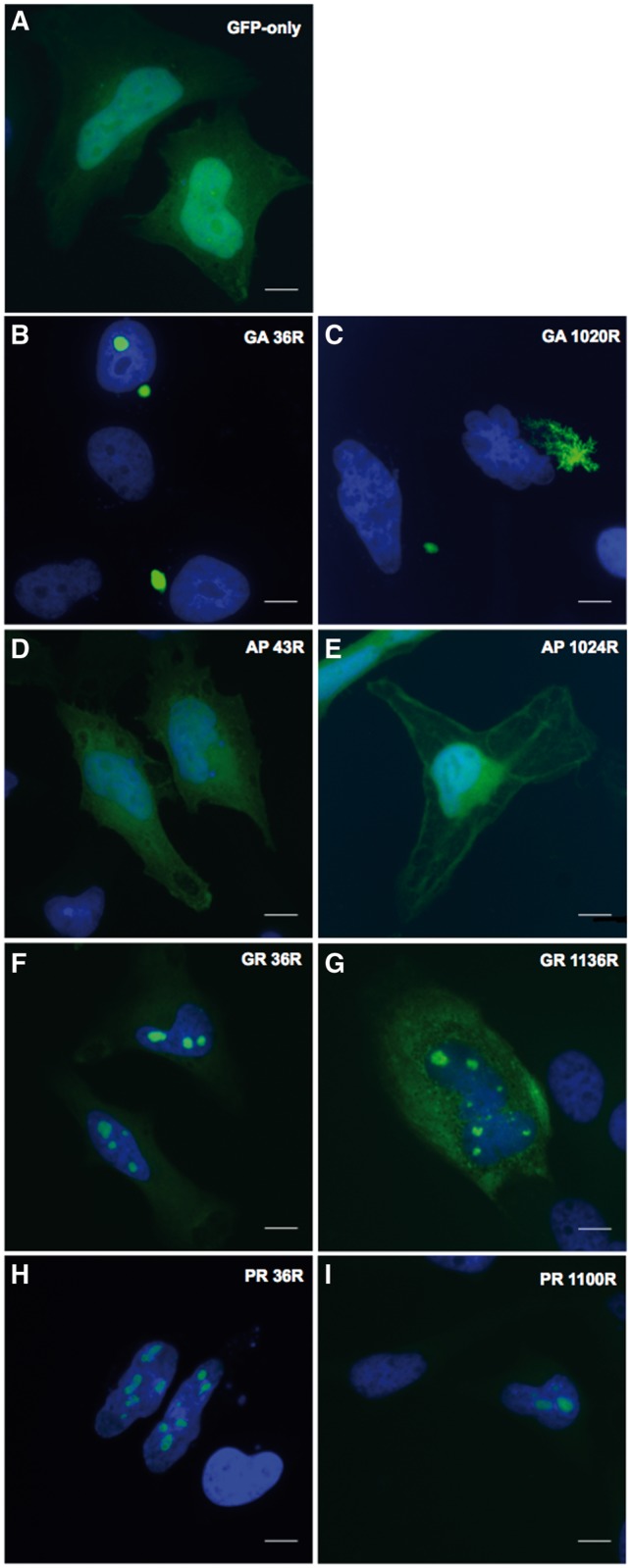
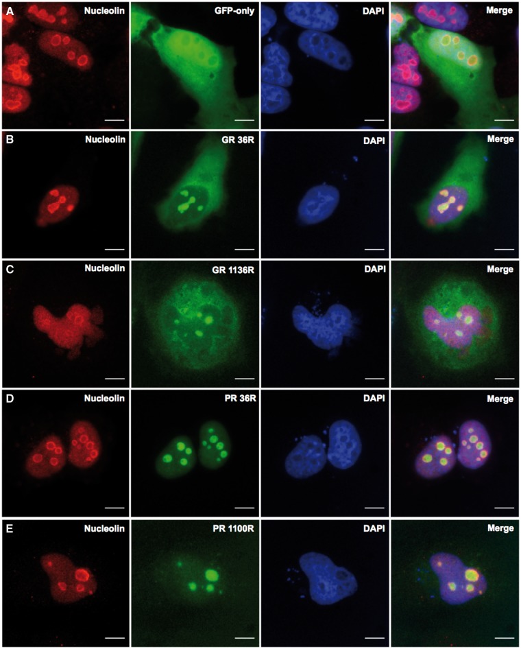
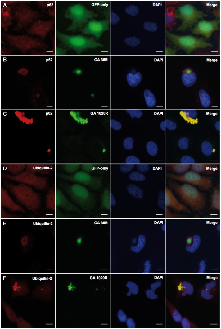
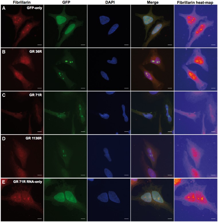
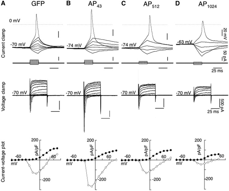
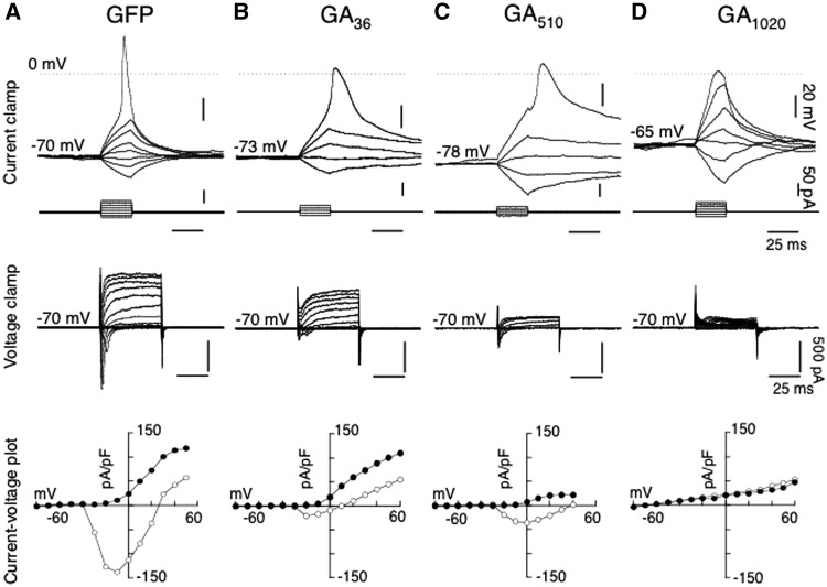
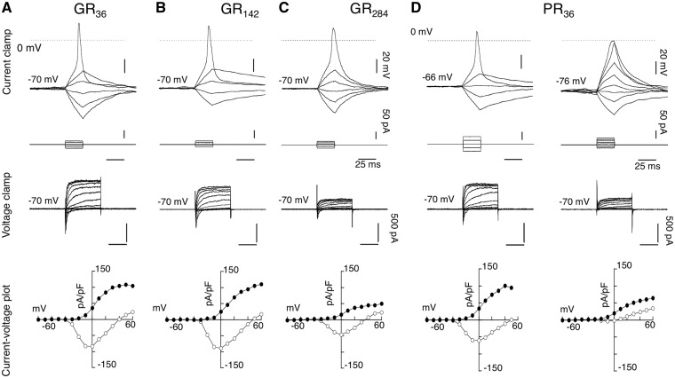
C9orf72 expansions are the most common genetic cause of FTLD and MND identified to date. Although being intronic, the expansion is translated into five different dipeptide repeat proteins (DPRs) that accumulate within patients' neurons. Attempts have been made to model DPRs in cell and animals. However, the majority of these use DPRs repeat numbers much shorter than those observed in patients. To address this we have generated a selection of DPR expression constructs with repeat numbers in excess of 1000 repeats, matching what is seen in patients. Small and larger DPRs produce inclusions with similar morphology but different cellular effects. We demonstrate a length dependent effect using electrophysiology with a phenotype only occurring with the longest DPRs. These data highlight the importance of using physiologically relevant repeat numbers when modelling DPRs.


Figures

Figure 1.
Figure 1.
Ethidium bromide-labelled agarose gels showing repeat sequences from GFP-tagged (A) poly-GA, (B) poly-AP, (C) poly-GR and (D) poly-PR constructs at a range of different repeat lengths. Repeat sequences were isolated from DPR constructs by double-digest with BamH1 and EcoR1 followed by gel purification. Band sizes shown as number of bases on left. Repeat-length indicated by numbers above. DNA ladders shown in left hand lanes are Bioline Hyperladders 1 kb+ and 100 bp. E-H Western blots confirming expression of full-length GFP-tagged DPR proteins in HeLa cells, 24 h post-transfection. Predicted sizes of GFP-tagged peptides are: GA1020 157.6 kDa; GR1136 269.2 kDa; AP1024 172.3 kDa and PR1100 278.7 kDa. Antibodies used were Santa-Cruz anti-GFP (E, F), ProteinTech anti-AP (G) and ProteinTech anti-PR (H). All antibodies diluted 1:1000 in 4% BSA.
Figure 2.
Figure 2.
Differential distribution of GFP-tagged DPRs (green) in HeLa cells at 24h post-transfection, or pEGFP-N1 expression as a GFP-only control (A). Representative images are shown at short and long repeat-lengths for Poly-GA (B,C), poly-AP (D-E), poly-GR (F-G) and poly-PR (H-I). Repeat lengths are denoted by the letter ‘R’. Nuclei are labelled in blue (DAPI). Images captured at x60 magnification. Scale bars represent 15 µm.
Figure 3.
Figure 3.
Arginine-rich DPRs translocate to the nucleolus in HeLa cells, as demonstrated by co-localization with the nucleolar marker protein, nucleolin (red). Nucleolin staining in GFP-only control cells expressing pGFP-N1 (green) is shown in A. Poly-GR (green) co-localizes with nucleolin at the short and long lengths of 36 and 1136 repeats (B and C, respectively). Poly-PR (green) also co-localizes with nucleolin at the short and long lengths of 36 and 1100 repeats (D and E). Images captured at x60 magnification, 24 h post-transfection. Scale bars represent 15 µm.
Figure 4.
Figure 4.
Co-localization of poly-GA with components of the ubiquitin-protease system in HeLa cells, 48h post-transfection. GFP-tagged GA36 and GA1020 (green) co-localised with (B, C) p62 (red) and (E, F) ubiquilin-2 (red). The distribution of p62 and ubiquilin-2 in GFP-only control cells transfected with pEGFP-N1 are shown in panels A and D respectively. Nuclei are labelled in blue (DAPI). Images captured at x60 magnification. Scale bars represent 15µm.
Figure 5.
Figure 5.
Poly-GR peptide, not RNA, causes a nucleolar stress phenotype. Heat-maps showing loss or re-distribution of fibrillarin in cells expressing nucleolar GR1136 compared to GFP-only control. Yellow ‘hot-spots’ indicate areas of high intensity fibrillarin staining, with lower signal areas displayed in red, purple and blue, in decreasing order. (A) Fibrillarin forms punctate nuclear ‘hot-spots’ corresponding to the nucleoli in cells expressing pEGFP-N1. Yellow hot-spots become more diffuse with increasing repeat-length of poly-GR from GR36(B) until no yellow is visible at GR1136(D). (E) Expression of an RNA-only poly-GR construct did not affect fibrillarin distribution. Images captured in HeLa cells at x60, 48 h post-transfection. Scales bars represent 15 µm.
Figure 6.
Figure 6.
Electrophysiological profiles from differentiated SH-SY5Y cells transfected with GFP alone and APn-GFP, under whole cell current and voltage clamp recording mode. Top traces depicted whole cell current clamp recordings in representative cells transfected with GFP (A), AP43-GFP (B), AP512-GFP (C) and PA1024-GFP (D), where voltage traces in response to hyperpolarizing and depolarizing steps (25 ms duration, 10 pA increment, corresponding traces shown below). Depolarizing steps to a threshold value elicited an action potential from their respective holding potentials. Middle traces show the current responses under whole cell voltage clamp mode of the corresponding cells shown in GFP (A), AP43-GFP (B), AP512-GFP (C) and PA1024-GFP (D) at a command holding potential of -70 mV to a series of hyperpolarizing and depolarizing pulses (50 ms, 10 mV increment), and (bottom traces), the resultant I-V plots of peak current (pA/pF) (open circle) and currents (pA/pF) at the end of 50 ms (closed circle).
Figure 7.
Figure 7.
Comparison of electrophysiological profiles from differentiated SH-SY5Y cells transfected with GFP alone and GAn-GFP, under whole cell current and voltage clamp recording mode. Top traces illustrated whole cell current clamp recordings in a control cell transfected with GFP (A, shown inFigure 1), and representative cells with cytoplasmic inclusions following transfection with GA36-GFP (B), GA510-GFP (C) and GA1020-GFP (D), where voltage traces in response to hyperpolarizing and depolarizing steps (25 ms duration, 10 pA increment, corresponding traces shown below), revealing the effect of the increasing repeat lengths of GAn-GFP on action potential waveform from their respective holding potentials. Middle traces show the current responses under whole cell voltage clamp mode of the corresponding cells shown in GFP (A), AP43-GFP (B), AP512-GFP (C) and PA1024-GFP (D) at a command holding potential of − 70 mV to a series of hyperpolarizing and depolarizing pulses (50 ms, 10 mV increment), and (bottom traces), the resultant I–V plots of peak current (pA/pF) (open circle) and currents (pA/pF) at the end of 50 ms (closed circle).
Figure 8.
Figure 8.
Electrophysiological profile of arginine-containing DPRs: Glycine-arginine (GR) and proline-arginine (PR) proteins in differentiated SH-SY5Y cells. (A), a representative cell expressing GR36-GFP, (B), a cell expressing GR142-GFP, (C) a cell expressing GR284-GFP and (D) representative cells expressing PR36-GFP. (Top traces), current clamp responses (25 ms, 10 pA increment) elicited action potential waveform in response to a threshold stimulation. (Middle traces) showing the corresponding current traces under voltage clamp from −70 mV (50 ms, 10 mV increment), and (bottom traces), the resultant I-V plots of peak current (pA/pF) (open circle) and currents (pA/pF) at the end of 50 ms (closed circle). Note, that recordings with PR even at the lowest repeats of 36 repeats evoked two types electrophysiological profiles, either ‘normal’ or ‘lack’ of action potential.

References

    1. Majounie E., Renton A.E., Mok K., Dopper E.G., Waite A., Rollinson S., Chio A., Restagno G., Nicolaou N., Simon-Sanchez J., et al. (2012) Frequency of the C9orf72 hexanucleotide repeat expansion in patients with amyotrophic lateral sclerosis and frontotemporal dementia: a cross-sectional study. Lancet Neurol., 11, 323–330. - PMC - PubMed
    1. Renton A.E., Majounie E., Waite A., Simon-Sanchez J., Rollinson S., Gibbs J.R., Schymick J.C., Laaksovirta H., van Swieten J.C., Myllykangas L., et al. (2011) A hexanucleotide repeat expansion in C9ORF72 is the cause of chromosome 9p21-linked ALS-FTD. Neuron, 72, 257–268. - PMC - PubMed
    1. DeJesus-Hernandez M., Mackenzie I.R., Boeve B.F., Boxer A.L., Baker M., Rutherford N.J., Nicholson A.M., Finch N.A., Flynn H., Adamson J., et al. (2011) Expanded GGGGCC hexanucleotide repeat in noncoding region of C9ORF72 causes chromosome 9p-linked FTD and ALS. Neuron, 72, 245–256. - PMC - PubMed
    1. Levine T.P., Daniels R.D., Gatta A.T., Wong L.H., Hayes M.J. (2013) The product of C9orf72, a gene strongly implicated in neurodegeneration, is structurally related to DENN Rab-GEFs. Bioinformatics, 29, 499–503. - PMC - PubMed
    1. Zhang D., Iyer L.M., He F., Aravind L. (2012) Discovery of novel DENN proteins: implications for the evolution of eukaryotic intracellular membrane structures and human disease. Front. Genet., 3, 283.. - PMC - PubMed

Publication types

MeSH terms