. 2016 Dec 15;27(25):4021-4032.
doi: 10.1091/mbc.E16-04-0239. Epub 2016 Oct 19.

Maintaining protein homeostasis: early and late endosomal dual recycling for the maintenance of intracellular pools of the plasma membrane protein Chs3

Affiliations

Maintaining protein homeostasis: early and late endosomal dual recycling for the maintenance of intracellular pools of the plasma membrane protein Chs3

Irene Arcones et al. Mol Biol Cell. .

Abstract

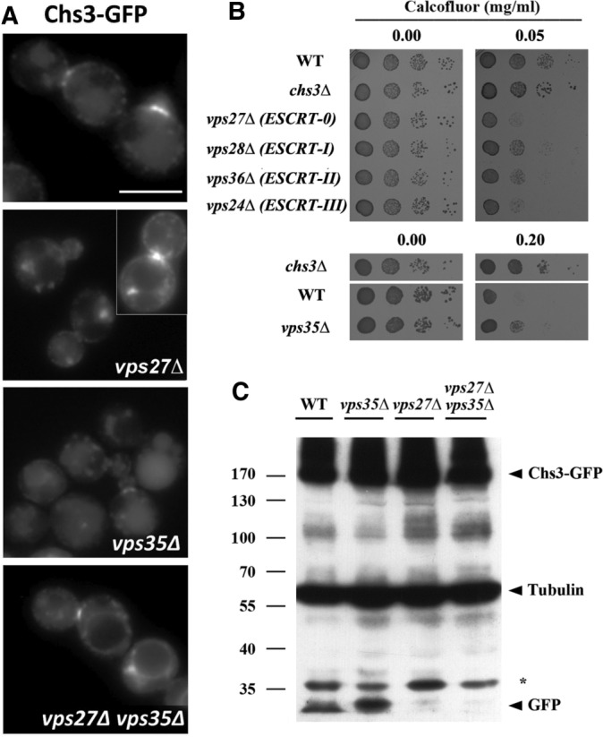
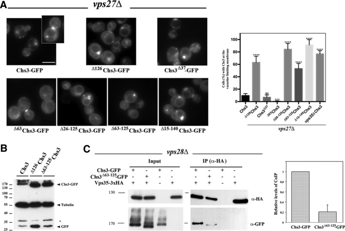
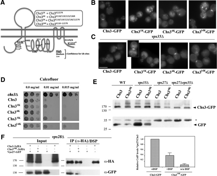
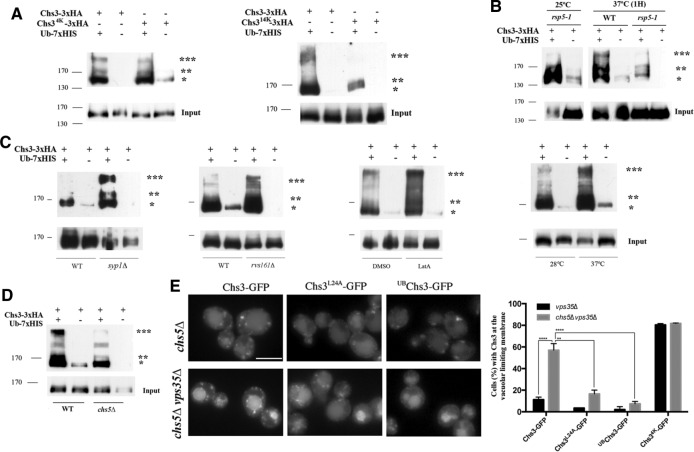
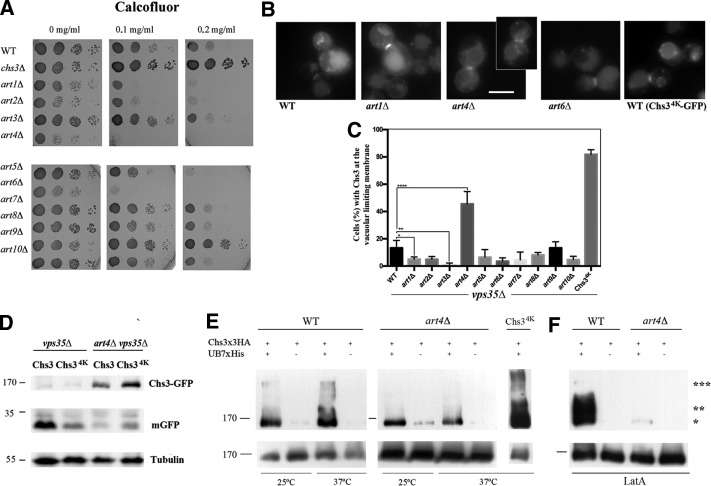
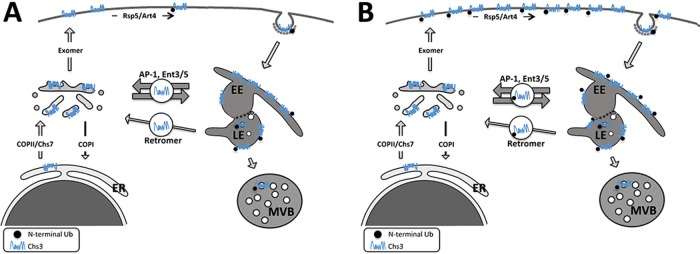
The major chitin synthase activity in yeast cells, Chs3, has become a paradigm in the study of the intracellular traffic of transmembrane proteins due to its tightly regulated trafficking. This includes an efficient mechanism for the maintenance of an extensive reservoir of Chs3 at the trans-Golgi network/EE, which allows for the timely delivery of the protein to the plasma membrane. Here we show that this intracellular reservoir of Chs3 is maintained not only by its efficient AP-1-mediated recycling, but also by recycling through the retromer complex, which interacts with Chs3 at a defined region in its N-terminal cytosolic domain. Moreover, the N-terminal ubiquitination of Chs3 at the plasma membrane by Rsp5/Art4 distinctly labels the protein and regulates its retromer-mediated recycling by enabling Chs3 to be recognized by the ESCRT machinery and degraded in the vacuole. Therefore the combined action of two independent but redundant endocytic recycling mechanisms, together with distinct labels for vacuolar degradation, determines the final fate of the intracellular traffic of the Chs3 protein, allowing yeast cells to regulate morphogenesis, depending on environmental constraints.


Figures

FIGURE 1:
FIGURE 1:
Chs3 transits through the late endosomal compartment. (A) Localization of Chs3-GFP in the indicated ESCRT and retromer mutants. Note the clear appearance of the vacuolar space in the vps27Δ mutant and the appearance of neat signals in the vacuolar limiting membrane in the double vps27Δ vps35Δ mutant. In both strains, the retention of Chs3 in the class E compartment can be observed. (B) Calcofluor resistance of the indicated mutants. Note the hypersensitivity of all ESCRT mutants but the moderate resistance of the retromer (vps35Δ) mutant. (C) Western blot showing the amounts of Chs3-GFP and tubulin in the indicated mutants. Note the band corresponding to the vacuolar free form of GFP.
FIGURE 2:
FIGURE 2:
The role of the N- and C-terminal regions of Chs3 in its trafficking through the LE pathway. (A) Localization of the indicated forms of Chs3-GFP in the vps27Δ mutant. In this strain, the different forms of Chs3 accumulate in the class E compartment and the vacuolar space is clear in all cases, but only some of the constructs stain the vacuolar limiting membrane. The graph represents the percentage of cells showing vacuolar membrane staining for each construct, including the numerical value for Chs3 in the double mutant vps27Δ vps35Δ. Note the absence of this signal for wild-type, Δ63Chs3, and Chs3Δ37 proteins. (B) Western blot showing the amounts of Chs3-GFP and tubulin for the indicated constructs. Note that the band corresponding to the vacuolar free form of GFP increased for proteins lacking the N-terminal domain. (C) CoIP assay after DSP cross-linking, showing Vps35/Chs3 interaction in the ESCRT mutant vps28Δ. Vps35 was immunoprecipitated with the anti-HA antibody, and the precipitate was developed with the anti-HA or anti-GFP as indicated. Note the significant levels of coIP between Chs3 and Vps35 proteins but the reduced coIP levels between Vps35 and Δ63-126Chs3. Quantitative analysis of the CoIP is presented in the graph representing the relative amounts of coimmunoprecipitated protein compared with the immunoprecipitated bait. The data included represent the average of four independent experiments.
FIGURE 3:
FIGURE 3:
Ubiquitination regulates Chs3 traffic. (A) Bioinformatic prediction of Chs3 ubiquitination sites (UbPred; www.ubpred.org). The asterisk marks the lysine sites confirmed experimentally to be ubiquitinated (Swaney et al., 2013). Replacement of the indicated lysine residues by arginines was carried out to create the different Chs3 mutants indicated. (B) Localization of the different Chs3 constructs in the wild-type strain. Note the similarity of the staining. (C) Localization of the different forms of Chs3-GFP in the vps35Δ mutant. Note the neat staining at the vacuolar limiting membrane in only the Chs34K- and Chs314K-containing strains. (D) Resistance to calcofluor of the strains carrying the different forms of Chs3 as a single copy in the pRS315 plasmid. (E) Western blot of the total extract from the indicated strains containing Chs3-GFP or Chs34K-GFP. Both are from the same Western blot, but the lower blot is overexposed to highlight the free GFP band. Note the reduced liberation of free GFP from Chs34K constructs. (F) CoIP between Chs3/Chs314K and Vps27. Protein extracts were obtained from cells of the vps28Δ mutant (Shi et al., 2011) containing the indicated construct. Experiments were performed after DSP cross-linking. Chs3-3xHA was immunoprecipitated with the anti-HA antibody and the immunoprecipitated developed with anti-HA or anti-GFP as indicated. Relative levels of CoIP are shown in the graph, and the values are the average of two independent experiments. Note the reduced level of CoIP between the Chs314K and Vps27 proteins, which is indicative of the poor recognition of the nonubiquitinated Chs3 protein by the ESCRT complex.
FIGURE 4:
FIGURE 4:
The ubiquitination status of Chs3. (A) Ubiquitination patterns of the indicated forms of Chs3. (B) Ubiquitinated forms of Chs3 in the rps5-1 mutant grown at permissive and restrictive temperatures. Note the increased ubiquitination of Chs3 at 37°C in the wild type but also the reduced ubiquitinated forms of Chs3 in the rps5-1 mutant at this temperature. (C) Ubiquitination of Chs3 in some endocytosis mutants, after LatA treatment and after thermal stress. Note the general increase of ubiquitination in all cases and also the distinct increased intensity of some bands. (D) Ubiquitination of Chs3 in the chs5Δ mutant. Note the significant reduction of bands indicated by double and triple asterisks. (E) Localization of the indicated forms of Chs3 in the chs5Δ or chs5Δ vps35Δ mutant. Note the significant accumulation of Chs3-GFP at the vacuolar limiting membrane, alleviated by the rerouting of the protein to the PM by the L24A mutation (Starr et al., 2012). A permanent ubiquitin tag in the N-terminal of Chs3 (UBChs3-GFP) also prevents its accumulation in the vacuolar limiting membrane. The graph on the right represents the quantitative average data from three independent experiments. The statistical significance of the differences is indicated. To determine Chs3 ubiquitination patterns, total ubiquitinated proteins were first enriched using the Co2+-TALON resin and separated in polyacrylamide gels, and blots were developed with the anti-HA antibody to visualize the tagged Chs3 protein. Note the occasional appearance of faint bands in the negative controls, most probably due to the nonspecific binding of Chs3-3xHA to the Co2+-TALON resin. Asterisks indicate the major forms of ubiquitinated Chs3 protein. Input images represent the relative levels of Chs3 protein in total cellular extracts, corresponding to one-fifth of the amount of protein enriched. See Materials and Methods and the text for more detailed explanations.
FIGURE 5:
FIGURE 5:
The role of Art4 in Chs3 trafficking. (A) Calcofluor resistance of the different arrestin (artΔ) mutants. Note the existence of hypersensitive and resistant mutants. (B) Localization of Chs3-GFP in the vps35Δ strain combined with different artΔ mutations as indicated and (C) quantitative analysis of the images; also see Supplemental Figure S4 for additional images. Note that only the art4Δ mutant shows significant accumulation of Chs3-GFP at the vacuolar limiting membrane. (D) Western blot showing Chs3-GFP levels in the art4Δ vps35Δ double mutant and the reduced liberation of free GFP. (E) Ubiquitination patterns of Chs3 in the art4Δ mutant after thermal stress at 37°C for 1 h. Note the lower amounts of ubiquitination in the art4Δ. Chs34K did not show apparent changes in the ubiquitination pattern compared with the control. (F) Ubiquitination patterns of Chs3 in the art4Δ mutant after LatA treatment for 1 h. Note the reduction of ubiquitination.
FIGURE 6:
FIGURE 6:
Chs3-GFP turnover after cell stress. (A) Microscopic localization of Chs3-GFP after temperature upshift or LatA treatment in the vps35Δ strain. Chs34K is included as a control. (B) Quantitative analysis of cells showing staining at the vacuolar limiting membrane after the indicated times. Cells were grown at 25°C (control) and shifted to 39°C for 30 or 60 min. (C) Quantitative vacuolar limiting membrane localization after LatA treatment for 60 min and further recovery in the absence of the drug for the indicated times. In both cases, the graph represents the average of three independent experiments, and in all cases, the localization of Chs34K was used as reference. Statistical analysis of the data (*p < 0.05).
FIGURE 7:
FIGURE 7:
Adaptation of the endosomal recycling of Chs3 to physiological conditions. (A) Under normal growth conditions, most Chs3 accumulates at the TGN/EE boundary, an intracellular reservoir replenished by the continuous supply of Chs3 from the ER. This reservoir is maintained by an extremely efficient endosomal recycling of Chs3 by the AP-1/Ent3/5 functions, preventing Chs3 degradation in the vacuole. The small amount of Chs3 that reaches the LE can be recycled back to TGN by the retromer, which acts as a safeguard mechanism. From this intracellular reservoir, a part of Chs3 is delivered by the exomer to the PM, where it is ubiquitinated at its N-terminal region by the Rsp5/Art4. This ubiquitin-tagged protein is endocytosed and travels to the LE compartment, where it is incorporated into ILVs for degradation in the vacuole. Accordingly, yeast cells distinguish the Chs3 protein that has been transported through the membrane from that retained at the TGN/EE. (B) Cellular stress triggers massive accumulation of Chs3 at the PM, depleting the TGN/EE reservoir. Under these conditions Chs3 is mostly ubiquitinated. On growth restoration, Chs3 is endocytosed, saturating the AP-1/Ent3/5 recycling mechanism and reaching the LE. Subsequently Chs3 is recycled by the retromer, which in this case contributes significantly to replenishing the TGN reservoir, avoiding massive Chs3 degradation. The color intensity and the width of the arrows reflect the relative contribution of these pathways to Chs3 trafficking. The model describes only the role of N-terminal ubiquitination of Chs3.

References

    1. Alvaro CG, O’Donnell AF, Prosser DC, Augustine AA, Goldman A, Brodsky JL, Cyert MS, Wendland B, Thorner J. Specific α-arrestins negatively regulate Saccharomyces cerevisiae pheromone response by down-modulating the G-protein-coupled receptor Ste2. Mol Cell Biol. 2014;34:2660–2681. - PMC - PubMed
    1. Barfield RM, Fromme JC, Schekman R. The exomer coat complex transports Fus1p to the plasma membrane via a novel plasma membrane sorting signal in yeast. Mol Biol Cell. 2009;20:4985–4996. - PMC - PubMed
    1. Becuwe M, Léon S. Integrated control of transporter endocytosis and recycling by the arrestin-related protein Rod1 and the ubiquitin ligase Rsp5. Elife. 2014;3:e03307. - PMC - PubMed
    1. Becuwe M, Vieira N, Lara D, Gomes-Rezende J, Soares-Cunha C, Casal M, Haguenauer-Tsapis R, Vincent O, Paiva S, Léon S. A molecular switch on an arrestin-like protein relays glucose signaling to transporter endocytosis. J Cell Biol. 2012;196:247–259. - PMC - PubMed
    1. Belgareh-Touzé N, Léon S, Erpapazoglou Z, Stawiecka-Mirota M, Urban-Grimal D, Haguenauer-Tsapis R. Versatile role of the yeast ubiquitin ligase Rsp5p in intracellular trafficking. Biochem Soc Trans. 2008;36:791–796. - PubMed

MeSH terms

Substances

LinkOut - more resources