. 2017 Aug;37(8):2806-2819.
doi: 10.1177/0271678X16675879. Epub 2016 Jan 1.

The rat cerebral vasculature exhibits time-of-day-dependent oscillations in circadian clock genes and vascular function that are attenuated following obstructive sleep apnea

Affiliations

The rat cerebral vasculature exhibits time-of-day-dependent oscillations in circadian clock genes and vascular function that are attenuated following obstructive sleep apnea

David J Durgan et al. J Cereb Blood Flow Metab. 2017 Aug.

Abstract

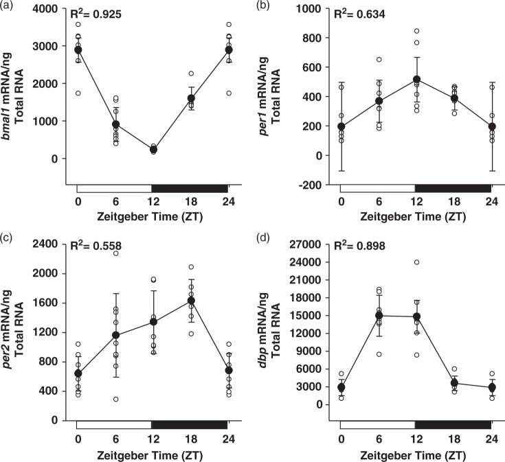
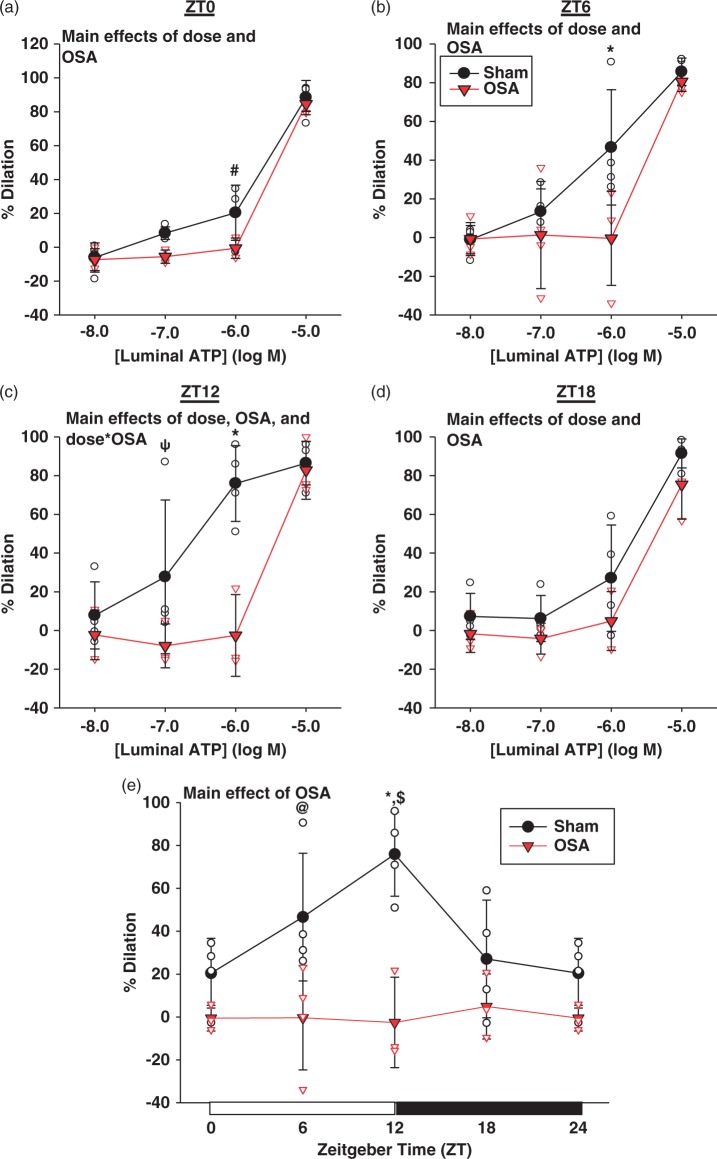
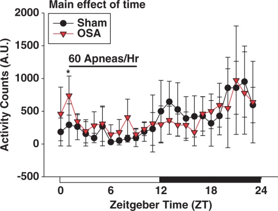
Circadian clock components oscillate in cells of the cardiovascular system. Disruption of these oscillations has been observed in cardiovascular diseases. We hypothesized that obstructive sleep apnea, which is associated with cerebrovascular diseases, disrupts the cerebrovascular circadian clock and rhythms in vascular function. Apneas were produced in rats during sleep. Following two weeks of sham or obstructive sleep apnea, cerebral arteries were isolated over 24 h for mRNA and functional analysis. mRNA expression of clock genes exhibited 24-h rhythms in cerebral arteries of sham rats (p < 0.05). Interestingly, peak expression of clock genes was significantly lower following obstructive sleep apnea (p < 0.05). Obstructive sleep apnea did not alter clock genes in the heart, or rhythms in locomotor activity. Isolated posterior cerebral arteries from sham rats exhibited a diurnal rhythm in sensitivity to luminally applied ATP, being most responsive at the beginning of the active phase (p < 0.05). This rhythm was absent in arteries from obstructive sleep apnea rats (p < 0.05). Rhythms in ATP sensitivity in sham vessels were absent, and not different from obstructive sleep apnea, following treatment with L-NAME and indomethacin. We conclude that cerebral arteries possess a functional circadian clock and exhibit a diurnal rhythm in vasoreactivity to ATP. Obstructive sleep apnea attenuates these rhythms in cerebral arteries, potentially contributing to obstructive sleep apnea-associated cerebrovascular disease.

Keywords: Cerebrovascular circulation; circadian clock; diurnal rhythm; obstructive sleep apnea; vasodilation.


Figures

Figure 1.
Figure 1.
Rhythmic expression of the circadian clock genes bmal1 (a), per1 (b), per2 (c), and the clock output gene dbp (d), in rat cerebral arteries isolated over the course of 24 h. R2 values resulting from cosinor analysis, assuming 24-h periodicity, are presented for each gene. Data are shown as the mean ± SD; n = 8 for ZT0/12/ 24, n = 9 for ZT6, and n = 7 for ZT18.
Figure 2.
Figure 2.
Rhythmic expression of the circadian clock genes bmal1 (a), per1 (b), per2 (c), and the clock output gene dbp (d), were not altered in the heart following two weeks of OSA. Data are shown as the mean ± SD; n = 3 for sham ZT0/6/24 and OSA ZT0/6/12/18/24, and n = 4 for sham ZT12/18. OSA: obstructive sleep apnea.
Figure 3.
Figure 3.
OSA significantly attenuated the rhythmic expression of clock and clock output genes in cerebral arteries. OSA reduced bmal1 expression at ZT6 (a). Peak expression of per1 (b), per2 (c), and dbp (d) were significantly attenuated at ZT12 in OSA cerebral arteries. Data are shown as the mean ± SD, n = 3 for sham ZT6/18 and OSA ZT0/12/18/24, n = 4 for sham ZT0/12/24 and OSA ZT6. *p = 0.039, #p = 0.046, ψp = 0.017 for sham vs. OSA at respective ZT. OSA: obstructive sleep apnea; ZT: zeitgeber.
Figure 4.
Figure 4.
Level of spontaneous tone developed by rat PCAs exhibits a diurnal variation, peaking during the active phase. OSA phase-shifts the rhythm in spontaneous tone developed, peaking during the middle of the sleep phase. Data are shown as the mean ± SD, n = 3 for sham ZT6/12/18 and OSA ZT0/12/18/24, n = 4 for sham ZT0/24 and OSA ZT6. *p < 0.001 and #p = 0.004 for sham vs. OSA at respective ZT. PCAs: posterior cerebral arteries; OSA: obstructive sleep apnea; ZT: zeitgeber.
Figure 5.
Figure 5.
Rat PCAs exhibit a diurnal rhythm in sensitivity to ATP induced vasodilation, peaking at the beginning of the active phase. OSA significantly decreases the sensitivity to ATP induced vasodilation (a–d), and abolishes the observed rhythm in ATP sensitivity (e). Data are shown as the mean ± SD, n = 3 for OSA ZT0/12/18/24, n = 4 for sham ZT0/6/12/18/24 and OSA ZT6. *p = 0.001, #p = 0.006, ψp = 0.041, @p = 0.003, for sham vs. OSA at respective ZT or ATP concentration, $p = 0.007 for sham ZT0 vs. ZT12. OSA: obstructive sleep apnea; ZT: zeitgeber.
Figure 6.
Figure 6.
Pretreatment of vessels with L-NAME and indomethacin abolished the diurnal rhythm in ATP sensitivity of sham PCAs. ATP sensitivity of OSA PCAs was not different than sham PCAs at any time point (a–e). Data are shown as the mean ± SD, n = 2 for OSA ZT0/24, n = 3 for sham ZT12 and OSA ZT6/12/18, n = 4 for sham ZT0/6/18/24. PCAs: posterior cerebral arteries; OSA: obstructive sleep apnea.
Figure 7.
Figure 7.
Two weeks of OSA, during the sleep cycle, did not significantly alter the diurnal variation in activity. Data are shown as the mean ± SD, n = 5 for sham, n = 6 for OSA, *p = 0.020 for sham vs. OSA at respective ZT. OSA: obstructive sleep apnea; ZT: zeitgeber.

References

    1. Edery I. Circadian rhythms in a nutshell. Physiol Genom 2000; 3: 59–74. - PubMed
    1. Wilsbacher LD, Takahashi JS. Circadian rhythms: Molecular basis of the clock. Curr Opin Genet Dev 1998; 8: 595–602. - PubMed
    1. Dunlap JC. Molecular bases for circadian clocks. Cell 1999; 96: 271–290. - PubMed
    1. Young ME. The circadian clock within the heart: Potential influence on myocardial gene expression, metabolism, and function. Am J Physiol Heart Circ Physiol 2006; 290: H1–H16. - PubMed
    1. Berson DM, Dunn FA, Takao M. Phototransduction by retinal ganglion cells that set the circadian clock. Science 2002; 295: 1070–1073. - PubMed

MeSH terms

Substances

LinkOut - more resources