Review
. 2017 Jan 18;28(1):23-32.
doi: 10.1021/acs.bioconjchem.6b00569. Epub 2016 Nov 16.

Biointerfacing and Applications of Cell Membrane-Coated Nanoparticles

Affiliations
Review

Biointerfacing and Applications of Cell Membrane-Coated Nanoparticles

Ashley V Kroll et al. Bioconjug Chem. .

Abstract

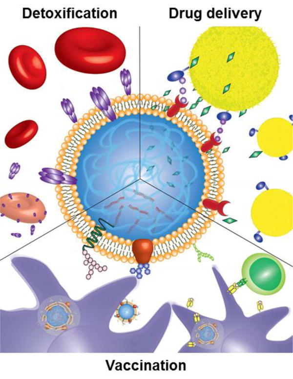
The cell membrane-coated nanoparticle is a biomimetic platform consisting of a nanoparticulate core coated with membrane derived from a cell, such as a red blood cell, platelet, or cancer cell. The cell membrane "disguise" allows the particles to be perceived by the body as the source cell by interacting with its surroundings using the translocated surface membrane components. The newly bestowed characteristics of the membrane-coated nanoparticle can be utilized for biological interfacing in the body, providing natural solutions to many biomedical issues. This Review will cover the interactions of these cell membrane-coated nanoparticles and their applications within three biomedical areas of interest, including (i) drug delivery, (ii) detoxification, and (iii) immune modulation.


Figures

Figure 1
Figure 1
Illustration showing three applications of cell membrane-coated nanoparticles. The particles can be used for targeted drug delivery to tumors, sites of inflammation, or pathogens via translocated surface markers (top right). They can also act as a decoy for toxins that damage cells through membrane interactions, safely detaining them and sparing their intended targets (top left). Finally, cell membrane-coated nanoparticles faithfully present antigens from their source cells and can be used for improved anticancer or antibacterial vaccination (bottom).
Figure 2
Figure 2
Stealth properties of RBC membrane-coated nanoparticles (RBC-NPs). (A) Schematic demonstrating transfer of CD47 onto RBC-NPs. (B) Transmission electron microscopy image of an RBC-NP and a bare NP stained with uranyl acetate (top) or immunostained for the extracellular domain of CD47 (bottom). (C) Quantitative flow cytometry of particle uptake into murine macrophage cells after a ten-minute incubation period. (D) CD47 density on RBC-NPs with different RBC membrane to polymer ratios. Adapted with permission from reference 42. Copyright 2013, Royal Society of Chemistry.
Figure 3
Figure 3
Platelet-mimicking nanovehicles (PM-NVs) for targeted cancer drug delivery. (A) Schematic of PM-NV targeting mechanisms to both circulating and primary tumor cells. (B) In vivo fluorescence imaging of PM-NV biodistribution in MDA-MB-231 tumor-bearing nude mice for (i) Cy5.5-labeled TRAIL-DOX-NVs (ii) and Cy5.5-labeled TRAIL-DOX-PM-NVs. (C) MDA-MB-231 tumor growth curves after intravenous injection of different TRAIL/DOX formulations. Error bars indicate s.d. (n = 5). Adapted with permission from reference 50. Copyright 2015, Wiley.
Figure 4
Figure 4
Platelet membrane-coated nanoparticles (PNPs) for biointerfacing. (A) Illustration demonstrating how PNPs are capable of multiple interactions in the body. (B) In vivo bacterial load in selected organs after treatment with free vancomycin at 10 mg/kg (Vanc-10), RBC-NP Vanc-10, PNP-Vanc-10, or free vancomycin at 60 mg/kg times the dosing (Vanc-60) in a systemic MRSA challenge model. (C) Histological cross-sections of rat arteries from a coronary restenosis model after different treatments (top; scale bar = 200 μm). Zoomed in images of arterial sections to highlight intima (I) to media (M) ratio (bottom; scale bar = 100 μm). Dtxl, docetaxel. Adapted with permission from reference 49. Copyright 2015, Macmillan Publishers Limited.
Figure 5
Figure 5
RBC membrane-coated supramolecular gelatin nanoparticles (SGNPs@RBC) for three-step antibiotic treatment. (A) Schematic showing nanoparticle formulation and antibiotic treatment steps. (B) Hemolysis activity by toxins from six different bacteria before (top) and after (bottom) treatment with SGNPs@RBC. (C) 2D (top) and 3D (bottom) confocal microscopy images of bacteria killing by vancomycin-loaded SGNPs@RBC when exposed to gelatinase-positive bacteria (S. aureus). Adapted with permission from reference 74. Copyright 2014, American Chemical Society.
Figure 6
Figure 6
Nanoparticle-detained toxins (nanotoxoid) for antivirulence vaccination. (A) Illustration of the nanotoxoid platform. (B) TUNEL assay on mouse skin 24 hours after injection with free α-hemolysin (Hla), heat-treated Hla (30 minutes), heat-treated Hla (60 minutes), or nanotoxoid(Hla). Scale bar = 400 μm. (C) Anti-Hla IgG titers over time for unvaccinated mice (black triangles) and mice immunized by nanotoxoid(Hla) with a single prime dose (blue circles) or a prime + boost (red circles). (D) Mouse survival rate over 15 days after bolus intravenous injection of Hla on day 21 after vaccination. Groups included no vaccination (black triangles), a single prime dose of heat-treated Hla (blue squares), a single prime dose of nanotoxoid(Hla) (blue circles), a prime + boost of heat-treated Hla (red squares), and a prime + boost of nanotoxoid(Hla) (red circles). Adapted with permission from reference 80. Copyright 2013, Macmillan Publishers Limited.
Figure 7
Figure 7
Cancer cell membrane-coated nanoparticles (CCNPs) for anticancer vaccination. (A) Depiction of CCNPs for antigen delivery to dendritic cells. (B) Quantitative flow cytometry data of dendritic cell maturation when incubated for 48 hours with CCNPs coated with membrane from B16-F10 mouse melanoma cancer cells (B16-F10 CCNPs), with or without the adjuvant MPLA. (C) Phase contrast microscopy images of splenocytes derived from pmel-1 transgenic mice when incubated with dendritic cells pulsed with B16-F10 CCNPs, with or without MPLA. Scale bar = 25 μm. (D) IFNγ ELISA of supernatant collected from co-culture at 24, 48, and 72 hours. UD, undetectable by ELISA. Adapted with permission from reference 56. Copyright 2014, American Chemical Society.

Similar articles

Cited by

References

    1. Kamaly N, Yameen B, Wu J, Farokhzad OC. Degradable Controlled-Release Polymers and Polymeric Nanoparticles: Mechanisms of Controlling Drug Release. Chem Rev. 2016;116:2602–2663. - PMC - PubMed
    1. Snipstad S, Westrom S, Morch Y, Afadzi M, Aslund AK, de Lange Davies C. Contact-Mediated Intracellular Delivery of Hydrophobic Drugs from Polymeric Nanoparticles. Cancer Nanotechnol. 2014;5:8. - PMC - PubMed
    1. Zhang S, Gao H, Bao G. Physical Principles of Nanoparticle Cellular Endocytosis. ACS Nano. 2015;9:8655–8671. - PMC - PubMed
    1. Blanco E, Shen H, Ferrari M. Principles of Nanoparticle Design for Overcoming Biological Barriers to Drug Delivery. Nat Biotechnol. 2015;33:941–951. - PMC - PubMed
    1. Bazak R, Houri M, Achy SE, Hussein W, Refaat T. Passive Targeting of Nanoparticles to Cancer: A Comprehensive Review of the Literature. Mol Clin Oncol. 2014;2:904–908. - PMC - PubMed

Publication types