. 2017;9(1):33-51.
doi: 10.1159/000449200. Epub 2016 Nov 1.

Vitronectin Binds to a Specific Stretch within the Head Region of Yersinia Adhesin A and Thereby Modulates Yersinia enterocolitica Host Interaction

Affiliations

Vitronectin Binds to a Specific Stretch within the Head Region of Yersinia Adhesin A and Thereby Modulates Yersinia enterocolitica Host Interaction

Melanie C Mühlenkamp et al. J Innate Immun. 2017.

Abstract

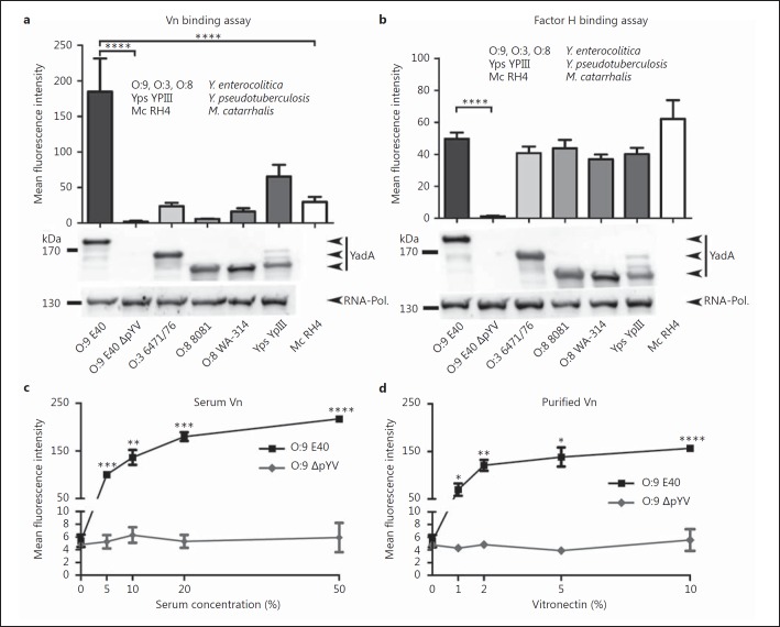
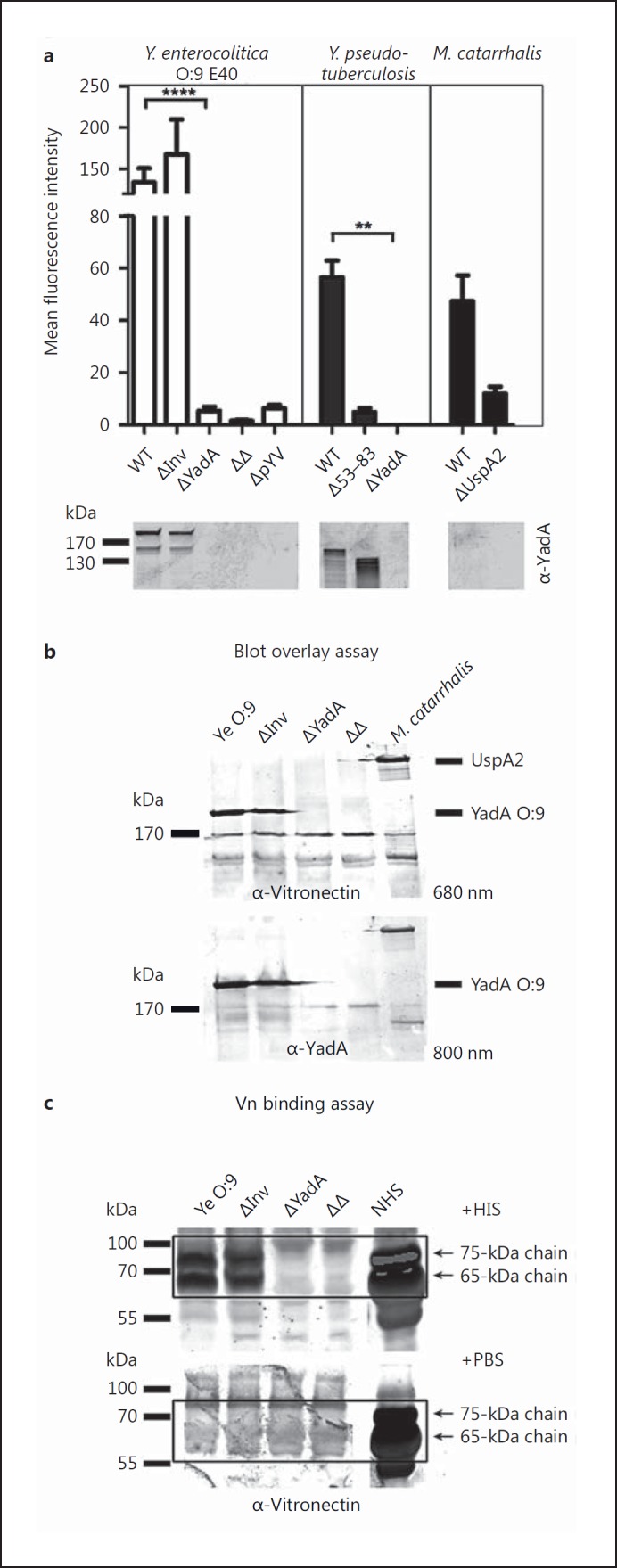
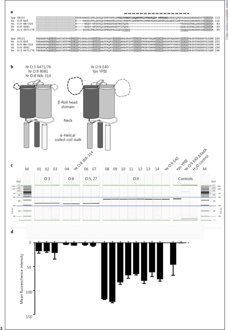
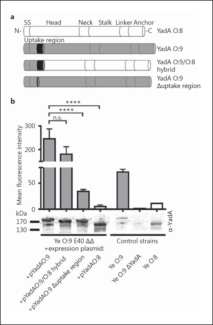
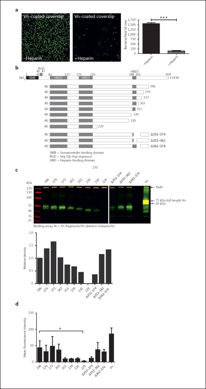
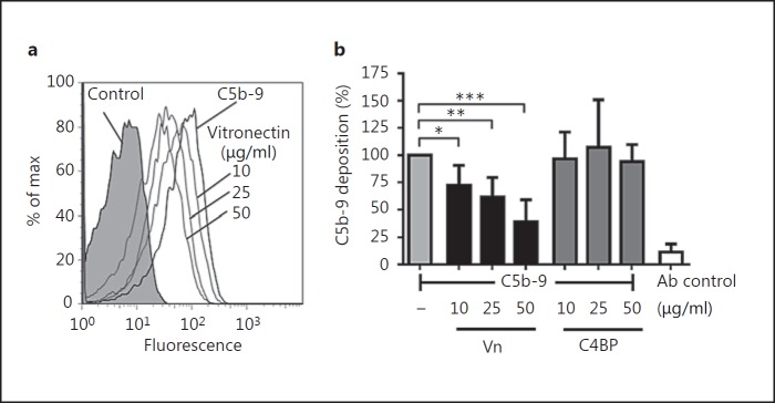
Complement resistance is an important virulence trait of Yersinia enterocolitica (Ye). The predominant virulence factor expressed by Ye is Yersinia adhesin A (YadA), which enables bacterial attachment to host cells and extracellular matrix and additionally allows the acquisition of soluble serum factors. The serum glycoprotein vitronectin (Vn) acts as an inhibitory regulator of the terminal complement complex by inhibiting the lytic pore formation. Here, we show YadA-mediated direct interaction of Ye with Vn and investigated the role of this Vn binding during mouse infection in vivo. Using different Yersinia strains, we identified a short stretch in the YadA head domain of Ye O:9 E40, similar to the 'uptake region' of Y. pseudotuberculosis YPIII YadA, as crucial for efficient Vn binding. Using recombinant fragments of Vn, we found the C-terminal part of Vn, including heparin-binding domain 3, to be responsible for binding to YadA. Moreover, we found that Vn bound to the bacterial surface is still functionally active and thus inhibits C5b-9 formation. In a mouse infection model, we demonstrate that Vn reduces complement-mediated killing of Ye O:9 E40 and, thus, improved bacterial survival. Taken together, these findings show that YadA-mediated Vn binding influences Ye pathogenesis.


Figures

Fig. 1
Fig. 1
Vn is efficiently bound by Ye O:9 E40 and Yps. a Several strains of Ye, serotype O:9 with and without virulence plasmid (O:9 E40 and O:9 E40 ΔpYV), serotype O:3 (O:3 6471/76) and serotype O:8 (O:8 8081; O:8 WA-314), and 1 Yps (Yps YPIII) WT strain were incubated with HIS, washed and subsequently analyzed for the presence of Vn on the bacterial surface by flow cytometry. Mc (Mc RH4), which is known to bind Vn and Yps, which we supposed also binds Vn, were included as a positive control for Vn binding. Ye O:9 E40, cured from the virulence plasmid (plasmid of Yersinia virulence; pYV) that encodes for the Ye T3SS, effector proteins and YadA, was included as a negative control because we surmised that Vn binding is pYV dependent. YadA protein levels were analyzed by Western blot analysis in whole-cell lysates and are shown below the bar chart (1 representative Western blot is shown). RNA polymerase protein (RNA-Pol.) was used as a loading control. YadAO:3 6471/76 has a calculated molecular weight of approximately 141 kDa (455 aa), YadAO:8 8081 of 132 kDa (422 aa), YadAO:8 WA-314 of 132 kDa (422 aa), YadAO:9 E40 of 153 kDa (487 aa), YadAYPIII of 135 kDa (434 aa) and UspA2H of approximately 272 kDa (876 aa). b To test if strain-specific differences in the binding of Vn are exclusive, we compared Vn binding levels to that of factor H. In contrast to Vn, factor H is bound in comparable amounts by all Yersinia strains tested, except for the negative control strain (O:9 E40 ΔpYV). The protein levels of YadA and the RNA polymerase as a loading control were analyzed by Western blots of whole-cell lysates and are shown below the bar chart (1 representative Western blot is shown). c Binding of serum-derived Vn to Ye O:9 E40 is dose dependent. Ye O:9 E40 and the pYV-cured version thereof were incubated with increasing serum concentrations. Afterwards, cell surface-associated Vn was quantified by flow cytometry. dYe O:9 E40 and the pYV-cured version thereof were incubated with increasing amounts of purified Vn. Afterwards, cell surface-associated Vn was quantified by flow cytometry. Binding of purified Vn to Ye O:9 E40 is dose-dependent. a–d Data are means ± SD of at least 4 individual experiments. a, b The main p values were determined by one-way ANOVA. p < 0.0001. Multiple comparisons were performed by one-way ANOVA with Dunnett's multiple-comparisons test. c, d The p values were determined by Student's t test. The error bars denote the SD. * p < 0.05, ** p < 0.01, *** p < 0.001, **** p < 0.0001.
Fig. 2
Fig. 2
Vn binding to Ye is YadA dependent. a Left panel: a Ye O:9 E40 WT strain or strains carrying individual deletions for the adhesins Invasin (ΔInv) or YadA (ΔYadA) and a respective double knockout strain (ΔΔ) as well as a virulence plasmid-cured strain (ΔpYV) were incubated with serum and washed, and then Vn binding was quantified by flow cytometry. Middle panel: Yps YPIII WT and corresponding strains lacking expression of ΔYadA or expressing a YadA version lacking part of the head domain (Δ53–83) were included as controls. Right panel: an Mc WT strain known to bind Vn via the surface adhesin UspA2 and a corresponding strain lacking expression of UspA2 (ΔUspA2) were included as positive and negative controls. YadA protein levels were analyzed by Western blot analysis of whole-cell lysates and are shown below the bar chart (1 representative blot is shown). b A selection of the strains used in (a) was tested for Vn binding in a blot overlay assay. Vn and YadA were detected on the identical blot with specific antibodies and differently labeled secondary antibodies (emission maximum at 680 and 800 nm, respectively) simultaneously. Vn is bound only in the presence of YadA (Ye) or UspA2, respectively. c In a direct binding assay, essentially performed as in a, Vn can be detected at the expected molecular weight (65 and 75 kDa) by Western blot only in those Ye strains expressing YadA. Data are means ± SD of at least 4 individual experiments (a) or 1/3 representative experiments is shown (b, c). The main p value was determined by one-way ANOVA (a: p < 0.0001). Multiple comparisons were performed by one-way ANOVA with Dunnett's multiple-comparisons test. The error bars denote the SD. ** p < 0.01, **** p > 0.0001.
Fig. 3
Fig. 3
A specific region in the YadA head domain is decisive for efficient binding of Vn. a Alignment of the head of various YadA variants. White letters on gray background: signal peptide. Black letters on gray background: the canonical ‘SVAIG’ head repeats of YadA. Italics: the neck region that links the head to the coiled-coil stalk of YadA. The insertion of Yps originally proposed by Heise and Dersch [18] is displayed in bold, and is slightly shifted towards the N-terminus of YadA. The dashed line on top shows the corrected position of the insertion, based on improved alignments and the structure of the YadA head from Ye O:3, where the short insertion is not resolved (underlined region). This and the unusually high number of prolines in this region suggest that it is not structured. The long version of the insertion carries a strongly positive net charge (+5 for Yps YPIII, +4 for the Ye O:9 E40), which probably plays a role in binding to fibronectin and Vn. b Schematic view of the differences in the YadA heads. The Yps YPIII and Ye O:9 E40 variants have long insertions in an unstructured loop region close to the N-terminus of the head. c PCR products comprising the YadA head region of Ye O:8 WA-314, Ye O:9 E40 with and without YadA, Yps YPIII and clinical isolates derived from fecal samples (Ye O:3, No. 01–03; Ye O:8, No. 04; Ye O:5,27, No. 06–07 and Ye O:9, No. 08–12) or blood (No. 13) were separated by capillary gel electrophoresis. The predicted length of PCR products was as follows: Ye O:3 346 bp; Ye O:8 337 bp; Ye O:9 451 bp; Ye O:5,27 346 bp, and Yps YPIII 442 bp. Water control and a YadA-deficient strain were included as negative controls. d The strains shown in c were tested for Vn binding. Cell surface-associated Vn after incubation in HIS was quantified by flow cytometry. One of 3 representative experiments is shown.
Fig. 4
Fig. 4
The uptake region is decisive for YadA-mediated Vn binding. Schematic representation of different YadA versions that were expressed from a plasmid in Ye O:9 E40 and analyzed for Vn binding capacity. The YadA versions tested comprise YadAO:8, YadAO:9, a YadAO:9/O:8 hybrid consisting of the O:9 head domain fused to the corresponding Ye O:8 head/stalk and membrane anchor domain (details: Material and Methods) and Ye YadAO:9 with the uptake region deleted (Δuptake region). b Flow cytometry analysis of Vn binding to different Ye strains carrying plasmids for inducible expression of the YadA versions depicted in a. As control strains, we used Ye strains expressing WT YadA from the endogenous pYV plasmid (Ye O:9 = positive control, Ye O:8) and a Ye O:9 E40 YadA-deficient strain (Ye O:9 ΔYadA = negative control). YadA protein levels were analyzed by Western blot analysis in whole-cell lysates and are shown below the bar chart. Data are means ± SD of at least 3 individual experiments (flow cytometry), or 1/3 representative experiments is shown (Western blot). The main p value was determined by one-way ANOVA (b, flow cytometry: p < 0.0001). Multiple comparisons were performed by one-way ANOVA with Dunnett's multiple-comparisons test. The error bars denote the SD. **** p > 0.0001.
Fig. 5
Fig. 5
Vn interacts with YadA via its C-terminal HBD-3. a Adhesion of Ye to Vn-coated coverslips can be blocked by heparin. b Schematic representation of Vn, the C-terminal-truncated Vn molecules [54] and the Vn molecules carrying deletions within and adjacent to the HBD-3 [35] that were used for a direct binding assay. c Western blot of a binding assay of Ye O:9 E40 with full-length Vn and all fragments depicted in b. Vn fragments appear in green; YadA, which was detected simultaneously, appears in yellow bands (trimer runs at approx. 200 kDa). d Flow cytometry analysis of Vn binding to Ye O:9 E40 with full-length Vn and all fragments depicted in b. Data are means ± SD of at least 3 individual experiments (a, d), or 1/3 representative experiments is shown. The p value for the comparison with and without heparin was determined by Student's t test. The main p value was determined by one-way ANOVA (d: p < 0.0001). Multiple comparisons were performed by one-way ANOVA with Dunnett's multiple-comparisons test. The error bars denote the SD. * p < 0.05, *** p < 0.001.
Fig. 6
Fig. 6
Vn is functionally active and inhibits the terminal pathway when bound to the surface of Ye. a Histogram overlay of flow cytometry analyses of TCC formation (detected by formation of the neoepitope C5b-9) on the surface of Ye O:9 E40 WT after preincubation of bacteria with PBS or different concentrations of Vn in PBS (10, 25 and 50 µg/ml). Preincubation with Vn reduces the amount of TCC that is formed. b Bar chart depicting C5b-9 deposition as percent of the amount of C5b-9 that was formed on the surface of bacteria preincubated with PBS only compared to bacteria preincubated with either Vn or C4BP at different concentrations (10, 25 and 50 µg/ml). Vn, but not C4BP, is able to reduce the formation of C5b-9. Antibody (Ab) control indicates background signal that was obtained using secondary antibody only for detection. Data are means ± SD of at least 3 individual experiments. The main p value was determined by one-way ANOVA (b: p < 0.001). Multiple comparisons were performed by one-way ANOVA with Dunnett's multiple-comparisons test. The error bars denote the SD. * p < 0.05; ** p < 0.01, *** p < 0.001.
Fig. 7
Fig. 7
Ye O:9 E40 is resistant to complement-mediated killing in vitro, and in an in vivo serum killing assay, Ye is more efficiently eliminated in the absence of Vn. a An in vitro serum killing assay using Ye O:9 E40, Ye O:9 E40 ΔYadA, Ye O:8 WA-314, Yps YPII, Ye O:9 E40 ΔΔ + pASK-IBA4C_yadAO:8, Ye O:9 E40 ΔΔ + pASK-IBA4C_yadAO:9, Ye O:9 E40 ΔΔ + pASK-IBA4C_yadAO:9/O:8 hybrid, Ye O:9 E40 ΔΔ + pASK-IBA4C_yadAO:9 Δuptake region. The serum bactericidal effect was calculated as the survival percentage. b WT and Vn−/− mice were infected intravenously with 1 × 10⁷Ye O:9 E40 for 30 min. After that, the mice were killed, and blood was withdrawn and plated on selective agar plates. CFU were determined by counting colonies the next day, shown as log10 CFU per gram of blood. a Data are means ± SD of at least 3 individual experiments. The main p value was determined by one-way ANOVA (p < 0.0001). Multiple comparisons were performed by one-way ANOVA with Dunnett's multiple-comparisons test. b The p value for the comparison of C57BL/6 and Vn−/− mice was determined by Student's t test. The horizontal lines denote the mean and the error bars denote the SD. * p < 0.05; ** p < 0.01, *** p < 0.001, **** p > 0.0001 (n = 6).

Similar articles

Cited by

References

    1. Bottone EJ. Yersinia enterocolitica: overview and epidemiologic correlates. Microbes Infect. 1999;1:323–333. - PubMed
    1. Cover TL, Aber RC. Yersinia enterocolitica. N Engl J Med. 1989;321:16–24. - PubMed
    1. Isberg RR. Mammalian cell adhesion functions and cellular penetration of enteropathogenic Yersinia species. Mol Microbiol. 1989;3:1449–1453. - PubMed
    1. Isberg RR. Determinants for thermoinducible cell binding and plasmid-encoded cellular penetration detected in the absence of the Yersinia pseudotuberculosis invasin protein. Infect Immun. 1989;57:1998–2005. - PMC - PubMed
    1. Miller VL, Falkow S. Evidence for two genetic loci in Yersinia enterocolitica that can promote invasion of epithelial cells. Infect Immun. 1988;56:1242–1248. - PMC - PubMed