

Toxicity of graphene-family nanoparticles: a general review of the origins and mechanisms
- PMID: 27799056
- PMCID: PMC5088662
- DOI: 10.1186/s12989-016-0168-y
Toxicity of graphene-family nanoparticles: a general review of the origins and mechanisms
Abstract
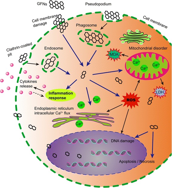
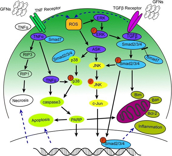
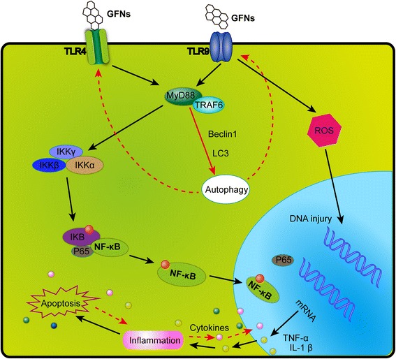
Due to their unique physicochemical properties, graphene-family nanomaterials (GFNs) are widely used in many fields, especially in biomedical applications. Currently, many studies have investigated the biocompatibility and toxicity of GFNs in vivo and in intro. Generally, GFNs may exert different degrees of toxicity in animals or cell models by following with different administration routes and penetrating through physiological barriers, subsequently being distributed in tissues or located in cells, eventually being excreted out of the bodies. This review collects studies on the toxic effects of GFNs in several organs and cell models. We also point out that various factors determine the toxicity of GFNs including the lateral size, surface structure, functionalization, charge, impurities, aggregations, and corona effect ect. In addition, several typical mechanisms underlying GFN toxicity have been revealed, for instance, physical destruction, oxidative stress, DNA damage, inflammatory response, apoptosis, autophagy, and necrosis. In these mechanisms, (toll-like receptors-) TLR-, transforming growth factor β- (TGF-β-) and tumor necrosis factor-alpha (TNF-α) dependent-pathways are involved in the signalling pathway network, and oxidative stress plays a crucial role in these pathways. In this review, we summarize the available information on regulating factors and the mechanisms of GFNs toxicity, and propose some challenges and suggestions for further investigations of GFNs, with the aim of completing the toxicology mechanisms, and providing suggestions to improve the biological safety of GFNs and facilitate their wide application.
Keywords: Future prospects; Graphene-family nanomaterials; Mechanisms; Physicochemical properties; Toxicity; Toxicokinetics.
Figures
Similar articles
-
Toxicology data of graphene-family nanomaterials: an update.Arch Toxicol. 2020 Jun;94(6):1915-1939. doi: 10.1007/s00204-020-02717-2. Epub 2020 Apr 2. Arch Toxicol. 2020. PMID: 32240330 Review.
-
Biological interactions of graphene-family nanomaterials: an interdisciplinary review.Chem Res Toxicol. 2012 Jan 13;25(1):15-34. doi: 10.1021/tx200339h. Epub 2011 Oct 21. Chem Res Toxicol. 2012. PMID: 21954945 Free PMC article. Review.
-
EPS-corona formation on graphene family nanomaterials (GO, rGO and graphene) and its role in mitigating their toxic effects in the marine alga Chlorella sp.Environ Pollut. 2024 Jan 15;341:123015. doi: 10.1016/j.envpol.2023.123015. Epub 2023 Nov 24. Environ Pollut. 2024. PMID: 38008250
-
Differential Toxicity of Graphene Family Nanomaterials Concerning Morphology.Adv Exp Med Biol. 2022;1351:23-39. doi: 10.1007/978-981-16-4923-3_2. Adv Exp Med Biol. 2022. PMID: 35175610
-
Graphene Family Nanomaterials in Ocular Applications: Physicochemical Properties and Toxicity.Chem Res Toxicol. 2021 Jun 21;34(6):1386-1402. doi: 10.1021/acs.chemrestox.0c00340. Epub 2021 May 27. Chem Res Toxicol. 2021. PMID: 34041903 Free PMC article. Review.
Cited by
-
Graphene oxide induces dose-dependent lung injury in rats by regulating autophagy.Exp Ther Med. 2021 May;21(5):462. doi: 10.3892/etm.2021.9893. Epub 2021 Mar 5. Exp Ther Med. 2021. PMID: 33747194 Free PMC article.
-
The Evaluation of Drug Delivery Nanocarrier Development and Pharmacological Briefing for Metabolic-Associated Fatty Liver Disease (MAFLD): An Update.Pharmaceuticals (Basel). 2021 Mar 4;14(3):215. doi: 10.3390/ph14030215. Pharmaceuticals (Basel). 2021. PMID: 33806527 Free PMC article. Review.
-
Potential role of calcium sulfate/β-tricalcium phosphate/graphene oxide nanocomposite for bone graft application_mechanical and biological analyses.J Orthop Surg Res. 2024 Oct 12;19(1):644. doi: 10.1186/s13018-024-05142-8. J Orthop Surg Res. 2024. PMID: 39396014 Free PMC article.
-
Molecular Dynamics Simulation of the Interaction between Graphene Oxide Quantum Dots and DNA Fragment.Materials (Basel). 2022 Nov 29;15(23):8506. doi: 10.3390/ma15238506. Materials (Basel). 2022. PMID: 36500001 Free PMC article.
-
Graphene Nanomaterials: Synthesis, Biocompatibility, and Cytotoxicity.Int J Mol Sci. 2018 Nov 12;19(11):3564. doi: 10.3390/ijms19113564. Int J Mol Sci. 2018. PMID: 30424535 Free PMC article. Review.
References
-
- Yang XY, Wang YS, Huang X, Ma YF, Huang Y, Yang RC, et al. Multi-functionalized graphene oxide based anticancer drug-carrier with dual-targeting function and pH-sensitivity. J Mat Chem. 2011;21(10):3448–54. doi: 10.1039/C0JM02494E. - DOI
Publication types
MeSH terms
Substances
LinkOut - more resources
Full Text Sources
Other Literature Sources
