. 2017 Jan 20;120(2):312-323.
doi: 10.1161/CIRCRESAHA.116.309006. Epub 2016 Oct 31.

Hypertension-Linked Pathophysiological Alterations in the Gut

Affiliations

Hypertension-Linked Pathophysiological Alterations in the Gut

Monica M Santisteban et al. Circ Res. .

Abstract

Rationale: Sympathetic nervous system control of inflammation plays a central role in hypertension. The gut receives significant sympathetic innervation, is densely populated with a diverse microbial ecosystem, and contains immune cells that greatly impact overall inflammatory homeostasis. Despite this uniqueness, little is known about the involvement of the gut in hypertension.

Objective: Test the hypothesis that increased sympathetic drive to the gut is associated with increased gut wall permeability, increased inflammatory status, and microbial dysbiosis and that these gut pathological changes are linked to hypertension.

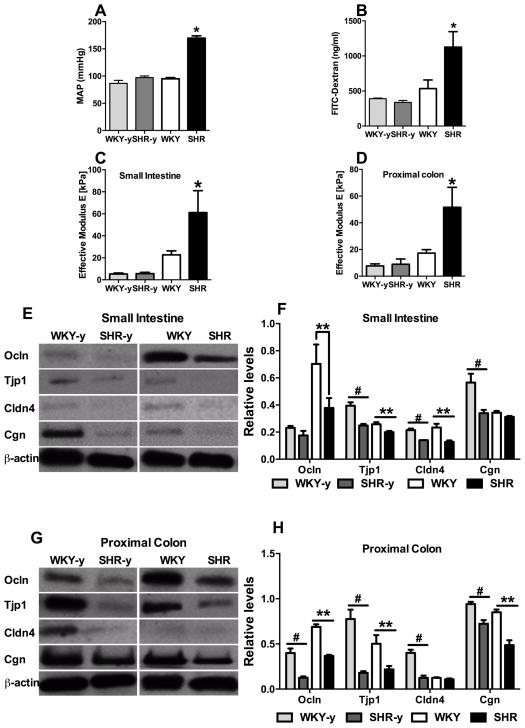
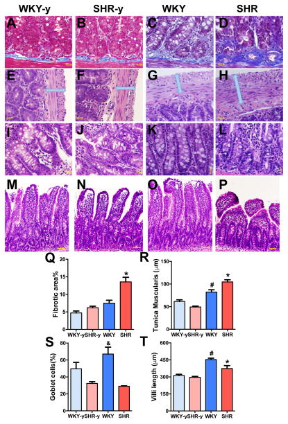
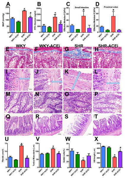
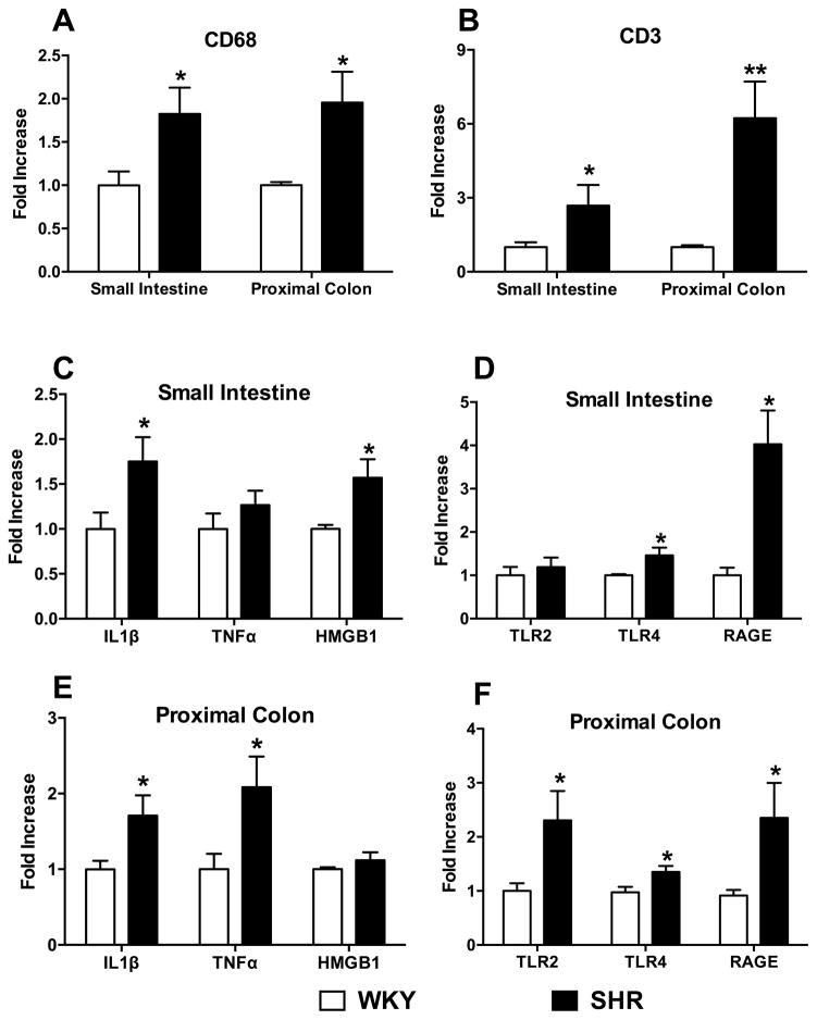
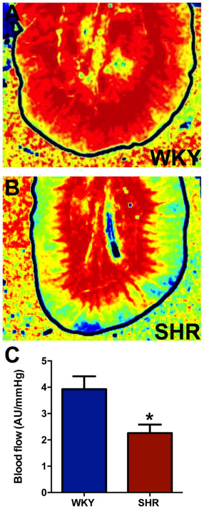
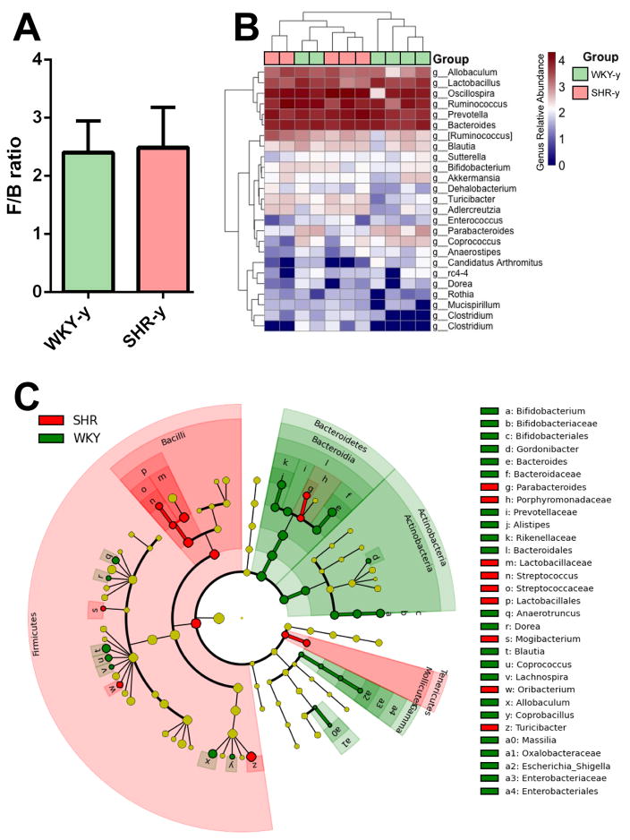
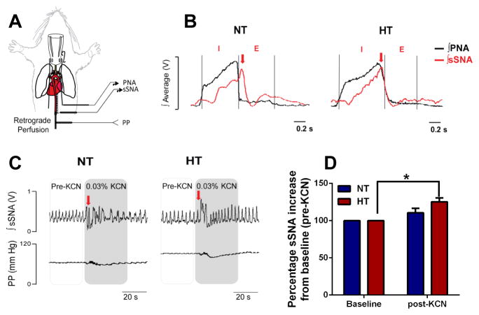
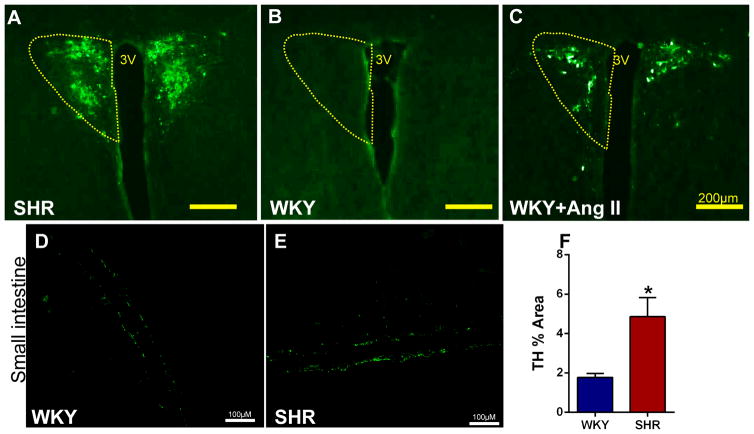
Methods and results: Gut epithelial integrity and wall pathology were examined in spontaneously hypertensive rat and chronic angiotensin II infusion rat models. The increase in blood pressure in spontaneously hypertensive rat was associated with gut pathology that included increased intestinal permeability and decreased tight junction proteins. These changes in gut pathology in hypertension were associated with alterations in microbial communities relevant in blood pressure control. We also observed enhanced gut-neuronal communication in hypertension originating from paraventricular nucleus of the hypothalamus and presenting as increased sympathetic drive to the gut. Finally, angiotensin-converting enzyme inhibition (captopril) normalized blood pressure and was associated with reversal of gut pathology.

Conclusions: A dysfunctional sympathetic-gut communication is associated with gut pathology, dysbiosis, and inflammation and plays a key role in hypertension. Thus, targeting of gut microbiota by innovative probiotics, antibiotics, and fecal transplant, in combination with the current pharmacotherapy, may be a novel strategy for hypertension treatment.

Keywords: autonomic nervous system; gut; hypertension; inflammation; intestines; microbiota.


Figures

Figure 1
Figure 1. Permeability and stiffness of small intestine and colon are increased in adult spontaneously hypertensive rat (SHR)
A, Mean arterial blood pressure (MAP) of young Wistar Kyoto rats WKY (WKY-y), young SHR (SHR-y), and adult WKY and SHR. B, Intestinal mucosa-to-blood permeability assessed by fluorescein isothiocyanate (FITC)-dextran 4kDa concentration in the plasma revealed increased FITC-dextran concentration in adult, but not young, SHR (n=4–7/group). C–D, Effective modulus measured by a Multi-Scale Indentation System is increased in both small intestine and proximal colon of WKY-y, SHR-y, and adult WKY, SHR (n=4–5/group). E–J, Western blotting for tight junction proteins in small intestine (E, G, I) and proximal colon (F, H, J) in WKY-y, SHR-y, and adult WKY, SHR. *p<0.05, SHR vs all other groups; # p<0.05, WKY-y vs SHR-y; **p<0.05, SHR vs WKY.
Figure 2
Figure 2. Pathological changes were observed in the small intestine of adult spontaneously hypertensive rat (SHR)
A–D, Small intestine was stained with Masson’s trichrome to quantify the fibrosis in young and adult WKY and SHR (Q, n=4/group). E–H, Cross sections of the small intestine of WKY and SHR were also stained with hematoxylin-eosin to measure the thickness of tunica muscularis layer (R). I–L, The number of goblet cells per 100 epithelial cells was decreased in SHR (S). M–P, Villi lengths were shorter in SHR (T). *p< 0.05 SHR vs all other groups; #p< 0.05 WKY vs all other groups; &p<0.05 WKY vs SHR-y and SHR.
Figure 3
Figure 3. Antihypertensive treatment with ACEi has beneficial effects on gut pathology
A, MAP of vehicle and captopril (ACEi) treated WKY and SHR (n=6–8/group). B, Intestinal mucosa-to-blood permeability assessed by FITC-dextran 4kDa concentration in the plasma revealed ACEi reverses increased permeability in the SHR. C–D, Mean effective modulus measured by a Multi-Scale Indentation System in both small intestine and proximal colon of vehicle and captopril treated WKY and SHR. E–H, Fibrotic area is decreased by ACEi in SHR (U). I–L, Cross sections of the small intestine of WKY and SHR shows protected tunica muscularis layer in ACEi treated SHR (V). M–P, The number of goblet cells per 100 epithelial cells was not improved by ACEi in SHR (W). Q–T, Villi lengths were improved by ACEi in SHR (X). *p<0.05 SHR vs all other groups; #p<0.05 SHR-ACEi vs SHR; +p<0.05 WKY-ACEi vs SHR-ACEi; φp<0.05 SHR vs WKY; ^p<0.05 SHR-ACEi vs WKY and WKY-ACEi.
Figure 4
Figure 4. Inflammatory cells, cytokines, and their receptors are increased in the SHR small intestine and colon
A–B, Small intestine and proximal colon of SHR show increased CD68 and CD3 mRNAs. C–D, Further mRNA revealed increased IL-1β, HMGB1, TLR4, and RAGE in the small intestine; and E–F, increased IL-1β, TNF-α, TLR2, TLR4, and RAGE in the proximal colon. *p<0.05, **p<0.01 vs WKY (n=5–7/group).
Figure 5
Figure 5. Blood perfusion to the gut is decreased in the SHR
A, Representative blood flow intensity maps during the last minute of the three-minute sample period. Blood flow levels are indicated by the standard “rainbow color map”, i.e. high flow is red and low flow is blue. B, Average blood perfusion normalized to blood pressure (i.e. AU (Arbitrary perfusion units) per mmHg of systolic pressure) over the three-minute sample period is decreased between WKY and SHR. *p<0.05 vs WKY (n=3/group).
Figure 6
Figure 6. Gut microbial community is altered in hypertensive SHR
A, Comparison of microbiota composition between young WKY and SHR. The Firmicutes/Bacteroidetes ratio (F/B ratio) was calculated as a biomarker of gut dysbiosis. B, Heat map indicating the genus-level changes in the young WKY and SHR groups. Each green or red box represent one animal (WKY-y green, SHR-y light red). The relative abundance of the bacteria in each genus is indicated by a gradient of color from blue (low abundance) to red (high abundance). C, Red circles indicate taxa enriched in the SHR, green circles indicate taxa enriched in WKY rats, while yellow circles indicate no difference between the groups. Each circle represents one bacterial taxa with circle size being proportional to abundance/effect size. Gamma on the bottom right in light green is an abbreviation for Gammaproteobacteria. (n=5–6/ group).
Figure 7
Figure 7. Elevated splanchnic sympathetic nerve activity in the SHR
A, In situ decerebrated artificially-perfused rat preparation (schematics on the left) reveals respiratory uncoupling of the B, sSNA (red trace) in the hypertensive (HT) SHR compared to normotensive (NT) control rats. Phrenic nerve activity (PNA, black trace) inspiratory (I) and expiratory (E) phases were used to determine respiratory uncoupling. Note the typical shift in the peak of sSNA burst from the E phase in the NT (left panel, red arrow) to the I phase of PNA in the HT (right panel, red arrow). C–B, Intra-arterial bolus KCN injection produced a ~25% higher sSNA burst from baseline (pre-KCN) in the HT compared to NT rats (n=5/group). *p<0.05 baseline HT vs post-KCN HT.
Figure 8
Figure 8. Higher PRV-GFP retrograde labeling from the small intestine to the PVN is associated with increased TH immunoreactivity in the small intestine of the SHR compared to WKY
A–B, GFP staining reveals robust PRV retrograde labeling from small intestine to the PVN in the SHR compared to WKY. C, Retrograde labeling in WKY is enhanced following chronic Ang II-infusion. D–E, Representative images of tyrosine hydroxylase (TH) immunoreactivity in WKY and SHR small intestinal tissue. F, Quantification of TH staining revealed increased immunoreactivity in the small intestine of SHR (n=3/group). *p<0.05 SHR vs WKY.

Comment in

Similar articles

Cited by

References

    1. Mozaffarian D, Benjamin EJ, Go AS, Arnett DK, Blaha MJ, Cushman M, de Ferranti S, Despres JP, Fullerton HJ, Howard VJ, Huffman MD, Judd SE, Kissela BM, Lackland DT, Lichtman JH, Lisabeth LD, Liu S, Mackey RH, Matchar DB, McGuire DK, Mohler ER, 3rd, Moy CS, Muntner P, Mussolino ME, Nasir K, Neumar RW, Nichol G, Palaniappan L, Pandey DK, Reeves MJ, Rodriguez CJ, Sorlie PD, Stein J, Towfighi A, Turan TN, Virani SS, Willey JZ, Woo D, Yeh RW, Turner MB. Heart disease and stroke statistics--2015 update: A report from the american heart association. Circulation. 2015;131:e29–322. - PubMed
    1. CDC. High blood pressure facts. 2016 cdc.Gov.
    1. Yoon SS, Carroll MD, Fryar CD. Hypertension prevalence and control among adults: United states, 2011–2014. NCHS Data Brief. 2015:1–8. - PubMed
    1. Modolo R, de Faria AP, Sabbatini AR, Barbaro NR, Ritter AM, Moreno H. Refractory and resistant hypertension: Characteristics and differences observed in a specialized clinic. J Am Soc Hypertens. 2015;9:397–402. - PubMed
    1. Judd E, Calhoun DA. Apparent and true resistant hypertension: Definition, prevalence and outcomes. J Hum Hypertens. 2014;28:463–468. - PMC - PubMed

MeSH terms

Substances