. 2017 Apr;28(4):1073-1078.
doi: 10.1681/ASN.2016070726. Epub 2016 Oct 31.

Renal Fanconi Syndrome and Hypophosphatemic Rickets in the Absence of Xenotropic and Polytropic Retroviral Receptor in the Nephron

Affiliations

Renal Fanconi Syndrome and Hypophosphatemic Rickets in the Absence of Xenotropic and Polytropic Retroviral Receptor in the Nephron

Camille Ansermet et al. J Am Soc Nephrol. 2017 Apr.

Abstract

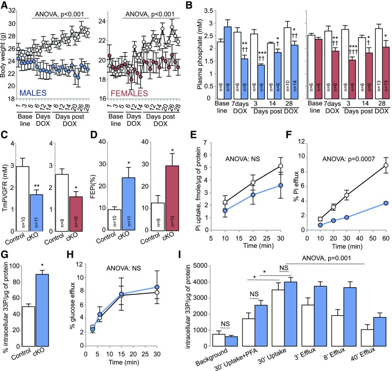
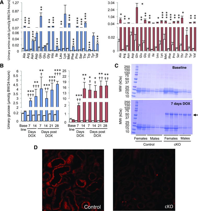
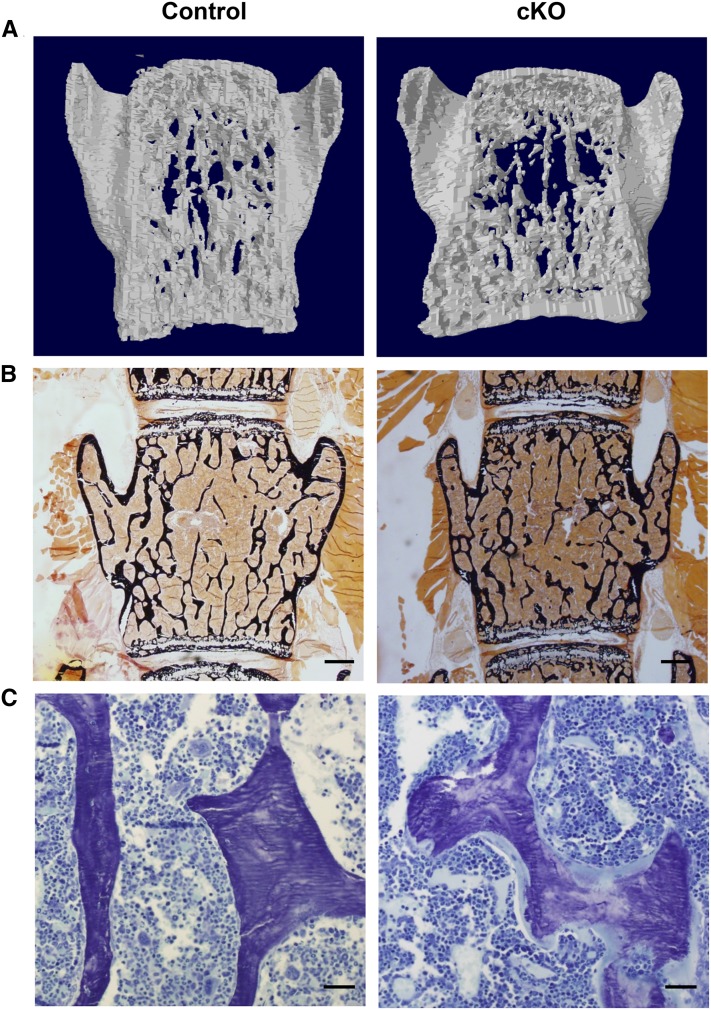
Tight control of extracellular and intracellular inorganic phosphate (Pi) levels is critical to most biochemical and physiologic processes. Urinary Pi is freely filtered at the kidney glomerulus and is reabsorbed in the renal tubule by the action of the apical sodium-dependent phosphate transporters, NaPi-IIa/NaPi-IIc/Pit2. However, the molecular identity of the protein(s) participating in the basolateral Pi efflux remains unknown. Evidence has suggested that xenotropic and polytropic retroviral receptor 1 (XPR1) might be involved in this process. Here, we show that conditional inactivation of Xpr1 in the renal tubule in mice resulted in impaired renal Pi reabsorption. Analysis of Pi transport in primary cultures of proximal tubular cells or in freshly isolated renal tubules revealed that this Xpr1 deficiency significantly affected Pi efflux. Further, mice with conditional inactivation of Xpr1 in the renal tubule exhibited generalized proximal tubular dysfunction indicative of Fanconi syndrome, characterized by glycosuria, aminoaciduria, calciuria, and albuminuria. Dramatic alterations in the renal transcriptome, including a significant reduction in NaPi-IIa/NaPi-IIc expression, accompanied these functional changes. Additionally, Xpr1-deficient mice developed hypophosphatemic rickets secondary to renal dysfunction. These results identify XPR1 as a major regulator of Pi homeostasis and as a potential therapeutic target in bone and kidney disorders.

Keywords: Fanconi syndrome; hypophosphatemic rickets; kidney; phosphate homeostasis; retroviral receptor XPR1.


Figures

Figure 1.
Figure 1.
Altered renal handling of Pi in cKO mice. White circles/bars indicate control mice. Blue and red circles/bars indicate male or female cKO mice, respectively. (A) Body weight in control and cKO male (left panel) or female (right panel) mice. The body weight was measured during 5 days preceding DOX treatment (baseline), during the 2-week period of DOX treatment (days DOX), and during 28 days after DOX withdrawal (days post DOX). n=6 in each group; ANOVA. (B) Plasma Pi levels in control and cKO male (left panel) or female (right panel) mice. Plasma Pi concentration was measured on the day preceding the 2-week period of DOX treatment (baseline), on day 7 of DOX treatment (7 days DOX), and on days 3, 14, 21, and 28 after DOX withdrawal (days post DOX). *P<0.05; **P<0.01; ***P<0.001; t test, statistical significance between control and cKO mice. P<0.05; ††P<0.01; †††P<0.001; t test, statistical significance between plasma Pi levels measured at baseline and plasma Pi levels measured on day 7 of DOX treatment or after DOX withdrawal (days 3, 14, and 28). (C) TmPi/GFR in control and cKO male (left panel) or female (right panel) mice. The TmPi/GFR was determined on day 28 after DOX withdrawal. *P<0.05; **P<0.01; t test. (D) FEPi in control and cKO male (left panel) or female (right panel) mice. The FEPi was determined on day 28 after DOX withdrawal. *P<0.05; t test. (E) [33Pi]phosphate uptake in primary cultures of proximal tubule cells isolated from DOX-untreated control or male cKO mice. Cells were exposed to DOX for 24 hours and the [33Pi]phosphate uptake was measured 24 hours after the end of DOX treatment (see Supplemental Material for details). n=4 in each group; ANOVA. (F) [33P]phosphate efflux from primary cultures of control or cKO proximal tubule cells. n=4 in each group; ANOVA. For (E), (F), and (H), white and blue circles indicate primary cultures of control or cKO proximal tubule cells, respectively. (G) [33P]phosphate remaining in primary cultures of control or cKO proximal tubule cells at the end of the efflux experiment (60 minutes of efflux); n=4 in each group. *P<0.05; t test. (H) [14C]glucose efflux from primary cultures of control or cKO proximal tubule cells. n=4 in each group; ANOVA. (I) [33P]phosphate uptake (30 minutes) and efflux (3 minutes, 8 minutes, and 40 minutes) from renal tubules freshly isolated from kidneys of control or cKO mice induced with DOX for 5 days (for efflux experiments, the 30-minute [33P]phosphate uptake was set as the zero time point). Pi uptake was determined in the presence or absence of 5 mmol PFA. Pi efflux was measured in the presence of 5 mmol PFA (see Supplemental Material for details). Background represents nonspecific binding of [33P]phosphate to the renal tubules. n=4 in each group. The difference in the efflux kinetics was evaluated by ANOVA (genotype–time interaction). The differences in background, 30’ uptake + PFA, and 30’ uptake conditions was evaluated by t test. Numbers inside of bars represent the number of animals. Data are mean±SEM. *P<0.05. FEPi, fractional excretion of Pi.
Figure 2.
Figure 2.
XPR1 deficiency in the nephron is associated with aminoaciduria, glucosuria, albuminuria, and impaired albumin reabsorption in the proximal tubule. (A) Aminoaciduria in cKO mice. The urinary excretion rate of 19 of 20 proteinogenic amino acids (at the exception of cysteine) was measured by mass spectrometry on urine collected on day 28 after DOX withdrawal. White bars indicate the urinary excretion rates of amino acids in control mice. Blue and red bars indicate the urinary excretion rates of amino acids in male or female cKO mice, respectively; n=6 in each group. *P<0.05; **P<0.01; ***P<0.001; t test. (B) Glucosuria in cKO mice. The urinary excretion rate of glucose was measured on urine collected on the day preceding the 2-week period of DOX treatment (baseline), on days 7 and 14 of DOX treatment (days DOX), and on days 7, 14, 21, and 28 after DOX withdrawal (days post DOX). White bars indicate the urinary excretion rates of glucose in control mice (n=6 for males and n=4 for females). Blue and red bars indicate the urinary excretion rates of glucose in male or female cKO mice, respectively (n=6 for males and n=6 for females). *P<0.05; **P<0.01; ***P<0.001; t test, statistical significance between control and cKO mice. P<0.05; ††P<0.01; †††P<0.001; t test, statistical significance between the urinary excretion rates of glucose measured at baseline and the urinary excretion rates of glucose measured during the period of DOX treatment or after DOX withdrawal. (C) Albuminuria associated with XPR1 deficiency. Urine (5 μl) was run on SDS-PAGE and stained with Coomassie blue. Urine was collected from the same mice on the day preceding the 2-week period of DOX treatment (baseline) or on day 7 of DOX treatment (7 days DOX). The albumin band (67 kDa) is indicated by an arrow. (D) Decreased tubular reabsorption of Texas Red (TR)-albumin in kidneys of cKO mice. Confocal microscopy analysis of kidney slices prepared from perfusion-fixed kidneys of TR-albumin–injected control (left panel) or cKO (right panel) mice. Mice were euthanized 5 minutes after intravenous injection of TR-albumin. Data are mean±SEM. Original magnification, ×40 in D.
Figure 3.
Figure 3.
XPR1 deficiency in the nephron causes vertebral osteomalacia in male mice. (A) Three-dimensional reconstructions of 400-μm thick coronal sections of L5 vertebral bodies scanned by microcomputed tomography revealed an impaired trabecular network in male cKO mice (images representative of two control and three cKO mice). (B) Calcium staining of vertebral sections by von Kossa revealed no significant change in trabecular bone of cKO animals. (C) Toluidine Blue staining of vertebral bone revealed a prominent osteoidosis (osteoid seam in light blue) in cKO mice. (B, C) Representative images of five control and five cKO mice; 4-μm thick sections of nondecalcified bone viewed under 2× (B) or 20× (C) magnification; scale bars, 500 μm (B) and 50 μm (C). Mice were euthanized on day 28 after DOX withdrawal.

References

    1. Hamburger D, Rezzonico E, MacDonald-Comber Petétot J, Somerville C, Poirier Y: Identification and characterization of the Arabidopsis PHO1 gene involved in phosphate loading to the xylem. Plant Cell 14: 889–902, 2002 - PMC - PubMed
    1. Stefanovic A, Arpat AB, Bligny R, Gout E, Vidoudez C, Bensimon M, Poirier Y: Over-expression of PHO1 in Arabidopsis leaves reveals its role in mediating phosphate efflux. Plant J 66: 689–699, 2011 - PubMed
    1. Giovannini D, Touhami J, Charnet P, Sitbon M, Battini JL: Inorganic phosphate export by the retrovirus receptor XPR1 in metazoans. Cell Reports 3: 1866–1873, 2013 - PubMed
    1. Wege S, Poirier Y: Expression of the mammalian Xenotropic Polytropic Virus Receptor 1 (XPR1) in tobacco leaves leads to phosphate export. FEBS Lett 588: 482–489, 2014 - PubMed
    1. Legati A, Giovannini D, Nicolas G, López-Sánchez U, Quintáns B, Oliveira JR, Sears RL, Ramos EM, Spiteri E, Sobrido MJ, Carracedo Á, Castro-Fernández C, Cubizolle S, Fogel BL, Goizet C, Jen JC, Kirdlarp S, Lang AE, Miedzybrodzka Z, Mitarnun W, Paucar M, Paulson H, Pariente J, Richard AC, Salins NS, Simpson SA, Striano P, Svenningsson P, Tison F, Unni VK, Vanakker O, Wessels MW, Wetchaphanphesat S, Yang M, Boller F, Campion D, Hannequin D, Sitbon M, Geschwind DH, Battini JL, Coppola G: Mutations in XPR1 cause primary familial brain calcification associated with altered phosphate export. Nat Genet 47: 579–581, 2015 - PMC - PubMed

Substances