

Fundus Findings in Dengue Fever: A Case Report
- PMID: 27800237
- PMCID: PMC5082246
- DOI: 10.4274/tjo.35761
Fundus Findings in Dengue Fever: A Case Report
Abstract
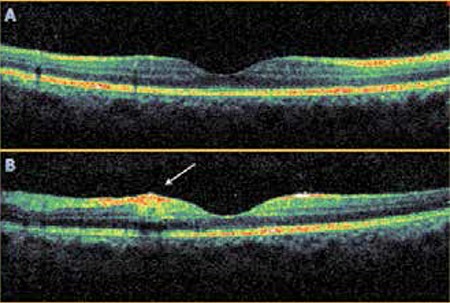
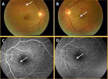
Dengue fever is a flavivirus infection transmitted through infected mosquitoes, and is endemic in Southeast Asia, Central and South America, the Pacific, Africa and the Eastern Mediterranean region. A 41-year-old male patient had visual impairment after travelling to Thailand, which is one of the endemic areas. Cotton wool spots were observed on fundus examination. Fundus fluorescein angiography showed minimal vascular leakage from areas near the cotton wool spots and dot hemorrhages in the macula. Dengue fever should be considered in patients with visual complaints who traveled to endemic areas of dengue fever.
Keywords: Dengue fever; Retina; flavivirus.
Conflict of interest statement
No conflict of interest was declared by the authors. Financial Disclosure: The authors declared that this study received no financial support.
Figures
Similar articles
-
[Ocular manifestation in dengue fever].Ophthalmologe. 2000 Jun;97(6):433-6. doi: 10.1007/s003470070094. Ophthalmologe. 2000. PMID: 10916388 German.
-
Retinal Vasculitis with Macular Infarction: A Dengue-related Ophthalmic Complication.Int Med Case Rep J. 2020 Aug 25;13:363-366. doi: 10.2147/IMCRJ.S264324. eCollection 2020. Int Med Case Rep J. 2020. PMID: 32943944 Free PMC article.
-
Ocular manifestations in Dengue fever.Ocul Immunol Inflamm. 2004 Dec;12(4):323-7. doi: 10.1080/092739490500345. Ocul Immunol Inflamm. 2004. PMID: 15621872
-
Dengue fever in the Western Hemisphere.Clin Lab Sci. 2003 Winter;16(1):34-8. Clin Lab Sci. 2003. PMID: 12587656 Review.
-
Dengue and other emerging flaviviruses.J Infect. 2001 Feb;42(2):104-15. doi: 10.1053/jinf.2001.0802. J Infect. 2001. PMID: 11531316 Review.
References
-
- Lim WK, Mathur R, Koh A, Yeoh R, Chee SP. Ocular manifestations of dengue fever. Ophthalmology. 2004;111:2057–2064. - PubMed
-
- Monath TP. Dengue and yellow fever--challenges for the development and use of vaccines. N Eng J Med. 2007;357:2222–2225. - PubMed
-
- Uyar Y, Aktaş E, Yağcı Çağlayık D, Ergönül Ö, Yüce A. An imported dengue fever case in Turkey and review of the literature. Mikrobiyol Bul. 2013;47:173–180. - PubMed
LinkOut - more resources
Full Text Sources
Other Literature Sources