

Uveal Melanoma: Current Trends in Diagnosis and Management
- PMID: 27800275
- PMCID: PMC5076295
- DOI: 10.4274/tjo.37431
Uveal Melanoma: Current Trends in Diagnosis and Management
Abstract
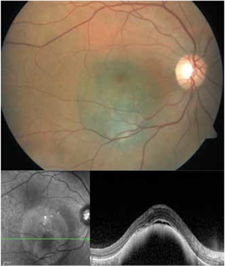
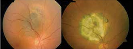
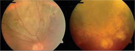
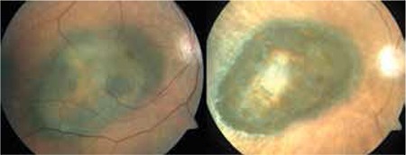
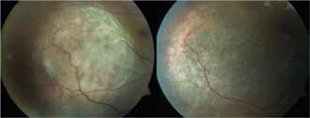
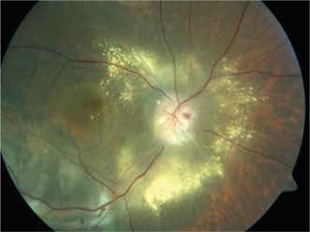
Uveal melanoma, which is the most common primary intraocular malignancy in adults, arises from melanocytes within the iris, ciliary body and choroid. The diagnosis is based principally on clinical examination of the tumor with biomicroscopy and indirect ophthalmoscopy and confirmed by diagnostic techniques such as ultrasonography, fundus fluorescein angiography and optical coherence tomography. The clinical diagnosis of posterior uveal melanomas can be made when the classical appearance of a pigmented dome-shaped mass is detected on dilated fundus exam. Uveal melanomas classically show low to medium reflectivity on A-scan ultrasonography and on B-scan ultrasonography the tumor appears as a hyperechoic, acoustically hollow intraocular mass. Management of a suspicious pigmented lesion is determined by its risk factors of transforming into a choroidal melanoma, such as documentation of growth, thickness greater than 2 mm, presence of subretinal fluid, symptoms and orange pigment, margin within 3 mm of the optic disc, and absence of halo and drusen. Advances in the diagnosis and local and systemic treatment of uveal melanoma have caused a shift from enucleation to eye-conserving treatment modalities including transpupillary thermotherapy and radiotherapy over the past few decades. Prognosis can be most accurately predicted by genetic profiling of fine needle aspiration biopsy of the tumor before the treatment, and high-risk patients can now be identified for clinical trials that may lead to target-based therapies for metastatic disease and adjuvant therapy which aims to prevent metastatic disease.
Keywords: Eye; neoplasm; uveal melanoma.
Conflict of interest statement
No conflict of interest was declared by the authors. Financial Disclosure: The authors declared that this study received no financial support.
Figures
References
-
- Chang AE, Karnell LH, Menck HR. The National Cancer Data Base report on cutaneous and noncutaneous melanoma: a summary of 84,836 cases from the past decade. The American College of Surgeons Commission on Cancer and the American Cancer Society. Cancer. 1998;83:1664–1678. - PubMed
-
- Shields CL, Kels JG, Shields JA. Melanoma of the eye: revealing hidden secrets, one at a time. Clin Dermatol. 2015;33:183–196. - PubMed
-
- Shields CL, Kaliki S, Furuta M, Mashayekhi A, Shields JA. Clinical spectrum and prognosis of uveal melanoma based on age at presentation in 8,033 cases. Retina. 2012;32:1363–1372. - PubMed
-
- Virgili G, Gatta G, Ciccolallo L, Capocaccia R, Biggeri A, Crocetti E, Lutz JM. Incidence of uveal melanoma in Europe. Ophthalmology. 2007;114:2309–2315. - PubMed
-
- Isager P, Osterlind A, Engholm G, Heegaard S, Lindegaard J, Overgaard J, Storm HH. Uveal and conjunctival malignant melanoma in Denmark, 1943-97: incidence and validation study. Ophthalmic Epidemiol. 2005;12:223–232. - PubMed
Publication types
LinkOut - more resources
Full Text Sources
Other Literature Sources
Molecular Biology Databases
Miscellaneous