

Fifteen days of 3,200 m simulated hypoxia marginally regulates markers for protein synthesis and degradation in human skeletal muscle
- PMID: 27800505
- PMCID: PMC5085286
- DOI: 10.2147/HP.S101133
Fifteen days of 3,200 m simulated hypoxia marginally regulates markers for protein synthesis and degradation in human skeletal muscle
Abstract
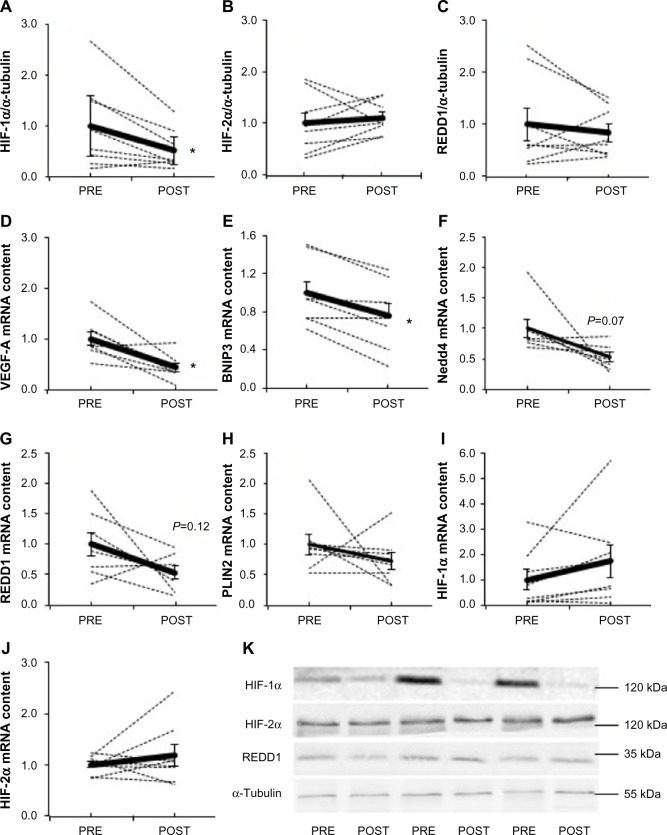
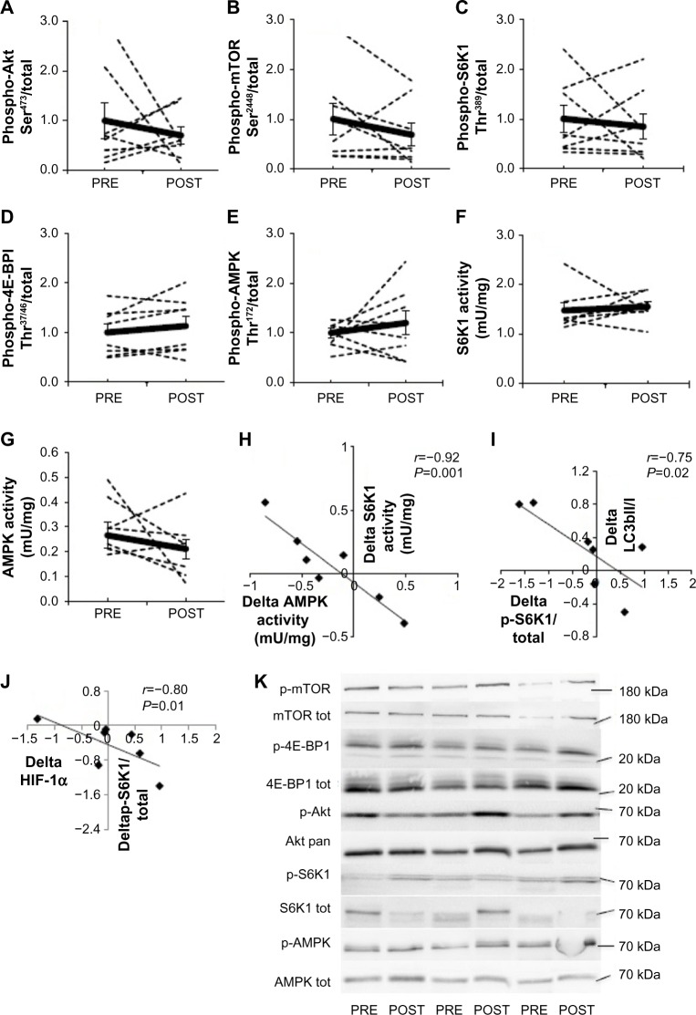
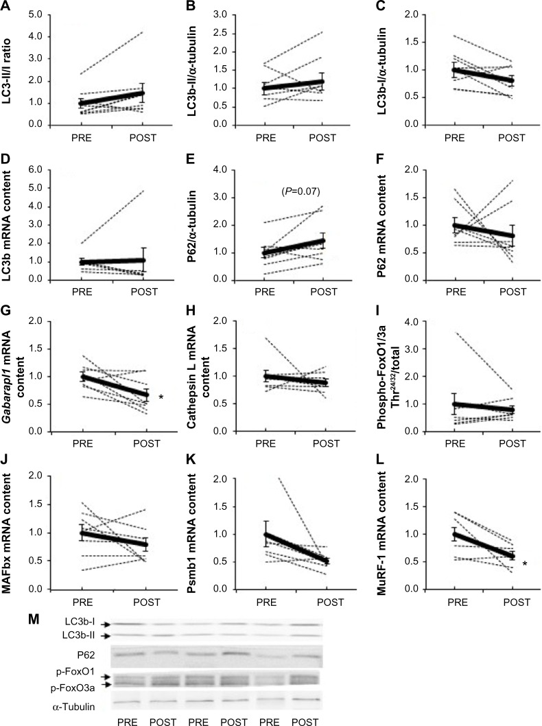
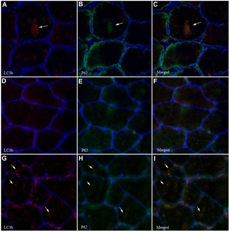
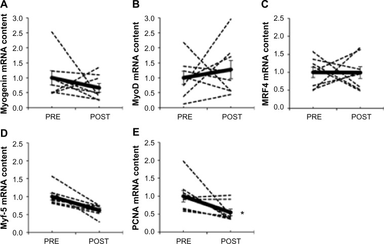
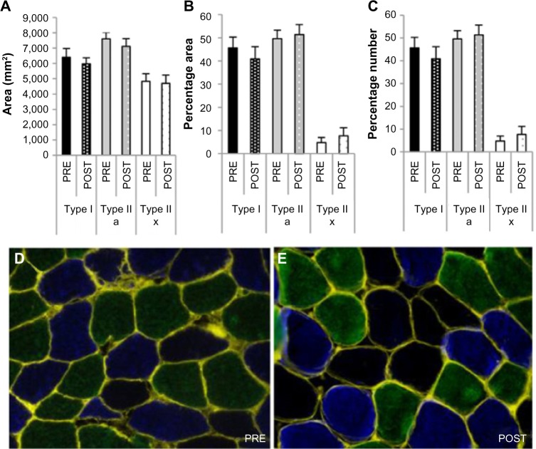
Chronic hypoxia leads to muscle atrophy. The molecular mechanisms responsible for this phenomenon are not well defined in vivo. We sought to determine how chronic hypoxia regulates molecular markers of protein synthesis and degradation in human skeletal muscle and whether these regulations were related to the regulation of the hypoxia-inducible factor (HIF) pathway. Eight young male subjects lived in a normobaric hypoxic hotel (FiO2 14.1%, 3,200 m) for 15 days in well-controlled conditions for nutrition and physical activity. Skeletal muscle biopsies were obtained in the musculus vastus lateralis before (PRE) and immediately after (POST) hypoxic exposure. Intramuscular hypoxia-inducible factor-1 alpha (HIF-1α) protein expression decreased (-49%, P=0.03), whereas hypoxia-inducible factor-2 alpha (HIF-2α) remained unaffected from PRE to POST hypoxic exposure. Also, downstream HIF-1α target genes VEGF-A (-66%, P=0.006) and BNIP3 (-24%, P=0.002) were downregulated, and a tendency was measured for neural precursor cell expressed, developmentally Nedd4 (-47%, P=0.07), suggesting lowered HIF-1α transcriptional activity after 15 days of exposure to environmental hypoxia. No difference was found on microtubule-associated protein 1 light chain 3 type II/I (LC3b-II/I) ratio, and P62 protein expression tended to increase (+45%, P=0.07) compared to PRE exposure levels, suggesting that autophagy was not modulated after chronic hypoxia. The mammalian target of rapamycin complex 1 pathway was not altered as Akt, mammalian target of rapamycin, S6 kinase 1, and 4E-binding protein 1 phosphorylation did not change between PRE and POST. Finally, myofiber cross-sectional area was unchanged between PRE and POST. In summary, our data indicate that moderate chronic hypoxia differentially regulates HIF-1α and HIF-2α, marginally affects markers of protein degradation, and does not modify markers of protein synthesis or myofiber cross-sectional area in human skeletal muscle.
Keywords: HIF-1α; autophagy; hypoxia; mTORC1.
Conflict of interest statement
The authors report no conflicts of interest in this work.
Figures
Similar articles
-
High-intensity interval training in hypoxia does not affect muscle HIF responses to acute hypoxia in humans.Eur J Appl Physiol. 2018 Apr;118(4):847-862. doi: 10.1007/s00421-018-3820-4. Epub 2018 Feb 8. Eur J Appl Physiol. 2018. PMID: 29423544
-
Muscle-specific expression of hypoxia-inducible factor in human skeletal muscle.Exp Physiol. 2010 Aug;95(8):899-907. doi: 10.1113/expphysiol.2010.052928. Epub 2010 May 21. Exp Physiol. 2010. PMID: 20494919
-
Suppression of the proliferation of hypoxia-Induced retinal pigment epithelial cell by rapamycin through the /mTOR/HIF-1α/VEGF/ signaling.IUBMB Life. 2015 Jun;67(6):446-52. doi: 10.1002/iub.1382. Epub 2015 May 19. IUBMB Life. 2015. PMID: 25988388
-
Multiplicity of hypoxia-inducible transcription factors and their connection to the circadian clock in the zebrafish.Physiol Biochem Zool. 2015 Mar-Apr;88(2):146-57. doi: 10.1086/679751. Epub 2015 Jan 14. Physiol Biochem Zool. 2015. PMID: 25730270 Review.
-
Skeletal Muscle Fiber Type in Hypoxia: Adaptation to High-Altitude Exposure and Under Conditions of Pathological Hypoxia.Front Physiol. 2018 Oct 12;9:1450. doi: 10.3389/fphys.2018.01450. eCollection 2018. Front Physiol. 2018. PMID: 30369887 Free PMC article. Review.
Cited by
-
Hemoglobin Changes After Long-Term Intermittent Work at High Altitude.Front Physiol. 2018 Nov 1;9:1552. doi: 10.3389/fphys.2018.01552. eCollection 2018. Front Physiol. 2018. PMID: 30443224 Free PMC article.
-
Altitude, Exercise, and Skeletal Muscle Angio-Adaptive Responses to Hypoxia: A Complex Story.Front Physiol. 2021 Sep 6;12:735557. doi: 10.3389/fphys.2021.735557. eCollection 2021. Front Physiol. 2021. PMID: 34552509 Free PMC article. Review.
-
The Impact of a High-Altitude Expedition on the Physical Performance and Nutritional Indices of Health Status of Alpinists.J Funct Morphol Kinesiol. 2025 Apr 25;10(2):143. doi: 10.3390/jfmk10020143. J Funct Morphol Kinesiol. 2025. PMID: 40407427 Free PMC article.
-
HIF-1α Negatively Regulates Irisin Expression Which Involves in Muscle Atrophy Induced by Hypoxia.Int J Mol Sci. 2022 Jan 14;23(2):887. doi: 10.3390/ijms23020887. Int J Mol Sci. 2022. PMID: 35055073 Free PMC article.
-
Hypoxia Aggravates Inactivity-Related Muscle Wasting.Front Physiol. 2018 May 15;9:494. doi: 10.3389/fphys.2018.00494. eCollection 2018. Front Physiol. 2018. PMID: 29867545 Free PMC article.
References
-
- Deldicque L, Francaux M. Acute vs chronic hypoxia: what are the consequences for skeletal muscle mass? Cell Mol Exerc Physiol. 2013;2(1):1–23.
-
- Kurobe K, Huang Z, Nishiwaki M, Yamamoto M, Kanehisa H, Ogita F. Effects of resistance training under hypoxic conditions on muscle hypertrophy and strength. Clin Physiol Funct Imaging. 2015;35(3):197–202. - PubMed
-
- Mizuno M, Savard GK, Areskog NH, Lundby C, Saltin B. Skeletal muscle adaptations to prolonged exposure to extreme altitude: a role of physical activity? High Alt Med Biol. 2008;9(4):311–317. - PubMed
-
- Hoppeler H, Kleinert E, Schlegel C, et al. Morphological adaptations of human skeletal muscle to chronic hypoxia. Int J Sport Med. 1990;11(suppl 1):S3–S9. - PubMed
-
- MacDougall JD, Green HJ, Sutton JR, et al. Operation Everest II: structural adaptations in skeletal muscle in response to extreme simulated altitude. Acta Physiol Scand. 1991;142(3):421–427. - PubMed
LinkOut - more resources
Full Text Sources
Other Literature Sources
Molecular Biology Databases
Research Materials
