Review
. 2016 Apr 7:4:29-39.
doi: 10.2147/HP.S83540. eCollection 2016.

Erythropoietin and the use of a transgenic model of erythropoietin-deficient mice

Affiliations
Review

Erythropoietin and the use of a transgenic model of erythropoietin-deficient mice

Aurélien Pichon et al. Hypoxia (Auckl). .

Abstract

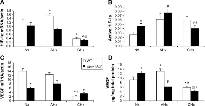
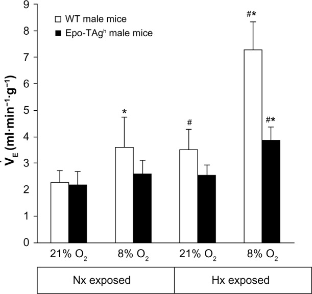
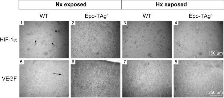
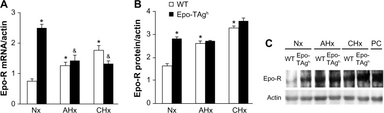
Despite its well-known role in red blood cell production, it is now accepted that erythropoietin (Epo) has other physiological functions. Epo and its receptors are expressed in many tissues, such as the brain and heart. The presence of Epo/Epo receptors in these organs suggests other roles than those usually assigned to this protein. Thus, the aim of this review is to describe the effects of Epo deficiency on adaptation to normoxic and hypoxic environments and to suggest a key role of Epo on main physiological adaptive functions. Our original model of Epo-deficient (Epo-TAgh) mice allowed us to improve our knowledge of the possible role of Epo in O2 homeostasis. The use of anemic transgenic mice revealed Epo as a crucial component of adaptation to hypoxia. Epo-TAgh mice survive well in hypoxic conditions despite low hematocrit. Furthermore, Epo plays a key role in neural control of ventilatory acclimatization and response to hypoxia, in deformability of red blood cells, in cerebral and cardiac angiogenesis, and in neuro- and cardioprotection.

Keywords: Epo-TAgh mice; hypoxia; mouse model; physiological functions.


Conflict of interest statement

The authors report no conflicts of interest in this work.

Figures

Figure 1
Figure 1
Cardiac angiogenesis in Epo-TAgh mice. Notes: HIF-1α and VEGF mRNA (A, C) and protein (B, D) expression in the heart of WT (white bar) and Epo-TAgh (black bar) mice in normoxia and after 48 hours (AHx) or 14 days (CHx) of hypoxic exposure. Epo deficiency led to a rise of angiogenesis through the HIF-1α and VEGF pathway activation. Epo-TAgh mice were not able to maintain cardiac adaptation to hypoxia during the long-term exposure. Values are expressed as mean ± SEM. *P<0.05 vs Nx WT; &P<0.05 vs Nx Epo-TAgh; #P<0.05 CHx vs AHx; @P<0.05 CHx Epo-TAgh vs CHx WT. μP<0.05 AHx Epo-TAgh vs AHx WT. Reprinted from Respir Physiol Neurobiol, Volume 186(2), El Hasnaoui-Saadani R, Marchant D, Pichon A, et al, Epo deficiency alters cardiac adaptation to chronic hypoxia, pages 146–154. Copyright 2013 with permission from Elsevier. Abbreviations: AHx, acute hypoxia; CHx, chronic hypoxia; Epo, erythropoietin; Epo-TAgh mice, Epo-deficient mice; HIF-1α, hypoxia-inducible factor-1α; Nx, normoxia; SEM, standard error of the mean; VEGF, vascular endothelial growth factor; WT, wild type.
Figure 2
Figure 2
Ventilatory response to hypoxia in Epo-TAgh mice. Notes: Minute ventilation measured in normoxia (FIO2 21%) or acute hypoxia (FIO2 8%) in WT (white bar) and Epo-TAgh (black bar) mice maintained in normoxic (Nx exposed) or hypoxic (14 days, Hx exposed) conditions. Epo-TAgh mice had a normal ventilation at rest, did not display ventilatory acclimatization to hypoxia, and did not respond to acute hypoxia even after the exposure to chronic hypoxia. Values are expressed as mean ± SD. *P<0.05 21% O2 vs 8% O2; #P<0.05 Nx exposed vs Hx exposed, same strain, same FIO2. Adapted from Voituron N, Jeton F, Cholley Y, et al. Catalyzing role of erythropoietin on the nitric oxide central pathway during the ventilatory responses to hypoxia. Physiol Rep. 2014;2(2):e00223. © 2014 Voituron N, Jeton F, Cholley Y, et al. Physiological Reports published by Wiley Periodicals, Inc. on behalf of the American Physiological Society and The Physiological Society. Abbreviations: Epo, erythropoietin; Epo-TAgh mice, Epo-deficient mice; Hx, hypoxia; Nx, normoxia; SD, standard deviation; WT, wild type.
Figure 3
Figure 3
Cerebral angiogenesis in Epo-TAgh mice. Notes: Immunohistological detection of HIF-1α and VEGF at the sensory cortex level in normoxia (Nx exposed) and after chronic hypoxia exposure (Hx exposed) in WT (1, 3, 5, 7) and Epo-TAgh mice (2, 4, 6, 8). Arrowheads and arrow indicate HIF-1α (1)- and VEGF (5)-positive cells, respectively. In normoxia, Epo-TAgh mice showed an increase in HIF-1α (2)- and VEGF (6)-positive cells suggesting an enhancement of cerebral angiogenesis through the HIF-1α/VEGF pathway. In WT mice, chronic hypoxia led to an increase in HIF-1α (3) and VEGF (7), while they led a decrease in Epo-TAgh mice (4, 8). Adapted from Am J Physiol Regul Integr Comp Physiol. Volume 296(3). El Hasnaoui-Saadani R, Pichon A, Marchant D, et al. Cerebral adaptations to chronic anemia in a model of erythropoietin-deficient mice exposed to hypoxia. Pages: R801–R811. Copyright 2009. Abbreviations: Epo, erythropoietin; Epo-TAgh mice, Epo-deficient mice; HIF-1α, hypoxia-inducible factor-1α; Hx, hypoxia; Nx, normoxia; VEGF, vascular endothelial growth factor; WT, wild type.
Figure 4
Figure 4
Effect of Epo deficiency on Epo-R expression in cerebral cortex. Notes: Quantitative determination of Epo-R in the cerebral cortex of WT and Epo-TAgh mice in Nx and following AHx and CHx. Epo-R mRNA (A) and protein level (B) are shown next to their corresponding protein bar graphs. Representative Western blot of Epo-receptor (Epo-R) (C). Values are expressed as mean ± SD. *P<0.05 vs Nx WT; &P<0.05 vs Nx Epo-TAgh. Adapted from Am J Physiol Regul Integr Comp Physiol. Volume 296(3). El Hasnaoui-Saadani R, Pichon A, Marchant D, et al. Cerebral adaptations to chronic anemia in a model of erythropoietin-deficient mice exposed to hypoxia. Pages: R801–R811. Copyright 2009. Abbreviations: AHx, acute hypoxia; CHx, chronic hypoxia; Epo, erythropoietin; Epo-R, Epo receptor; Epo-TAgh mice, Epo-deficient mice; Nx, normoxia; SD, standard deviation; WT, wild type; PC, peptide control.

References

    1. Semenza GL. Oxygen homeostasis. Wiley Interdiscip Rev Syst Biol Med. 2010;2(3):336–361. - PubMed
    1. Gassmann M, Soliz J. Erythropoietin modulates the neural control of hypoxic ventilation. Cell Mol Life Sci. 2009;66(22):3575–3582. - PMC - PubMed
    1. Fandrey J, Gorr TA, Gassmann M. Regulating cellular oxygen sensing by hydroxylation. Cardiovasc Res. 2006;71(4):642–651. - PubMed
    1. Yeo EJ, Chun YS, Park JW. New anticancer strategies targeting HIF-1. Biochem Pharmacol. 2004;68(6):1061–1069. - PubMed
    1. Forsythe JA, Jiang BH, Iyer NV, et al. Activation of vascular endothelial growth factor gene transcription by hypoxia-inducible factor 1. Mol Cell Biol. 1996;16(9):4604–4613. - PMC - PubMed