. 2016 Aug 16:4:121-133.
doi: 10.2147/HP.S104377. eCollection 2016.

Erythropoietin and a nonerythropoietic peptide analog promote aortic endothelial cell repair under hypoxic conditions: role of nitric oxide

Affiliations

Erythropoietin and a nonerythropoietic peptide analog promote aortic endothelial cell repair under hypoxic conditions: role of nitric oxide

Lamia Heikal et al. Hypoxia (Auckl). .

Abstract

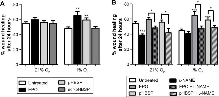
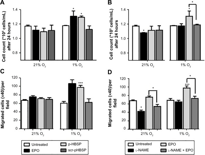
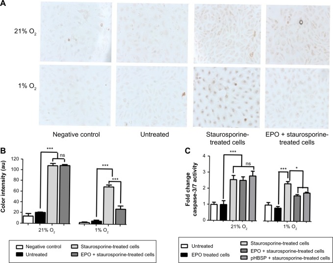
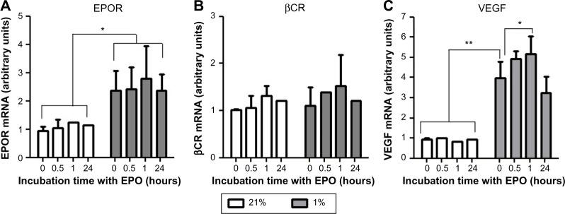
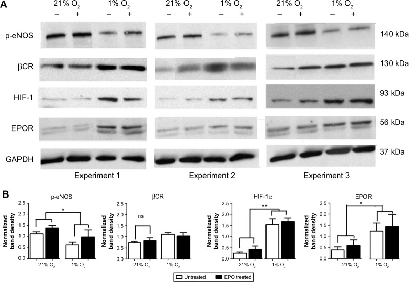
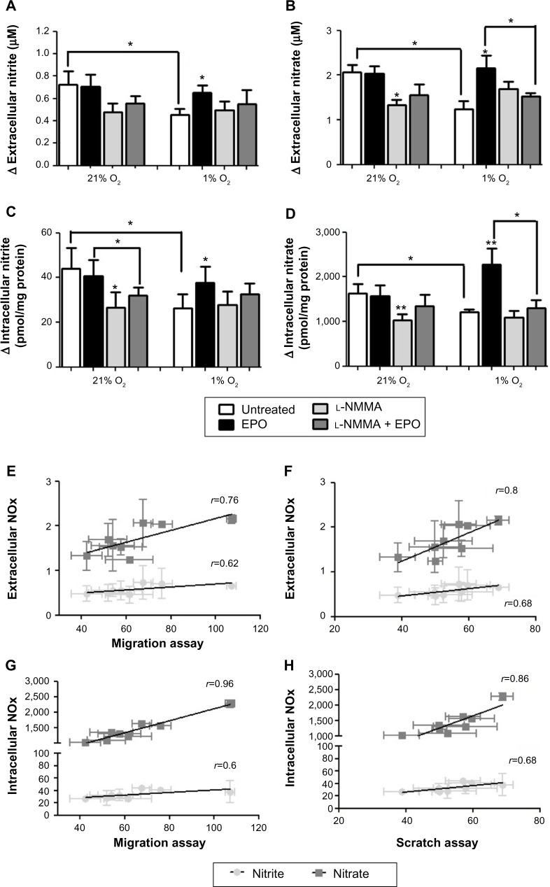
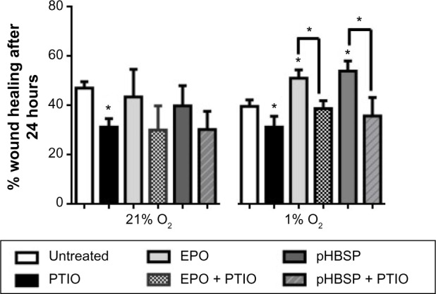
The cytoprotective effects of erythropoietin (EPO) and an EPO-related nonerythropoietic analog, pyroglutamate helix B surface peptide (pHBSP), were investigated in an in vitro model of bovine aortic endothelial cell injury under normoxic (21% O2) and hypoxic (1% O2) conditions. The potential molecular mechanisms of these effects were also explored. Using a model of endothelial injury (the scratch assay), we found that, under hypoxic conditions, EPO and pHBSP enhanced scratch closure by promoting cell migration and proliferation, but did not show any effect under normoxic conditions. Furthermore, EPO protected bovine aortic endothelial cells from staurosporine-induced apoptosis under hypoxic conditions. The priming effect of hypoxia was associated with stabilization of hypoxia inducible factor-1α, EPO receptor upregulation, and decreased Ser-1177 phosphorylation of endothelial nitric oxide synthase (NOS); the effect of hypoxia on the latter was rescued by EPO. Hypoxia was associated with a reduction in nitric oxide (NO) production as assessed by its oxidation products, nitrite and nitrate, consistent with the oxygen requirement for endogenous production of NO by endothelial NOS. However, while EPO did not affect NO formation in normoxia, it markedly increased NO production, in a manner sensitive to NOS inhibition, under hypoxic conditions. These data are consistent with the notion that the tissue-protective actions of EPO-related cytokines in pathophysiological settings associated with poor oxygenation are mediated by NO. These findings may be particularly relevant to atherogenesis and postangioplasty restenosis.

Keywords: apoptosis; erythropoietin; migration; proliferation; pyroglutamate helix B surface peptide; scratch assay.


Conflict of interest statement

The authors report no conflicts of interest in this work.

Figures

Figure 1
Figure 1
EPO and pHBSP stimulate repair in a scratch assay model in BAEC under hypoxia. Notes: (A) EPO and pHBSP (1 ng/mL each) enhanced wound closure in BAECs when incubated in 1% O2 for 24 hours, but not under 21% O2 (**P<0.01 for EPO and pHBSP treated cells versus untreated cells). The scrambled peptide control (scr-pHBSP) had no significant effect on wound closure in BAECs under either 21% or 1% O2. (B)The NOs inhibitor; l-NAME (300 μM) decreased wound closure for untreated cells under 21% O2 (*** P<0.001), but showed no significant effect under 1% O2. l-NAME (300 μM) inhibited wound closure in EPO or pHBSP treated cells under both 21% and 1% O2 (* P<0.05 for EPO and pHBSP treated cells in presence versus absence of l-NAME). Results are expressed as % wound healing after 24 hours. Each data point represents the mean value ± SEM (n=6). Statistical analysis was carried out using one-way ANOVA followed by Bonferroni posthoc test. Abbreviations: BAEC, bovine aortic endothelial cell; EPO, erythropoietin; l-NAME, NG-nitro-l-arginine methyl ester; pHBSP, pyroglutamate helix B surface peptide; scr-pHBSP, scrambled pHBSP; SEM, standard error of the mean.
Figure 2
Figure 2
EPO and pHBSP stimulate proliferation and migration of BAECs under hypoxia. BAECs were treated with EPO, pHBSP, and scr-pHBSP at 1 ng/mL and were then incubated under either 21% or 1% O2 for 24 hours. Notes: The effects of EPO, pHBSP, and scr-pHBSP, as well as the effect of l-NAME (300 μM) in the presence and absence of EPO on proliferation of BAECs were analyzed by cell counting using the trypan blue exclusion assay (A and B) and on migration of BAECs using a micro-Boyden chamber chemotactic assay (C and D). EPO and pHBSP (1 ng/mL each) enhanced proliferation (*P<0.05) and migration (***P<0.001) in BAECs when incubated in 1% O2 for 24 hours, but not under 21% O2. l-NAME (300 μM) inhibited proliferation and migration in EPO treated cells (*P<0.05). Each data point represents mean ± SEM (n=3). statistical analysis was carried out using one-way ANOVA followed by Bonferroni posthoc test. Abbreviations: BAEC, bovine aortic endothelial cell; EPO, erythropoietin; l-NAME, NG-nitro-l-arginine methyl ester; pHBSP, pyroglutamate helix B surface peptide; scr-pHBSP, scrambled pHBSP; SEM, standard error of the mean.
Figure 3
Figure 3
Antiapoptotic effects of EPO and pHBSP under hypoxic, but not normoxic conditions. Apoptosis was induced in BAECs by treatment with 500 nM staurosporine for 18 hours after incubation with or without EPO or pHBSP for 3 hours. Notes: (A) representative micrographs of the TUNEL staining under 20× magnification. (B) Quantification of TUNEL staining. EPO caused a decrease in apoptosis-stimulated cells under 1% O2 (***P<0.001) but not under 21% O2 (ns, P>0.05) (C) Caspase-3/7 activity where EPO and pHBSP showed anti-apoptotic effect under 1% O2 (*P<0.05) but not under 21% O2 (ns P>0.05). Each data point represents mean ± SEM (n=3). Statistical analysis was carried out using one-way ANOVA followed by Bonferroni posthoc test. Abbreviations: BAEC, bovine aortic endothelial cell; EPO, erythropoietin; ns, non-significant; pHBSP, pyroglutamate helix B surface peptide; SEM, standard error of the mean; TUNEL, terminal deoxynucleotidyl transferase dUTP nick end labeling.
Figure 4
Figure 4
Hypoxia increased gene expression of EPOR, but not the βCR in BAECs. Cells were cultured under 21% or 1% O2 for 24 hours and treated with EPO (1 ng/mL) for 0, 0.5, 1, and 24 hours. Notes: (A) EPOR and (B) βCR expression were then analyzed by qPCR. (C) in parallel, VEGF, a known hypoxia-induced gene, was measured as a positive control. The data are plotted in arbitrary units versus one of the samples incubated in 21% O2 at time 0 and expressed as the mean ± SEM of nine samples. EPOR expression but not βCR increased under 1% O2 compared to 21% O2 (*P<0.05). Addition of EPO had no effect on the expression of both EPOR and βCR at the time points indicated. Hypoxia increase the expression of VEGF (positive control) (**P<0.01). Each data point represents mean ± SEM (n=3). Statistical analysis was carried out using t-test and one-way ANOVA to compare expression under different O2 level conditions. Abbreviations: βCR, β common receptor; BAECs, bovine aortic endothelial cells; EPO, erythropoietin; EPOR, EPO receptor; qPCR, quantitative polymerase chain reaction; SEM, standard error of the mean; VEGF, vascular endothelial growth factor.
Figure 5
Figure 5
EPOR but not βCR protein expression increases under hypoxic conditions. Notes: (A) Western blot analysis of BAECs cultured under 21% O2 and 1% O2 for 24 hours in the absence (−) and presence (+) of EPO, showing the expression of p-eNOS (140 kDa), βCR(130 kDa), HIF-1α (93 kDa), and EPOR(56 kDa). GAPDH (37 kDa) was used as loading control for the samples. (B) Densitometric quantification following normalization against GAPDH. Each data point represents mean ± SEM of three independent experiments (n=3). Expression of EPOR and HIF-1α increased under hypoxia (*P<0.05 and **P<0.01). On the other hand, expression of p-eNOS decreased under hypoxia (*P<0.05). Expression of βCR was not affected by change in O2 levels. Each data point represents the mean value ± SEM (n=3). Statistical analysis was carried out using t-test and one-way ANOVA to compare expression under different O2 level conditions. Abbreviations: βCR, β common receptor; BAECs, bovine aortic endothelial cells; EPO, erythropoietin; EPOR, EPO receptor; HIF-1, hypoxia inducible factor-1; p-eNOS, phosphorylated endothelial nitric oxide synthase; SEM, standard error of the mean.
Figure 6
Figure 6
Effect of EPO on NO production in 1% or 21% O2. Notes: Nitrite and nitrate were measured extracellularly (A, B) and intracellularly (C, D). NO production was lower in 1% O2 compared to 21% O2 (*P<0.05). EPO caused an increase in NO production in 1% O2 but not under 21% O2 (**P<0.01). l-NMMA caused a decrease in NO production in EPO treated cells under hypoxic conditions (*P<0.05). Each data point represents the mean ± SEM (n=5). Association analyses revealed that 75% (cell migration) and 80% (scratch assay) of the variability in biological responses can be explained by the observed changes in nitrate concentration (EH); correlation coefficients for nitrite with the same biological read-outs were somewhat weaker, possibly due to its very low levels. Abbreviations: EPO, erythropoietin; l-NMMA, l-NG-monomethyl-l-arginine; NO, nitric oxide; SEM, standard error of the mean.
Figure 7
Figure 7
PTIO (150 μM) inhibits repair in a scratch assay model in BAECs under both normoxia and hypoxia both in untreated cells (*P<0.05) and in EPO/pHBSP-treated cells (*P<0.05). Note: Each data point represents the mean value ± SEM (n=3). Statistical analysis was carried out using one-way ANOVA followed by Bonferroni post-hoc test. Abbreviations: BAECs, bovine aortic endothelial cells; EPO, erythropoietin; pHBSP, pyroglutamate helix B surface peptide; PTIO, 2-phenyl-4,4,5,5-tetramethylimidazoline-1-oxyl 3-oxide; SEM, standard error of the mean.

Similar articles

Cited by

References

    1. Kleemann R, Zadelaar S, Kooistra T. Cytokines and atherosclerosis: a comprehensive review of studies in mice. Cardiovasc Res. 2008;79(3):360–376. - PMC - PubMed
    1. Silvestre JS, Mallat Z, Tedgui A, Levy BI. Post-ischaemic neovascularization and inflammation. Cardiovasc Res. 2008;78(2):242–249. - PubMed
    1. Heikal L, Ghezzi P, Mengozzi M, Ferns G. Low oxygen tension primes aortic endothelial cells to the reparative effect of tissue-protective cytokines. Mol Med. 2015;21:709–716. - PMC - PubMed
    1. Ahluwalia A, Tarnawski AS. Critical role of hypoxia sensor – HIF-1 alpha in VEGF gene activation. Implications for angiogenesis and tissue injury healing. Curr Med Chem. 2012;19(1):90–97. - PubMed
    1. Sanchis-Gomar F, Perez-Quilis C, Lippi G. Erythropoietin receptor (EpoR) agonism is used to treat a wide range of disease. Mol Med. 2013;19:62–64. - PMC - PubMed