. 2016 Dec;13(12):1197-1203.
doi: 10.1080/15476286.2016.1240140. Epub 2016 Nov 1.

Translocation as continuous movement through the ribosome

Affiliations

Translocation as continuous movement through the ribosome

Riccardo Belardinelli et al. RNA Biol. 2016 Dec.

Abstract

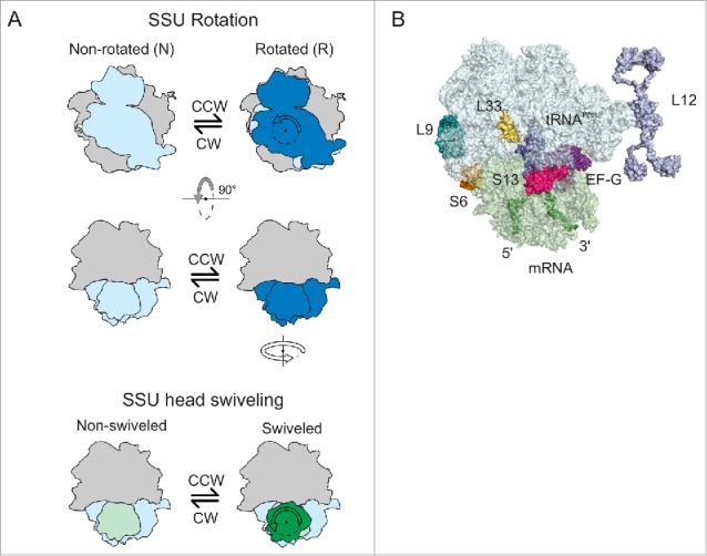
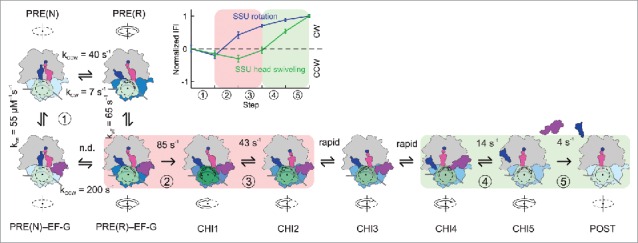
In each round of translation elongation, tRNAs and mRNA move within the ribosome by one codon at a time. tRNA-mRNA translocation is promoted by elongation factor G (EF-G) at the cost of GTP hydrolysis. The key questions for understanding translocation are how and when the tRNAs move and how EF-G coordinates motions of the ribosomal subunits with tRNA movement. Here we present 2 recent papers which describe the choreography of movements over the whole trajectory of translocation. We present the view that EF-G accelerates translocation by promoting the steps that lead to GTPase-dependent ribosome unlocking. EF-G facilitates the formation of the rotated state of the ribosome and uncouples the backward motions of the ribosomal subunits, forming an open conformation in which the tRNAs can rapidly move. Ribosome dynamics are important not only in translocation, but also in recoding events, such as frameshifting and bypassing, and mediate sensitivity to antibiotics.

Keywords: EF-G; mRNA; molecular machines; protein synthesis; ribosome; tRNA; translation; translation elongation; translocation.


Figures

Figure 1.
Figure 1.
Ribosome dynamics (A) Modes of SSU movements. The rotation states of the SSU relative to the LSU (gray) are indicated by the color intensity of the SSU body (light blue for N, dark blue for R). The swiveling motions of the SSU head are illustrated by a color change from light green (classical non-swiveled SSU head position) to forest green (maximum degree of swiveling relative to the SSU body). (B) The toolbox of fluorescence reporters. Positions of fluorescent reporter groups on ribosomal proteins, tRNA, mRNA, and EF-G are indicated. The fluorescence change of the label on protein S13 reports on EF-G binding. FRET changes monitor the SSU rotation relative to the LSU (S6–L9), SSU head swiveling (S13–L33), or EF-G binding and dissociation (EF-G–L12). FRET changes between the P-site tRNA and SSU or LSU monitor the transit of the deacylated tRNA through the E site and its dissociation from the ribosome. SSU, light green; LSU, light cyan.
Figure 2.
Figure 2.
Translocation model The rotation states of the SSU relative to the LSU (gray) are indicated by color intensity of the SSU body (light blue for N, dark blue for R). The swiveling motions of the SSU head are depicted by a color gradient from light green (classical non-swiveled SSU head position) to forest green (maximum degree of swiveling relative to the SSU body). Peptidyl- and deacylated-tRNA in the PRE complex are shown in magenta and blue, respectively. EF-G (purple) is depicted in 2 conformations, a compact and an extended one after engagement with the ribosome. The rates of transitions between PRE(N) and PRE(R) and PRE(N)–EF-G and PRE(R)–EF-G are from ref. The rates of EF-G binding and dissociation (step ①) are ensemble rate constants obtained for a mixture of N and R states in which the PRE(R) state is predominant. All other rate constants for the kinetically defined steps ②, ③, ④, and ⑤ are from ensemble kinetics studies with the PRE(fMF) complexes at 37°C. The existence of rapid steps between steps ③ and ④ was demonstrated previously. Translocation intermediates (CHI1 to CHI4) are adopted from an smFRET study and are consistent with other smFRET data, ensemble kinetics and structural studies. An additional intermediate, CHI5, was identified by ensemble kinetics and smFRET. The POST state may entail further conformational sub-states. Kinetics of GTP hydrolysis and Pi release were described earlier. The light red background indicates complexes undergoing unlocking; the light green background shows complexes that move toward relocking. Inset: Distinct timing of CCW and CW movements of the SSU body relative to LSU (blue symbols) and of the SSU head (green symbols) as indicated by normalized intrinsic fluorescence intensities (IFI).
Figure 3.
Figure 3.
Effect of GTP hydrolysis (A) Rate constants of translocation steps measured with GTP (black bars) and GTPγS (red bars). Note the logarithmic scale used for the rate constants. (B, C) Examples of different trajectories of translocation with GTP (black symbols) and GTPγS (red symbols) for the SSU rotation (B) and SSU head swiveling (C). Intrinsic fluorescence intensities (IFI) reflect structural differences of the intermediates formed in steps ① to ⑤ (Fig. 2).

References

    1. Frank J, Agrawal RK. A ratchet-like inter-subunit reorganization of the ribosome during translocation. Nature 2000; 406:318-22; PMID:10917535; http://dx.doi.org/10.1038/35018597 - DOI - PubMed
    1. Moazed D, Noller HF. Intermediate states in the movement of transfer RNA in the ribosome. Nature 1989; 342:142-8; PMID:2682263; http://dx.doi.org/10.1038/342142a0 - DOI - PubMed
    1. Brilot AF, Korostelev AA, Ermolenko DN, Grigorieff N. Structure of the ribosome with elongation factor G trapped in the pretranslocation state. Proc Natl Acad Sci U S A 2013; 110:20994-9; PMID:24324137; http://dx.doi.org/10.1073/pnas.1311423110 - DOI - PMC - PubMed
    1. Pulk A, Cate JH. Control of ribosomal subunit rotation by elongation factor G. Science 2013; 340:1235970; PMID:23812721; http://dx.doi.org/10.1126/science.1235970 - DOI - PMC - PubMed
    1. Zhou J, Lancaster L, Donohue JP, Noller HF. Crystal structures of EF-G-ribosome complexes trapped in intermediate states of translocation. Science 2013; 340:1236086; PMID:23812722; http://dx.doi.org/10.1126/science.1236086 - DOI - PMC - PubMed

MeSH terms

LinkOut - more resources