. 2016 Oct 28;8(11):314.
doi: 10.3390/toxins8110314.

Enterotoxin Gene Cluster-Encoded SEI and SElN from Staphylococcus aureus Isolates are Crucial for the Induction of Human Blood Cell Proliferation and Pathogenicity in Rabbits

Affiliations

Enterotoxin Gene Cluster-Encoded SEI and SElN from Staphylococcus aureus Isolates are Crucial for the Induction of Human Blood Cell Proliferation and Pathogenicity in Rabbits

Andreas Roetzer et al. Toxins (Basel). .

Abstract

Among the toxin family of bacterial superantigens, the six members of the enterotoxin gene cluster (egc) seem to have unusual characteristics. They are present in the majority of Staphylococcus aureus strains, but their role in disease remains uncertain. We assessed secretion levels, immunogenicity, and toxicity of native and recombinant egc proteins. After having developed enzyme-linked immunosorbent assays, we found different quantities of egc proteins secreted by bacterial isolates. Supernatants induced proliferation of human peripheral blood mononuclear cells. However, purified recombinant egc proteins were shown to have differing superantigenicity potentials. Immunization with identical amounts of all members of egc, and the prominent toxic agent SEB, resulted in neutralizing antisera. Two egc proteins, SEI and SElN, were found to play a predominant role within the cluster. Both displayed the highest potential to activate blood cells, and were essential to be neutralized in supernatants. The application of a supernatant of a strain bearing only egc was sufficient for a lethal outcome in a rabbit model. Again, neutralization of SEI and SElN led to the survival of all tested animals. Finally, nanogram amounts of purified rSEI and rSElN led to lethality in vivo, pointing out the importance of both as virulence determinants among egc superantigens.

Keywords: Staphylococcus; enterotoxin gene cluster; immunogenicity; toxicity.


Conflict of interest statement

The authors declare no conflict of interest.

Figures

Figure 1
Figure 1
Western blot analysis of specificity of polyclonal antisera raised against egc proteins. Fifty nanograms of recombinant proteins were employed, antiserum against SEI (first blot), against SElM (second blot), and against SElN (third blot), were tested. PageRulerTM Plus (Thermo Scientific, Vilnius, Lithuania) was added as ladder to identify correct bands (lanes 1 and 7).
Figure 2
Figure 2
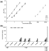
Quantification of proteins of the egc cluster through a polyclonal ELISA system. (a) Standard curves of ELISA systems for the detection of SEI, SElM, SElN, and SEB in a lin-log scale. Microtiter plates were coated with the polyclonal murine IgG anti-SE antibody in a dilution of 1:3,000 as the capture antibody. As the detecting antibody, the polyclonal rabbit IgG anti-SE antibody was used in dilution of 1:1,000. The conjugate, a polyclonal horseradish peroxidase-conjugated goat anti-rabbit antibody, was used in a dilution of 1:5,000. Standard curve samples ranged from 0.125 ng/mL to 5 ng/mL for SEI, from 0.5 ng/mL to 25 ng/mL for SElM and SElN, and from 0.5 µg/mL to 10 µg/mL for SEB. The standard curve graphs were obtained by logarithmic regression fit with six (five for SEB) data points made up of the average of duplicate values (SEI R2 = 0.9925; SElM R2 = 0.9912; SElN R2 = 0.9962; SEB R2 = 0.9948). Error bars indicate the standard error of the mean of five independent experiments; and (b) quantification of SEI, SElM, SElN, and SEB in late stationary phase supernatants through polyclonal ELISA system evaluated in (a). Samples were 10-fold diluted in 1× PBS containing 2% BSA and 0.1% Tween 20. ATCC 10832 was used as the negative control strain, since it harbors no egc cluster.
Figure 3
Figure 3
Potentials of recombinant SEG, SEI, SElM, SElN, and SEB to induce proliferation of human peripheral blood mononuclear cells (MNC). Induction of proliferation was quantified by counting incorporated [3H] thymidine after incubation for 4 d in a humidified atmosphere (37 °C, 5% CO2). Experiments were done with blood from two independent donors. Recombinant wild-type proteins were diluted in RPMI 1640 complete medium. MNCs were adjusted to 1 × 10⁶ cells per mL. Phytohaemagglutinin (PHA) was used as a control.
Figure 4
Figure 4
Proliferation of human peripheral blood mononuclear cells (MNC) is stimulated by egc superantigens and can be neutralized by antisera against SEI and SElN. (a) Induced proliferation was measured by the counting of incorporated [3H] thymidine after incubation for 4 d in a humidified atmosphere (37 °C, 5% CO2). Each experiment was performed in triplicate. Supernatants were diluted in RPMI 1640 medium, phytohaemagglutinin (PHA) was used as a control. The supernatant of strain B7709 was additionally incubated with alpha toxin antiserum in a dilution of 1:50 for 1 h at 37 °C (900 rpm); (b) Diluted supernatants of Rv52825I or Rv51379 were neutralized with antisera (x-axis annotation) in a dilution of 1:50 for 1 h at 37 °C (900 rpm). Different suspensions of egc antisera (left panel), suspensions of anti-SEB/SEH or plus-favored anti-SEI/SElN antisera (right panel), were applied to specific 10−4-fold diluted supernatants of tested strains. After incubation, mixes were added to isolated blood cells, and incorporation of [3H] thymidine was measured. Experiments were performed as described in (a).

References

    1. Salgado-Pabón W., Breshears L., Spaulding A.R., Merriman J.A., Stach C.S., Horswill A.R., Peterson M.L., Schlievert P.M. Superantigens are critical for Staphylococcus aureus Infective endocarditis, sepsis, and acute kidney injury. mBio. 2013;4 doi: 10.1128/mBio.00494-13. - DOI - PMC - PubMed
    1. Singer A.J., Talan D.A. Management of skin abscesses in the era of methicillin-resistant Staphylococcus aureus. N. Engl. J. Med. 2014;370:1039–1047. doi: 10.1056/NEJMra1212788. - DOI - PubMed
    1. Peacock S.J., Moore C.E., Justice A., Kantzanou M., Story L., Mackie K., O’Neill G., Day N.P.J. Virulent combinations of adhesin and toxin genes in natural populations of Staphylococcus aureus. Infect. Immun. 2002;70:4987–4996. doi: 10.1128/IAI.70.9.4987-4996.2002. - DOI - PMC - PubMed
    1. Chavakis T., Preissner K.T., Herrmann M. The anti-inflammatory activities of Staphylococcus aureus. Trends Immunol. 2007;28:408–418. doi: 10.1016/j.it.2007.07.002. - DOI - PubMed
    1. Foster T.J., Geoghegan J.A., Ganesh V.K., Höök M. Adhesion, invasion and evasion: The many functions of the surface proteins of Staphylococcus aureus. Nat. Rev. Microbiol. 2014;12:49–62. doi: 10.1038/nrmicro3161. - DOI - PMC - PubMed

Publication types

MeSH terms