. 2016 Dec 1;143(23):4474-4485.
doi: 10.1242/dev.141978. Epub 2016 Oct 17.

Neural specificity of the RNA-binding protein Elav is achieved by post-transcriptional repression in non-neural tissues

Affiliations

Neural specificity of the RNA-binding protein Elav is achieved by post-transcriptional repression in non-neural tissues

Piero Sanfilippo et al. Development. .

Erratum in

Abstract

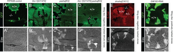
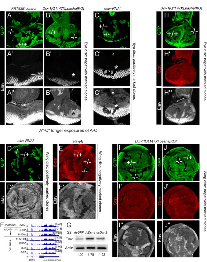
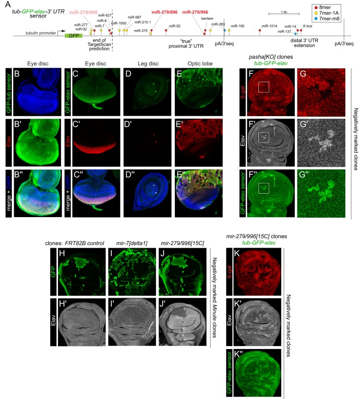
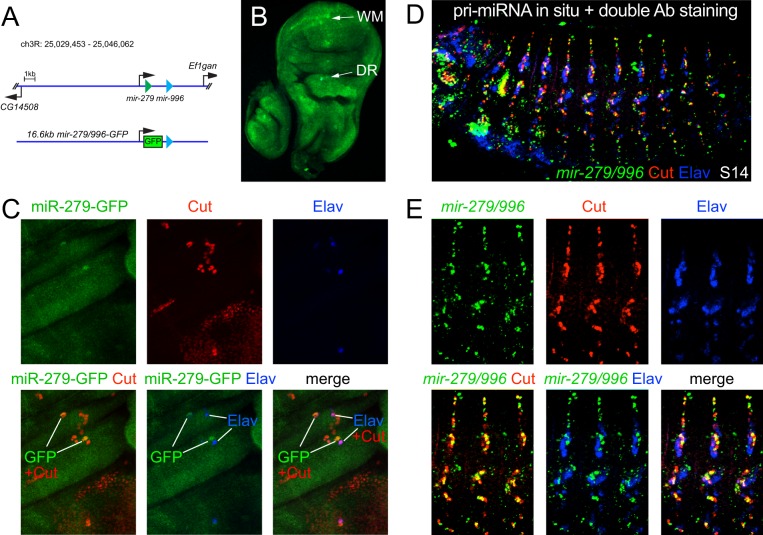
Drosophila Elav is the founding member of the conserved family of Hu RNA-binding proteins (RBPs), which play crucial and diverse roles in post-transcriptional regulation. Elav has long served as the canonical neuronal marker. Surprisingly, although Elav has a well-characterized neural cis-regulatory module, we find endogenous Elav is also ubiquitously transcribed and post-transcriptionally repressed in non-neural settings. Mutant clones of multiple miRNA pathway components derepress ubiquitous Elav protein. Our re-annotation of the elav transcription unit shows not only that it generates extended 3' UTR isoforms, but also that its universal 3' UTR isoform is much longer than previously believed. This longer common 3' UTR includes multiple conserved, high-affinity sites for the miR-279/996 family. Of several miRNA mutants tested, endogenous Elav and a transgenic elav 3' UTR sensor are derepressed in mutant clones of mir-279/996 We also observe cross-repression of Elav by Mei-P26, another RBP derepressed in non-neural miRNA pathway clones. Ubiquitous Elav has regulatory capacity, since derepressed Elav can stabilize an Elav-responsive sensor. Repression of Elav in non-neural territories is crucial as misexpression here has profoundly adverse consequences. Altogether, we define unexpected post-transcriptional mechanisms that direct appropriate cell type-specific expression of a conserved neural RBP.

Keywords: Drosophila; Elav; Mei-P26; MicroRNA; Neuron; RNA-binding protein.


Conflict of interest statement

The authors declare no competing or financial interests.

Figures

Fig. 1.
Fig. 1.
Loss of the miRNA pathway derepresses the canonical neural marker Elav in non-neural territories. Shown are regions of the wing imaginal disc pouch, co-stained for a clonal marker (A-F; GFP, green or β-Gal, red) and Elav (A′-F′; grayscale). Negatively marked clones were generated using the Minute technique in A-D or conventional technique in E. Positively marked clones using the MARCM technique are in F. Example wild-type territories are indicated by +/+ and example clones are noted by −/−; heterozygous (+/−) tissue was only generated in E. Elav protein is derepressed in mutant clones lacking diverse core miRNA pathway factors (Dcr-1, pasha or drosha), or that are depleted for the miRNA effector (GW182). At least five imaginal discs were assayed for each genotype, and each set of stainings was performed at least twice; representative clones are shown.
Fig. 2.
Fig. 2.
Elav is expressed outside of the nervous system and is specifically derepressed in miRNA pathway clones. Shown are eye imaginal discs (A-C,H) and pouch regions of wing imaginal discs (D,E,I,J) stained for clonal markers (GFP, green or β-Gal, red), Elav (grayscale) and/or other pan-neural markers (red). (A-B″) Comparison of control wild-type clones (A) and Dcr-1, pasha double-mutant clones (B), all made using the Minute system. ‘Conventional' imaging technique for Elav shows typical photoreceptor (R) expression posterior to the morphogenetic furrow (MF), which is not substantially affected by miRNA pathway loss (A′,B′). Longer exposure (A″,B″) emphasizes ectopic Elav anterior to the MF and in the antennal region. (C-C″) Clonal expression of elav-RNAi eliminates Elav in R cells (hash mark), and longer exposure also shows loss of basal Elav in non-neural disc regions (asterisk). (D,D′) Clonal expression of elav-RNAi eliminates basal Elav in the wing disc. (E,E′) Mitotic clonal analysis of null allele elav[4] shows graded expression of basal, non-neural Elav in heterozygous and mutant regions. (F) RNA-seq data across an embryo timecourse show clear maternal expression and zygotic expression of elav long before the earliest presence of neurons (8-10 h). (G) Western blot of S2 cells shows that Elav is specifically derepressed upon Dcr-1 knockdown. Values beneath lanes indicate fold derepression compared with dsGFP control (H-J″) Analysis of canonical neural factors Scratch (Scrt; H,I) and Deadpan (Dpn; J) shows that they are not derepressed in miRNA pathway Minute clones in eye (H) or wing (I,J) discs. At least five imaginal discs were assayed for each genotype, and each set of stainings was performed at least twice; representative clones are shown.
Fig. 3.
Fig. 3.
Direct repression of the elav 3′ UTR via the miRNA pathway and miR-279/996. (A) The tub-GFP-elav 3′ UTR sensor transgene. We find that the 3′ terminus used in public miRNA annotations (e.g. TargetScan) is not detected in vivo. Rather, the genuine proximal 3′ UTR isoform is nearly 3 kb longer, and we identify an extended 3′ UTR isoform, both of which are supported by 3′-seq tags (as annotated). (B-K″) Images depict eye imaginal discs (B,C), leg imaginal disc (D), brain optic lobe (E) and pouch regions of wing imaginal discs (F-K) stained for reporter GFP (B-E,F,G,K), clonal markers (GFP, green or β-Gal, red) and Elav (grayscale). (B-E″) Comparison of tub-GFP-tubulin-3′ UTR and tub-GFP-elav-3′ UTR sensor transgenes. (B) The tub 3′ UTR sensor is broadly expressed in the eye disc, as well as other tissues (not shown). (C-E) The elav 3′ UTR restricts ubiquitously expressed GFP into the pattern of endogenous Elav protein, as seen by their colocalization in several tissues. (F-G″) Mitotic pasha[KO] clones (marked by absence of β-Gal staining) show coincident derepression of Elav and the elav 3′ UTR sensor. Boxed regions in F-F″ are magnified in G-G″. (H-J′) Tests of individual miRNA loci on post-transcriptional repression of Elav. (H) Control clones and (I) mir-7 null clones do not affect Elav, whereas clones of mir-279/996 derepress Elav protein (J). (K-K″) Clonal deletion of mir-279/996 causes concomitant elevation of Elav and the elav 3′ UTR sensor. At least five imaginal discs were assayed for each genotype, and each set of stainings was performed at least twice; representative clones are shown.
Fig. 4.
Fig. 4.
Complementary expression of mir-279/996 and Elav. (A) The bicistronic mir-279/996 locus, and a 16.6 kb knock-in reporter that contains all regulatory elements for genetic rescue (Sun et al., 2015), in which GFP replaces the mir-279 hairpin. (B) Wing imaginal disc carrying the mir-279-GFP transcriptional reporter shows ubiquitous expression, as well as upregulation in presumptive neural territories: the wing margin (WM) and dorsal radius (DR). (C) Magnification of the dorsal radius region co-stained for miR-279-GFP, the pan-sensory organ marker Cut, and Elav. The cells that upregulate mir-279-GFP are Cut+ Elav sensory organ cells. (D) Stage 14 embryo subjected to in situ hybridization for primary mir-279/996 transcripts and double labeling for Cut and Elav proteins. (E) Magnification of three lateral segments bearing PNS structures. In the embryo, Cut accumulates to a higher level in non-neural PNS cells. Endogenous mir-279/996 transcripts accumulate in Cut+ Elav cells. At least five imaginal discs or embryos were assayed for each genotype; representative images are shown.
Fig. 5.
Fig. 5.
Mei-P26 represses elav via its 3′ UTR. Shown are (A) eye imaginal disc and (B-G) pouch regions of wing imaginal discs. (A-B‴) Clonal activation of Mei-P26 leads to cell-autonomous decrease in Elav protein in both a high-expression domain (photoreceptors, A) and a low-expression domain (wing pouch, B, arrows). (C-D‴) Ectopic Mei-P26 represses the tub-GFP-elav-3′ UTR sensor. Although the effect is quantitatively mild, it is more clearly observed in higher magnification clones (D, outlined regions). (E) Clonal knockdown of mei-P26 causes cell-autonomous increase in basal Elav. (F-G″) MARCM analysis of pasha[KO] clones. (F) Control pasha clones derepress both Mei-P26 and Elav proteins. (G) Knockdown of mei-P26 reverses the accumulation of Mei-P26 protein in pasha clones, but does not reliably superactivate Elav. At least five imaginal discs were assayed for each genotype, and each set of stainings was performed at least twice; representative clones are shown.
Fig. 6.
Fig. 6.
Functional consequences of elevating Elav in wing imaginal discs. (A) The UGgH Elav sensor consists of ubiquitously expressed GFP followed by the AU-rich element (ARE)-rich 3′ UTR of Hsp70Ab, a sensor previously shown to be stabilized by Elav. (B,B′) Clonal expression of UAS-elav increases UGgH expression (arrowheads). (C-D″) Dcr-1 (C) or pasha (D) mutant clones that derepress Elav also stabilize the UGgH Elav activity sensor. (E) Summary of elav misexpression tests shows that it induces lethality when activated with multiple non-neuronal drivers, but not when activated neuronally. (F-G‴) Flp-out expression clones in eye discs, marked by activation of UAS-lacZ (β-Gal, green). (F) Control GFP clones are recovered throughout the eye disc. (G) Elav-expressing clones are preferentially recovered in photoreceptors, and do not induce cell death as marked by cleaved caspase 3 (c-casp3; G″). (H-K′) Flp-out expression clones in wing discs, marked by activation of UAS-lacZ (β-Gal, green). (H) Control GFP clones are recovered throughout the wing disc and do not induce c-casp3 activity. (I) Elav-expressing clones are recovered in the wing pouch (WP) and prospective notum (N), but cells in the latter region are not in the disc epithelium but rather reside in the adepithelial layer (AE). Elav-expressing cells in the wing pouch accumulate high levels of c-casp3 (I″, arrowhead), whereas Elav-expressing adepithelial cells do not (I″, arrow). (J) High magnification of wing pouch region shows that Elav-expressing clones are fragmented. Lack of continuous DAPI signal is due to visualization of a narrow z-section, and the pyknotic nuclei (J″, arrows) delaminate from the epithelium. (K) Transverse section through the wing pouch illustrates how dying, Elav+ c-casp-3+ clones (asterisks) in the center of the wing pouch are removed from the epithelium, whereas laterally located clones remain integrated and express little or no c-casp3+ (hash marks). At least five imaginal discs were assayed for each genotype, and each set of stainings was performed at least twice; representative clones are shown.

Similar articles

Cited by

References

    1. Aboobaker A. A., Tomancak P., Patel N., Rubin G. M. and Lai E. C. (2005). Drosophila microRNAs exhibit diverse spatial expression patterns during embryonic development. Proc. Natl. Acad. Sci. USA 102, 18017-18022. 10.1073/pnas.0508823102 - DOI - PMC - PubMed
    1. Albert M. L. and Darnell R. B. (2004). Paraneoplastic neurological degenerations: keys to tumour immunity. Nat. Rev. Cancer 4, 36-44. 10.1038/nrc1255 - DOI - PubMed
    1. Andersson T., Rahman S., Sansom S. N., Alsiö J. M., Kaneda M., Smith J., O'Carroll D., Tarakhovsky A. and Livesey F. J. (2010). Reversible block of mouse neural stem cell differentiation in the absence of dicer and microRNAs. PLoS ONE 5, e13453 10.1371/journal.pone.0013453 - DOI - PMC - PubMed
    1. Barolo S., Walker R., Polyanovsky A., Freschi G., Keil T. and Posakony J. W. (2000). A Notch-independent activity of Suppressor of Hairless is required for normal mechanoreceptor physiology. Cell 103, 957-970. 10.1016/S0092-8674(00)00198-7 - DOI - PubMed
    1. Bartel D. P. (2009). MicroRNAs: target recognition and regulatory functions. Cell 136, 215-233. 10.1016/j.cell.2009.01.002 - DOI - PMC - PubMed

Publication types

MeSH terms

LinkOut - more resources