

Different imaging techniques for the detection of pelvic lymph nodes metastasis from gynecological malignancies: a systematic review and meta-analysis
- PMID: 27802186
- PMCID: PMC5355166
- DOI: 10.18632/oncotarget.12959
Different imaging techniques for the detection of pelvic lymph nodes metastasis from gynecological malignancies: a systematic review and meta-analysis
Abstract
Objective: This study aimed to evaluate the diagnostic performance of different imaging techniques and the corresponding diagnostic criteria for preoperative detection of pelvic lymph node metastasis from gynecological carcinomas.
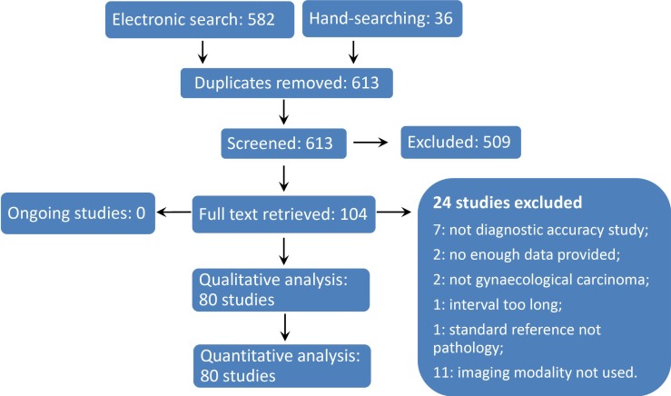
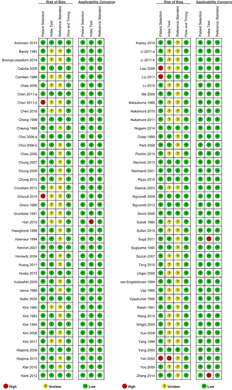
Methods: Six databases were systematically searched for retrieving eligible studies. Study inclusion, data extraction and risk of bias assessment were performed by 2 reviewers independently. STATA 14.0 was used to perform the meta-analysis.
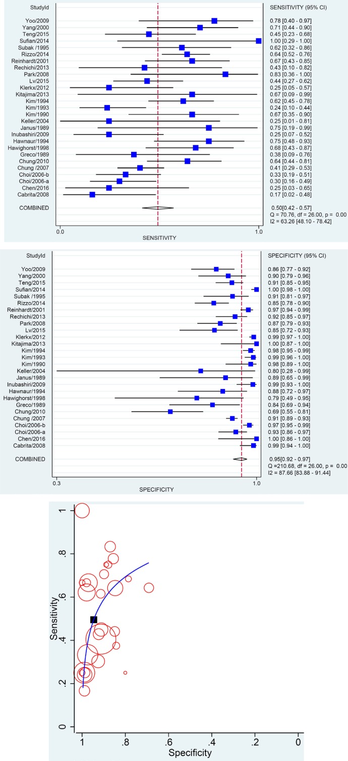
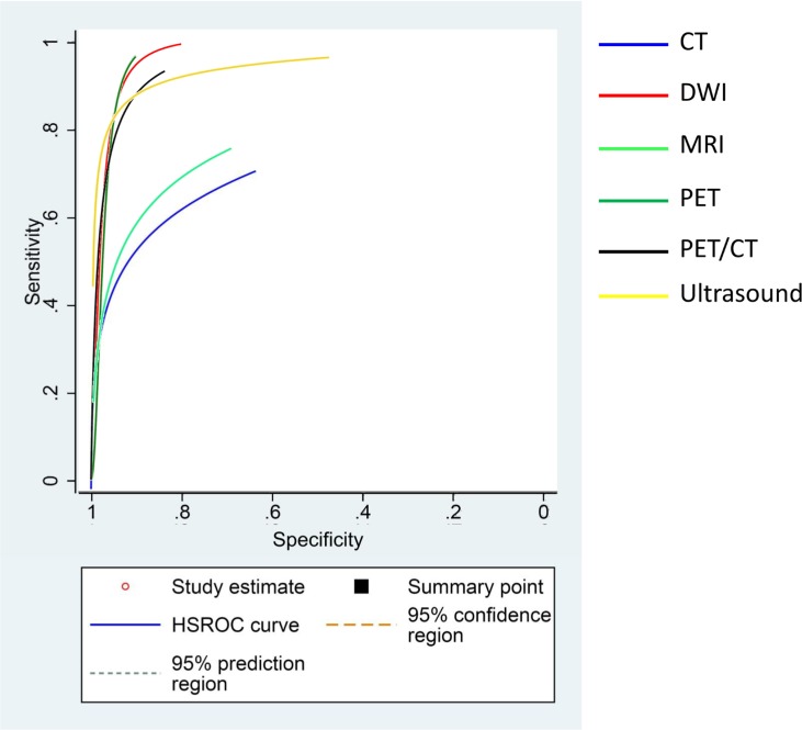
Results: Eighty eligible studies were collected. The pooled sensitivity, specificity, and area under curve (AUC) of CT, MRI and DWI were 47%, 93%, 0.7424; 50%, 95%, 0.8039 and 84%, 95%, 0.9523 respectively. As regards PET, PET-CT and US, the pooled sensitivity, specificity and AUC were 56%, 97%, 0.9592; 68%, 97%, 0.9363 and 71%, 99%, 0.9008 respectively. The summary receiver operating characteristic (SROC) curve indicated that the systematic diagnostic performances of PET, PET-CT, DWI were superior to other imaging modalities.
Conclusions: The present work demonstrated that DWI, PET, PET-CT were the top-priority consideration of imaging modalities for detecting metastatic pelvic lymph node in gynecological carcinoma. DWI was recommended as the first choice for metastasis exclusion and all the other imaging techniques including CT and MRI were suitable for metastasis conformation. However, for the early stage lymph node malignancy, PET or PET-CT could represent a better choice. More studies exploring the diagnostic efficacy of detailed criteria are required in the future.
Keywords: gynecological malignance; imaging technique; metastasis; pelvic lymph node; systematic review and meta-analysis.
Conflict of interest statement
The authors declare that there are no conflicts of interest.
Figures
References
-
- Kamura T, Tsukamoto N, Tsuruchi N, Saito T, Matsuyama T, Akazawa K, Nakano H. Multivariate analysis of the histopathologic prognostic factors of cervical cancer in patients undergoing radical hysterectomy. Cancer. 1992;69:181–186. - PubMed
-
- Lewin SN, Herzog TJ, Medel NIB, Deutsch I, Burke WM, Sun X, Wright JD. Comparative performance of the 2009 international Federation of gynecology and obstetrics’ staging system for uterine corpus cancer. Obstetrics & Gynecology. 2010;116:1141–1149. - PubMed
-
- Sakuragi N, Satoh C, Takeda N, Hareyama H, Takeda M, Yamamoto R, Fujimoto T, Oikawa M, Fujino T, Fujimoto S. Incidence and distribution pattern of pelvic and paraaortic lymph node metastasis in patients with stages IB, IIA, and IIB cervical carcinoma treated with radical hysterectomy. Cancer. 1999;85:1547–1554. - PubMed
-
- Michel G, Morice P, Castaigne D, Leblanc M, Rey A, Duvillard P. Lymphatic spread in stage Ib and II cervical carcinoma: anatomy and surgical implications. Obstetrics & Gynecology. 1998;91:360–363. - PubMed
-
- Park SO, Kim JK, Kim KA, Park BW, Kim N, Cho G, Choi HJ, Cho KS. Relative apparent diffusion coefficient: determination of reference site and validation of benefit for detecting metastatic lymph nodes in uterine cervical cancer. Journal of Magnetic Resonance Imaging. 2009;29:383–390. - PubMed
Publication types
MeSH terms
LinkOut - more resources
Full Text Sources
Other Literature Sources
Medical
Research Materials
