. 2016 Dec 16;291(51):26262-26272.
doi: 10.1074/jbc.M116.760017. Epub 2016 Nov 1.

The Neural Cell Adhesion Molecule (NCAM) Promotes Clustering and Activation of EphA3 Receptors in GABAergic Interneurons to Induce Ras Homolog Gene Family, Member A (RhoA)/Rho-associated protein kinase (ROCK)-mediated Growth Cone Collapse

Affiliations

The Neural Cell Adhesion Molecule (NCAM) Promotes Clustering and Activation of EphA3 Receptors in GABAergic Interneurons to Induce Ras Homolog Gene Family, Member A (RhoA)/Rho-associated protein kinase (ROCK)-mediated Growth Cone Collapse

Chelsea S Sullivan et al. J Biol Chem. .

Abstract

Establishment of a proper balance of excitatory and inhibitory connectivity is achieved during development of cortical networks and adjusted through synaptic plasticity. The neural cell adhesion molecule (NCAM) and the receptor tyrosine kinase EphA3 regulate the perisomatic synapse density of inhibitory GABAergic interneurons in the mouse frontal cortex through ephrin-A5-induced growth cone collapse. In this study, it was demonstrated that binding of NCAM and EphA3 occurred between the NCAM Ig2 domain and EphA3 cysteine-rich domain (CRD). The binding interface was further refined through molecular modeling and mutagenesis and shown to be comprised of complementary charged residues in the NCAM Ig2 domain (Arg-156 and Lys-162) and the EphA3 CRD (Glu-248 and Glu-264). Ephrin-A5 induced co-clustering of surface-bound NCAM and EphA3 in GABAergic cortical interneurons in culture. Receptor clustering was impaired by a charge reversal mutation that disrupted NCAM/EphA3 association, emphasizing the importance of the NCAM/EphA3 binding interface for cluster formation. NCAM enhanced ephrin-A5-induced EphA3 autophosphorylation and activation of RhoA GTPase, indicating a role for NCAM in activating EphA3 signaling through clustering. NCAM-mediated clustering of EphA3 was essential for ephrin-A5-induced growth cone collapse in cortical GABAergic interneurons, and RhoA and a principal effector, Rho-associated protein kinase, mediated the collapse response. This study delineates a mechanism in which NCAM promotes ephrin-A5-dependent clustering of EphA3 through interaction of the NCAM Ig2 domain and the EphA3 CRD, stimulating EphA3 autophosphorylation and RhoA signaling necessary for growth cone repulsion in GABAergic interneurons in vitro, which may extend to remodeling of axonal terminals of interneurons in vivo.

Keywords: EphA3; NCAM; Ras homolog gene family, member A (RhoA); growth cone collapse; interneuron; neurobiology; neurodevelopment; oligomerization; synaptic plasticity.


Figures

FIGURE 1.
FIGURE 1.
The EphA3 CRD binds the NCAM Ig2 domain. A, a candidate pose of Ig1-Ig3 of NCAM docked with LBD-CRD of EphA3. The predicted interface is labeled with arrows. B, Fc pulldowns of NCAM-EC and EphA3 or EphA3 deletion mutants. C, Fc pulldowns of NCAM-EC or truncation mutants and EphA3. D, densitometry of C. The amount of bound EphA3 for each Fc protein was normalized to control NCAM-EC-bound EphA3 (n = 3; *, p < 0.05). E, co-immunoprecipitation (IP) of WT NCAM or mutants of NCAM and EphA3 from transfected HEK293T cells. IB, immunoblot. F, densitometry of E. The amount of co-immunoprecipitated EphA3 for each NCAM immunoprecipitation was normalized to control WT NCAM-bound EphA3 (n = 3; *, p < 0.05). G, co-immunoprecipitation of NCAM and WT EphA3 or EphA3 mutants from transfected HEK293T cells. H, densitometry of G. The amount of co-immunoprecipitated EphA3 for each NCAM immunoprecipitation was normalized to control WT EphA3 (n = 3; *, p < 0.05). Student's t test was performed for each of the binding experiments.
FIGURE 2.
FIGURE 2.
Ephrin-A5 induces clustering of NCAM and EphA3 in cortical interneurons. A, NCAM KO cortical neurons were co-transfected with WT NCAM or NCAM mutants and pCAG-IRES-EGFP. Cells were treated with preclustered Fc or ephrin-A5-Fc, and localization of NCAM (pseudocolored green) and EphA3 (red) was assessed by confocal microscopy. Axons of GABA-immunoreactive interneurons were imaged, and colocalization of NCAM and EphA3 was assessed using ImageJ to generate a heat map of colocalization (fourth column). Scale bar = 5 μm. Thresholded Manders coefficients (tM1, B; tM2, C) and Pearson correlation coefficients (R-Total, D) were generated using ImageJ colocalization software (n = 3; *, p < 0.05; n.s., not significant; Student's t test was performed between Fc and ephrinA5-Fc treated cells for each vector).
FIGURE 3.
FIGURE 3.
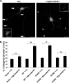
NCAM binding to EphA3 promotes ephrin-A5-induced growth cone collapse in GABAergic interneurons. A, NCAM KO neurons were co-transfected with empty vector, WT NCAM, or mutant NCAM together with pCAG-IRES-EGFP and stained for GFP and GABA. Representative images of GABA-positive interneurons (DIV 12) treated with preclustered control Fc or ephrin-A5-Fc and stained for GABA are shown. Spread growth cones are indicated with arrows, and collapsed growth cones are marked with arrowheads. Scale bars = 10 μm and 2.5 μm in the magnified insets. B, the percentage of collapsed growth cones was determined (300 growth cones/condition; n = 3 experiments; *, p < 0.05; n.s., not significant). Student's t test was performed between Fc- and ephrinA5-Fc-treated cells for each vector.
FIGURE 4.
FIGURE 4.
NCAM stimulates EphA3 autophosphorylation in response to ephrin-A5 treatment in cortical neurons. A, cortical neuron cultures from WT or NCAM KO mice (DIV 12) were treated with preclustered control Fc or ephrin-A5-Fc, and EphA3 was immunoprecipitated (IP). EphA3 autophosphorylation was assessed using phosphotyrosine antibodies, and total levels of immunoprecipitated EphA3 were assessed by reprobing with EphA3 antibody. IB, immunoblot. B, densitometry of A. The graph indicates the ratio of phosphotyrosine to EphA3 values for each condition (n = 3; *, p < 0.05; n.s., not significant). Student's t test was performed between Fc- and ephrinA5-Fc-treated cells for each genotype.
FIGURE 5.
FIGURE 5.
NCAM-dependent EphA3 activation promotes activation of RhoA, required for growth cone collapse. A, activation of RhoA, Rac1, and Cdc42 was assessed in WT or NCAM KO cultures after treatment with preclustered ephrin-A5-Fc or control Fc. B, densitometry of active RhoA (in WT and NCAM KO cultures), Rac1 (WT), and Cdc42 (WT) normalized to control Fc. The graph shows mean with S.E. A two-way ANOVA with Bonferroni post hoc testing was performed between NCAM KO and WT cells for RhoA activation after Fc or ephrinA5-Fc treatment (***, p < 0.001). For Cdc42 and Rac1 activation in WT cells, Student's t test was performed between Fc- and ephrinA5-Fc-treated cells for each GTPase. C, growth cone collapse of WT neurons pretreated with C3, Y-27632, or control untreated and preclustered Fc or ephrin-A5-Fc. The graph shows mean with S.E., two-way ANOVA with Bonferroni post hoc testing (n = 3 experiments; >500 growth cones/condition; ***, p < 0.001).
FIGURE 6.
FIGURE 6.
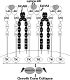
Model of NCAM clustering and activation of EphA3. Ephrin-A5 dimers bind the LBD domains of an EphA3 dimer. NCAM homodimerizes through Ig1-Ig2 interactions and clusters EphA3 receptors through binding to the EphA3 CRD. EphA3 clustering activates tyrosine kinase signaling through RhoA and ROCK1/2, leading to axonal growth cone collapse. NCAM dimers are shown in black, and EphA3 dimers are shown in white.

References

    1. Maness P. F., and Schachner M. (2007) Neural recognition molecules of the immunoglobulin superfamily: signaling transducers of axon guidance and neuronal migration. Nat. Neurosci. 10, 19–26 - PubMed
    1. Brennaman L. H., Zhang X., Guan H., Triplett J. W., Brown A., Demyanenko G. P., Manis P. B., Landmesser L., and Maness P. F. (2013) Polysialylated NCAM and EphrinA/EphA regulate synaptic development of GABAergic interneurons in prefrontal cortex. Cereb. Cortex 23, 162–177 - PMC - PubMed
    1. Tremblay R., Lee S., and Rudy B. (2016) GABAergic interneurons in the neocortex: from cellular properties to circuits. Neuron 91, 260–292 - PMC - PubMed
    1. Mann E. O., Kohl M. M., and Paulsen O. (2009) Distinct roles of GABAA and GABAB receptors in balancing and terminating persistent cortical activity. J. Neurosci. 29, 7513–7518 - PMC - PubMed
    1. Isaacson J. S., and Scanziani M. (2011) How inhibition shapes cortical activity. Neuron 72, 231–243 - PMC - PubMed

MeSH terms

LinkOut - more resources