. 2016 Nov 1;7(6):e00831-16.
doi: 10.1128/mBio.00831-16.

A Class 1 Histone Deacetylase with Potential as an Antifungal Target

Affiliations

A Class 1 Histone Deacetylase with Potential as an Antifungal Target

Ingo Bauer et al. mBio. .

Abstract

Histone deacetylases (HDACs) remove acetyl moieties from lysine residues at histone tails and nuclear regulatory proteins and thus significantly impact chromatin remodeling and transcriptional regulation in eukaryotes. In recent years, HDACs of filamentous fungi were found to be decisive regulators of genes involved in pathogenicity and the production of important fungal metabolites such as antibiotics and toxins. Here we present proof that one of these enzymes, the class 1 type HDAC RpdA, is of vital importance for the opportunistic human pathogen Aspergillus fumigatus Recombinant expression of inactivated RpdA shows that loss of catalytic activity is responsible for the lethal phenotype of Aspergillus RpdA null mutants. Furthermore, we demonstrate that a fungus-specific C-terminal region of only a few acidic amino acids is required for both the nuclear localization and catalytic activity of the enzyme in the model organism Aspergillus nidulans Since strains with single or multiple deletions of other classical HDACs revealed no or only moderate growth deficiencies, it is highly probable that the significant delay of germination and the growth defects observed in strains growing under the HDAC inhibitor trichostatin A are caused primarily by inhibition of catalytic RpdA activity. Indeed, even at low nanomolar concentrations of the inhibitor, the catalytic activity of purified RpdA is considerably diminished. Considering these results, RpdA with its fungus-specific motif represents a promising target for novel HDAC inhibitors that, in addition to their increasing impact as anticancer drugs, might gain in importance as antifungals against life-threatening invasive infections, apart from or in combination with classical antifungal therapy regimes.

Importance: This paper reports on the fungal histone deacetylase RpdA and its importance for the viability of the fungal pathogen Aspergillus fumigatus and other filamentous fungi, a finding that is without precedent in other eukaryotic pathogens. Our data clearly indicate that loss of RpdA activity, as well as depletion of the enzyme in the nucleus, results in lethality of the corresponding Aspergillus mutants. Interestingly, both catalytic activity and proper cellular localization depend on the presence of an acidic motif within the C terminus of RpdA-type enzymes of filamentous fungi that is missing from the homologous proteins of yeasts and higher eukaryotes. The pivotal role, together with the fungus-specific features, turns RpdA into a promising antifungal target of histone deacetylase inhibitors, a class of molecules that is successfully used for the treatment of certain types of cancer. Indeed, some of these inhibitors significantly delay the germination and growth of different filamentous fungi via inhibition of RpdA. Upcoming analyses of clinically approved and novel inhibitors will elucidate their therapeutic potential as new agents for the therapy of invasive fungal infections-an interesting aspect in light of the rising resistance of fungal pathogens to conventional therapies.


Figures

FIG 1
FIG 1
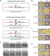
Heterokaryon rescue of an A. fumigatus Ku70 recipient strain transformed with an rpdA deletion cassette. After transformation, heterokaryotic strains t1 to t6 were grown under selective conditions (pyrithiamine) and uninucleate conidia were streaked onto sectors on agar plates with and without selection and grown for 48 h at 37°C (A). Molecular size markers (M) are shown on the left and right. Genomic DNA from putative heterokaryons was prepared and subjected to a diagnostic PCR. DNA of the recipient strain served as a control (B). The scheme of the homologous integration of the deletion cassette, the annealing sites of the primers, and the sizes of the fragments amplified are shown in panel C. Ao ptrA, pyrithiamine resistance gene of A. oryzae, Af rpdA, coding sequence of A. fumigatus RpdA. UTR, untranslated region; wt, wild type.
FIG 2
FIG 2
Phenotypic analysis of Aspergillus strains expressing different RpdA variants. A two-promoter system was used to determine the biological function of mutated RpdA fragments. Recipient strains RIB214 and TSG5 expressing endogenous rpdA (full length, FL) under the control of the alcohol dehydrogenase promoter (alcAp) were transformed with expression cassettes comprising the coding regions of different RpdA variants under the control of the xylanase promoter (xylPp) (A). Transformed protoplasts were regenerated under alcAp induction (LT), and RpdA variants H158A and D193A (B), del-N18 and del-C18 (C), and del-C12 and del-C6 (D) were analyzed for the ability to compensate for wild-type RpdA depletion under alcAp repressive and xylPp inductive conditions (GX). Expression of the recombinant variants was verified by immunoblotting of whole protein extracts under GX conditions with anti-RpdA antibodies. Recipient strain TSG5 (no xylPp expression cassette) and a strain expressing wild-type RpdA under the control of xylPp (FL) were used as negative and positive controls, respectively (E).
FIG 3
FIG 3
Germination of spores and hyphal growth of A. fumigatus under TSA treatment. Conidia (1 × 10⁵/ml) were incubated into 24-well plates with RPMI medium supplied with 10 µM TSA. Spores were incubated for 10 or 15 h at 37°C before wells were examined under a light microscope (A). Growth retardation of hyphae was observed in liquid medium 24 h after the addition of 10 µM TSA to a culture grown for 10 h at 37°C without an inhibitor (B). DMSO, the solvent of TSA, was used in the corresponding concentration as a negative control.
FIG 4
FIG 4
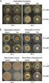
Mycelial growth and sporulation of A. fumigatus (A), A. nidulans, A. terreus, N. crassa, and P. chrysogenum (B) at different TSA concentrations. Spores (1 × 10³) were dotted into the middle of each agar well, and strains were grown overnight to allow germination. Subsequently, colonies were overlaid with 100 µl of liquid medium containing 2.5 or 10 µm of the inhibitor. A corresponding concentration of DMSO was used as a negative control. After incubation for different periods of time at 37°C (A. fumigatus, A. nidulans, A. terreus, and N. crassa) or 25°C (P. chrysogenum), the size of the colony and conidiation of the mycelium were assessed.
FIG 5
FIG 5
Comparison of RPD3-type HDACs of fungi and higher eukaryotes. A schematic representation of S. cerevisiae (Sce) RPD3, Homo sapiens (Hsa) HDAC1, and A. nidulans (Ani) RpdA is shown. The highly conserved region comprising amino residues essential for catalytic activity is green, the N-terminal fungus-specific region (N18) and the acidic C-terminal stretch conserved in filamentous fungi and higher eukaryotes (C18 and C6) are black and red, respectively. Putative nuclear localization sequences in enzymes of higher eukaryotes are yellow. The C-terminal tail (C-ter) expressed as a Venus-tagged peptide is indicated (A). aa, amino acids. A detailed alignment of the region adjacent to the acidic C-terminal motif essential for RpdA-type enzymes of filamentous fungi is shown for different fungal species, amphibians (Xenopus), humans, and plants (Zea) in panel B. Stretches conserved in filamentous fungi (C12) and in all eukaryotes except yeasts (C6) are shown as black lines at the bottom. Residues are shaded red (acidic), blue (basic), or gray (uncharged). Deletions or alanine substitutions of the RpdA variants tested are shown at the top. Gray lines represent mutations with no effect on the biological function of RpdA, and black lines depict mutations leading to a lethal phenotype of the corresponding expression strains. Afu, A. fumigatus; Ate, A. terreus; Ncr, N. crassa; Pch, P. chrysogenum; Cca, C. carbonum; Xla, Xenopus laevis; Zma, Zea mays; Hsa, Homo sapiens.
FIG 6
FIG 6
(A) Phenotypic analysis of Aspergillus strains expressing RpdA-type enzymes of different filamentous fungi and human HDAC1. Strains TSG5 and RIB214 expressing nonmutated RpdA under the control of the alcohol dehydrogenase promoter (alcAp) (Fig. 2A) were transformed with expression cassettes comprising the coding regions of the different RPD3-type HDACs under the control of the xylanase promoter (xylPp). Transformed protoplasts were regenerated under alcAp induction (LT), and recombinant heterologous HDACs were analyzed for the ability to compensate for RpdA under alcAp repressive and xylPp inductive conditions (GX). A strain of A. nidulans (Ani) expressing wild-type RpdA under the control of xylPp was used as a positive control. (B) Transcription of noncomplementing HDAC1 (H. sapiens [Hsa]) was verified by Northern analysis with 10 and 30 µg of total RNA. rRNA was used as a loading and quality control. (C) Expression of HDAC1 was further confirmed by immunoblotting. Crude protein extract of Hsa grown under HDAC1 inductive (GX) conditions was blotted and probed with an anti-HDAC1 antibody. A 70-kDa marker protein (lane M) is shown. As a negative control for both Northern and Western analyses, the corresponding strains were grown under xylPp repressive conditions (LT). Cca, C. carbonum; Pch, P. chrysogenum; Ncr, N. crassa.
FIG 7
FIG 7
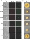
RpdA localization and phenotypic analysis of Aspergillus strains expressing Venus-tagged RpdA variants and mRFP-tagged histone H2A. Venus-tagged RpdA variants were expressed under the control of the xylPp promoter in strain TSG5 expressing endogenous RpdA under the control of alcAp and mRFP-tagged H2A under the control of the constitutive gpdA promoter. For microscopic analysis, strains were grown in eight-well plates (A), and for phenotypic analysis, they were grown on agar plates (B) under xylPp inductive conditions (GX). Hyphae were viewed under a light microscope (LM) at a magnification of ×630, and for subcellular localization of RpdA, they were examined by confocal laser scanning microscopy. Nuclei (mRFP-tagged H2A, H2A-mRFP) are red, and the distribution of expressed Venus-tagged RpdA variants (RpdA-Venus) is green.
FIG 8
FIG 8
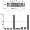
(A) HDAC activities of purified recombinant RpdA variants. IgG Sepharose-purified recombinant TAP-tagged RpdA variants were eluted by cleavage with TEV protease, and 10-µl aliquots of the eluates were subjected to SDS-PAGE, followed by silver staining of the proteins for quality control. For Western blotting, 5-µl volumes of the eluates were separated by SDS-PAGE and blotted onto a nitrocellulose membrane. RpdA expression products were detected with an anti-CBP antibody. An identically treated protein extract of a strain expressing Venus-tagged RpdA under the control of xylPp served as a mock-treated control (RpdAm). The molecular masses of relevant marker proteins (lanes M) are indicated. (B) Twenty-five-microliter volumes of the eluates were used for HDAC activity assays. The activity of wild-type RpdA was set to 100%, catalytic RpdA mutants (H158A, D193A) and RpdAm served as negative controls. Elution buffer (bu, with and without TEV protease) was used to determine background activity. The error bars represent standard deviations of three independent replicates. Asterisks indicate statistically significant differences from the buffer control (*, P < 0.001; **, P < 0.00001).

Comment in

References

    1. Wolffe AP, Kurumizaka H. 1998. The nucleosome: a powerful regulator of transcription. Prog Nucleic Acid Res Mol Biol 61:379–422. doi:10.1016/S0079-6603(08)60832-6. - DOI - PubMed
    1. Robertson KD. 2005. DNA methylation and human disease. Nat Rev Genet 6:597–610. doi:10.1038/nrg1655. - DOI - PubMed
    1. Grimaldi B, Coiro P, Filetici P, Berge E, Dobosy JR, Freitag M, Selker EU, Ballario P. 2006. The Neurospora crassa White Collar-1 dependent blue light response requires acetylation of histone H3 lysine 14 by NGF-1. Mol Biol Cell 17:4576–4583. doi:10.1091/mbc.E06-03-0232. - DOI - PMC - PubMed
    1. Turner BM. 2007. Defining an epigenetic code. Nat Cell Biol 9:2–6. doi:10.1038/ncb0107-2. - DOI - PubMed
    1. Bhaumik SR, Smith E, Shilatifard A. 2007. Covalent modifications of histones during development and disease pathogenesis. Nat Struct Mol Biol 14:1008–1016. doi:10.1038/nsmb1337. - DOI - PubMed

MeSH terms

LinkOut - more resources