Case Reports
. 2016 Oct 16;4(10):344-350.
doi: 10.12998/wjcc.v4.i10.344.

Surgeon-performed point-of-care ultrasound in severe eye trauma: Report of two cases

Affiliations
Case Reports

Surgeon-performed point-of-care ultrasound in severe eye trauma: Report of two cases

Fikri M Abu-Zidan et al. World J Clin Cases. .

Abstract

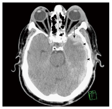
The indications of point-of-care ultrasound (POCUS) in the management of multiple trauma patients have been expanding. Although computed tomography (CT) scan of the orbit remains the gold standard for imaging orbital trauma, ultrasound is a quick, safe, and portable tool that can be performed bedside. Here we report two patients who had severe eye injuries with major visual impairment where surgeon-performed POCUS was very useful. One had a foreign body injury while the other had blunt trauma. POCUS was done using a linear probe under sterile conditions with minimum pressure on the eyes. Ultrasound showed a foreign body at the back of the left eye globe touching the eye globe in the first patient, and was normal in the second patient. Workup using CT scan, fundsocopy, optical coherence tomography, and magnetic resonance imaging of the orbits confirmed these findings. The first patient had vitreous and sub retinal haemorrhage and a full thickness macular hole of the left eye, while the second had traumatic optic neuropathy. POCUS gave accurate information concerning severe eye injuries. Trauma surgeons and emergency physicians should be trained in performing ocular ultrasound for eye injuries.

Keywords: Eye; Injury; Point-of-care; Trauma; Ultrasound.


Conflict of interest statement

Conflict-of-interest statement: None declared by all authors.

Figures

Figure 1
Figure 1
A metallic foreign body (white arrow) embedded in the lateral side of the left orbit.
Figure 2
Figure 2
Surgeon performed point-of-care ultrasound of the left eye using a linear probe under sterile conditions with minimum pressure. It showed a foreign body (yellow arrow heads) which was most probably touching the eye globe without penetration because the left eye moved freely. Gas bubbles were seen as shiny white dots within the wound (black arrows). The foreign body caused a mirror artefact (M) of the eye globe on its other side.
Figure 3
Figure 3
Plain computed tomography scan of the orbits showing the foreign body touching the left eye globe (yellow arrow).
Figure 4
Figure 4
Fundoscopy showing retinal haemorrhages (yellow arrows) and a possible macular hole (black arrow head).
Figure 5
Figure 5
Optical coherence tomography showing a full thickness macular hole of the retina of the left eye (yellow arrow).
Figure 6
Figure 6
Brain computed tomography scan showing thin left subdural haematoma (arrow heads) associated with left parenchymal haemorrhagic contusion (arrow).
Figure 7
Figure 7
Examination of the left eye showed raccoon eye, an oedematous left eye lid, and severe ecchymosis of the conjunctiva. The left pupil was dilated and not reactive to light.
Figure 8
Figure 8
Bedside surgeon-performed point-of-care ultrasound of the left eye was normal. There was no intraocular bleeding, the optic nerve was intact (arrow heads), and there was not retro-bulbar haematoma.
Figure 9
Figure 9
Magnetic resonance imaging confirmed that both optic nerves were intact. Parenchymal haemorrhagic contusion is seen on the left side (arrow).
Figure 10
Figure 10
There are three common views to examine the eye. The transverse antero-posterior view (A) is useful for examining the optic nerve (B, arrow heads). The sagittal antero-posterior view (C) is useful in visualizing the anterior and posterior chambers of the eye (D, V: Vitreous; p: Pupil). The coronal view (E) is useful for examining the pupil (F, p: Pupil, white arrows: Edge of the iris). The marker of the probe (yellow arrows) should point to the right side of the patient or upwards.
Figure 11
Figure 11
Coronal eye view (A) showing the pupil and edge of the iris (white arrows). The pupil (P) constricts when light is applied to the closed eye (B). M Mode (C) accurately measures the size of the pupil which constricted from 4.2 mm to 1.6 mm to light reflex.

References

    1. Mohammad A, Hefny AF, Abu-Zidan FM. Focused Assessment Sonography for Trauma (FAST) training: a systematic review. World J Surg. 2014;38:1009–1018. - PubMed
    1. Dunkin JM, Crum AV, Swanger RS, Bokhari SA. Globe trauma. Semin Ultrasound CT MR. 2011;32:51–56. - PubMed
    1. Poon A, McCluskey PJ, Hill DA. Eye injuries in patients with major trauma. J Trauma. 1999;46:494–499. - PubMed
    1. Sung EK, Nadgir RN, Fujita A, Siegel C, Ghafouri RH, Traband A, Sakai O. Injuries of the globe: what can the radiologist offer? Radiographics. 2014;34:764–776. - PubMed
    1. Abu-Zidan FM. Point-of-care ultrasound in critically ill patients: Where do we stand? J Emerg Trauma Shock. 2012;5:70–71. - PMC - PubMed

Publication types

LinkOut - more resources