. 2016 Dec;80(6):909-923.
doi: 10.1002/ana.24815. Epub 2016 Nov 23.

Efficacy of phosphodiesterase-4 inhibitors in juvenile Batten disease (CLN3)

Affiliations

Efficacy of phosphodiesterase-4 inhibitors in juvenile Batten disease (CLN3)

Amy Aldrich et al. Ann Neurol. 2016 Dec.

Abstract

Objective: Juvenile neuronal ceroid lipofuscinosis (JNCL), or juvenile Batten disease, is a pediatric lysosomal storage disease caused by autosomal recessive mutations in CLN3, typified by blindness, seizures, progressive cognitive and motor decline, and premature death. Currently, there is no treatment for JNCL that slows disease progression, which highlights the need to explore novel strategies to extend the survival and quality of life of afflicted children. Cyclic adenosine monophosphate (cAMP) is a second messenger with pleiotropic effects, including regulating neuroinflammation and neuronal survival. Here we investigated whether 3 phosphodiesterase-4 (PDE4) inhibitors (rolipram, roflumilast, and PF-06266047) could mitigate behavioral deficits and cell-specific pathology in the Cln3Δex7/8 mouse model of JNCL.

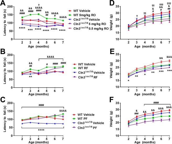
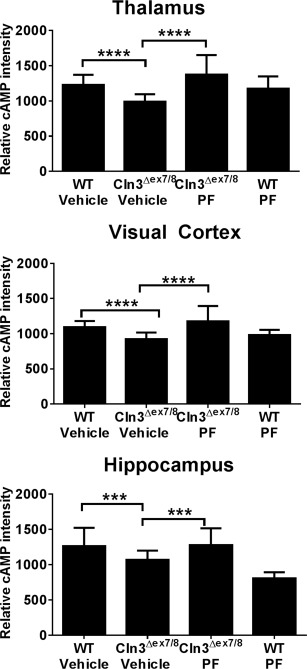
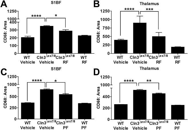
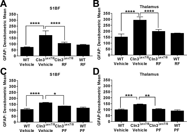
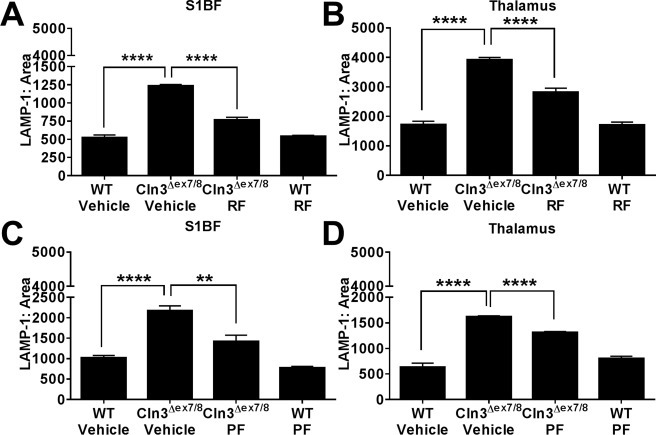
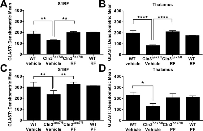
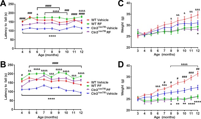
Methods: In a randomized, blinded study, wild-type (WT) and Cln3Δex7/8 mice received PDE4 inhibitors daily beginning at 1 or 3 months of age and continuing for 6 to 9 months, with motor deficits assessed by accelerating rotarod testing. The effect of PDE4 inhibitors on cAMP levels, astrocyte and microglial activation (glial fibrillary acidic protein and CD68, respectively), lysosomal pathology (lysosomal-associated membrane protein 1), and astrocyte glutamate transporter expression (glutamate/aspartate transporter) were also examined in WT and Cln3Δex7/8 animals.

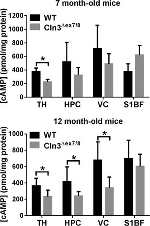
Results: cAMP levels were significantly reduced in the Cln3Δex7/8 brain, and were restored by PF-06266047. PDE4 inhibitors significantly improved motor function in Cln3Δex7/8 mice, attenuated glial activation and lysosomal pathology, and restored glutamate transporter expression to levels observed in WT animals, with no evidence of toxicity as revealed by blood chemistry analysis.

Interpretation: These studies reveal neuroprotective effects for PDE4 inhibitors in Cln3Δex7/8 mice and support their therapeutic potential in JNCL patients. Ann Neurol 2016;80:909-923.


Figures

Figure 1
Figure 1
Cyclic adenosine monophosphate (cAMP) levels are decreased in vulnerable brain regions of Cln3Δex7/8 mice. Cln3Δex7/8 and wild‐type (WT) mice (n = 5–10/group) were sacrificed at 7 (top panel) or 12 months of age (bottom panel), whereupon cAMP levels were quantified in the thalamus (TH), hippocampus (HPC), visual cortex (VC), and somatosensory barrel field cortex (S1BF) by enzyme‐linked immunosorbent assay and normalized to total protein to account for differences in tissue size. Results are combined from 2 independent experiments, and significant differences between WT and Cln3Δex7/8 mice are denoted by asterisks (p < 0.05).
Figure 2
Figure 2
Phosphodiesterase 4 (PDE4) inhibitors improve motor function in Cln3Δex7/8 mice. Cln3Δex7/8 and wild‐type (WT) mice (n = 8/group) received vehicle or (A, D) rolipram (RO; 0.5 or 5mg/kg), (B, E) roflumilast (RF; 5mg/kg), or (C, F) PF‐06266047 (PF; 0.5mg/kg) beginning at 1 month of age, after which motor activity was assessed by accelerating rotarod testing at monthly intervals. Across all comparisons, significant differences between vehicle‐treated WT and Cln3Δex7/8 animals are denoted by asterisks. (A) Significant differences between Cln3Δex7/8 vehicle‐treated versus Cln3Δex7/8 5mg/kg/day rolipram are indicated by hash signs, Cln3Δex7/8 vehicle‐treated versus Cln3Δex7/8 0.5mg/kg/day rolipram are denoted by daggers, and WT vehicle‐treated versus WT 5mg/kg/day rolipram are indicated by ampersand signs. (B) Cln3Δex7/8 vehicle‐treated versus Cln3Δex7/8 roflumilast‐treated mice are indicated by hash signs, and WT vehicle‐treated versus WT roflumilast are indicated by ampersand signs. (C) Cln3Δex7/8 vehicle‐treated versus Cln3Δex7/8 PF‐06266047–treated mice are indicated by hash signs, and WT vehicle‐treated versus WT PF‐06266047–treated mice are indicated by ampersand signs. (D–F) Weights of Cln3Δex7/8 and WT mice during PDE4 inhibitor treatment. (D) Significant differences between Cln3Δex7/8 vehicle‐treated versus Cln3Δex7/8 5mg/kg/day rolipram are indicated by hash signs, Cln3Δex7/8 vehicle‐treated versus Cln3Δex7/8 0.5mg/kg/day rolipram are denoted by daggers, and WT vehicle‐treated versus WT 5mg/kg/day rolipram are indicated by ampersand signs. (E) Significant differences between WT vehicle‐treated and WT roflumilast are indicated by ampersand signs. (F) Significant differences between Cln3Δex7/8 vehicle‐treated versus Cln3Δex7/8 PF‐06266047–treated mice are indicated by hash signs, and WT vehicle‐treated versus WT PF‐06266047–treated mice are indicated by ampersand signs. For all comparisons, *, #, &, † reflect p < 0.05; **, ##, &&, †† denote p < 0.01; ***, ###, &&&, ††† reflect p < 0.001; and ****, ####, &&&& represent p < 0.0001.
Figure 3
Figure 3
PF‐06266047 restores cyclic adenosine monophosphate (cAMP) levels in Cln3Δex7/8 mice. Cln3Δex7/8 and wild‐type (WT) mice received PF‐06266047 (PF; 0.5mg/kg) or vehicle via daily oral gavage beginning at 1 month of age, whereupon animals were sacrificed 6 months later for cAMP quantitation by immunofluorescence staining in the thalamus, visual cortex, and hippocampus (n = 5/group). Significant differences between groups are denoted by asterisks (***p < 0.001; ****p < 0.0001).
Figure 4
Figure 4
Phosphodiesterase 4 inhibitors attenuate microglial activation in Cln3Δex7/8 mice. Cln3Δex7/8 and wild‐type (WT) mice (n = 4–8/group) received vehicle, (A, B) 5mg/kg roflumilast (RF), or (C, D) 0.5mg/kg PF‐06266047 (PF) via daily oral gavage beginning at 1 month of age, whereupon animals were sacrificed 6 months later for quantitative assessments of microglial activation in the somatosensory barrel field cortex (S1BF) and thalamus by immunofluorescence staining for CD68. Significant differences between groups are denoted by asterisks (*p < 0.05; **p < 0.01; ***p < 0.001; ****p < 0.0001).
Figure 5
Figure 5
Phosphodiesterase 4 inhibitors reduce astrocyte activation in Cln3Δex7/8 mice. Cln3Δex7/8 and wild‐type (WT) mice (n = 4–8/group) received vehicle, (A, B) 5mg/kg roflumilast (RF), or (C, D) 0.5mg/kg PF‐06266047 (PF) via daily oral gavage beginning at 1 month of age, whereupon animals were sacrificed 6 months later for quantitative assessments of astrocyte activation in the somatosensory barrel field cortex (S1BF) and thalamus by immunofluorescence staining for glial fibrillary acidic protein (GFAP). Significant differences between groups are denoted by asterisks (*p < 0.05; **p < 0.01; ***p < 0.001; ****p < 0.0001).
Figure 6
Figure 6
Phosphodiesterase 4 inhibitors attenuate lysosomal pathology in the Cln3Δex7/8 brain. Cln3Δex7/8 and wild‐type (WT) mice (n = 4–8/group) received vehicle, (A, B) 5mg/kg roflumilast (RF), or (C, D) 0.5mg/kg PF‐06266047 (PF) via daily oral gavage beginning at 1 month of age, whereupon animals were sacrificed 6 months later for quantitative assessments of lysosomal‐associated membrane protein 1 (LAMP‐1) expression in the somatosensory barrel field cortex (S1BF) and thalamus. Significant differences between groups are denoted by asterisks (**p < 0.01; ****p < 0.0001).
Figure 7
Figure 7
Phosphodiesterase 4 inhibitors restore the loss of astrocytic glutamate/aspartate transporter (GLAST) expression in Cln3Δex7/8 mice. Cln3Δex7/8 and wild‐type (WT) mice (n = 4–8/group) received vehicle, (A, B) 5mg/kg roflumilast (RF), or (C, D) 0.5mg/kg PF‐06266047 (PF) via daily oral gavage beginning at 1 month of age, whereupon animals were sacrificed 6 months later for quantitative assessments of GLAST expression in the somatosensory barrel field cortex (S1BF) and thalamus. Significant differences between groups are denoted by asterisks (*p < 0.05; **p < 0.01; ****p < 0.0001).
Figure 8
Figure 8
Delayed treatment with phosphodiesterase 4 (PDE4) inhibitors improves motor function in Cln3Δex7/8 mice. Cln3Δex7/8 and wild‐type (WT) mice (n = 8/group) received vehicle, (A, C) 5mg/kg roflumilast (RF), or (B, D) 0.5mg/kg PF‐06266047 (PF) via daily oral gavage beginning at 3 months of age, after which motor activity was assessed by accelerating rotarod testing at monthly intervals. Across all comparisons, significant differences between vehicle‐treated WT and Cln3Δex7/8 animals are denoted by asterisks, Cln3Δex7/8 vehicle‐treated versus Cln3Δex7/8 PDE4 inhibitor‐treated mice are indicated by hash signs, and WT vehicle‐treated versus WT PDE4 inhibitor‐treated mice are indicated by ampersand signs. (C, D) Weights of Cln3Δex7/8 and WT mice receiving delayed PDE4 inhibitor treatment. Significant differences between Cln3Δex7/8 vehicle‐treated versus Cln3Δex7/8 PDE4 inhibitor‐treated mice are indicated by hash signs, and WT vehicle‐treated versus WT PDE4 inhibitor‐treated mice are indicated by ampersand signs. For all comparisons, *, #, & reflect p < 0.05; **, ##, && denote p < 0.01; ###, &&& reflect p < 0.001; and ****, ####, &&&& represent p < 0.0001.

References

    1. International Batten Disease Consortium . Isolation of a novel gene underlying Batten disease, CLN3. Cell 1995;82:949–957. - PubMed
    1. Williams RE, Mole SE. New nomenclature and classification scheme for the neuronal ceroid lipofuscinoses. Neurology 2012;79:183–191. - PubMed
    1. Schultz ML, Tecedor L, Chang M, Davidson BL. Clarifying lysosomal storage diseases. Trends Neurosci 2011;34:401–410. - PMC - PubMed
    1. Autti T, Hamalainen J, Aberg L, et al. Thalami and corona radiata in juvenile NCL (CLN3): a voxel‐based morphometric study. Eur J Neurol 2007;14:447–450. - PubMed
    1. Autti TH, Hamalainen J, Mannerkoski M, et al. JNCL patients show marked brain volume alterations on longitudinal MRI in adolescence. J Neurol 2008;255:1226–1230. - PubMed

Publication types

MeSH terms