. 2016 Nov 2:6:36033.
doi: 10.1038/srep36033.

miR-19b Regulates Ventricular Action Potential Duration in Zebrafish

Affiliations

miR-19b Regulates Ventricular Action Potential Duration in Zebrafish

Alexander Benz et al. Sci Rep. .

Abstract

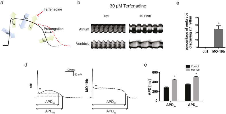
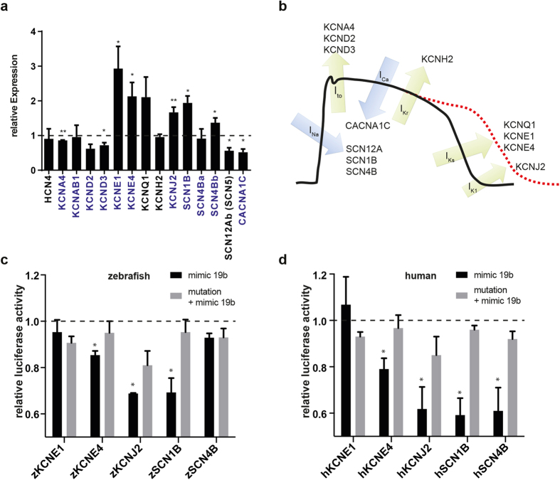
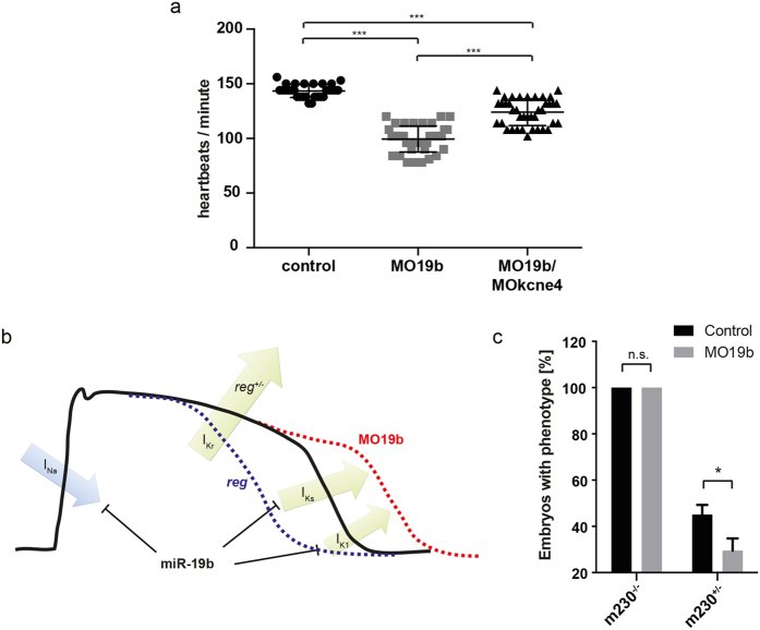
Sudden cardiac death due to ventricular arrhythmias often caused by action potential duration (APD) prolongation is a common mode of death in heart failure (HF). microRNAs, noncoding RNAs that fine tune gene expression, are frequently dysregulated during HF, suggesting a potential involvement in the electrical remodeling process accompanying HF progression. Here, we identified miR-19b as an important regulator of heart function. Zebrafish lacking miR-19b developed severe bradycardia and reduced cardiac contractility. miR-19b deficient fish displayed increased sensitivity to AV-block, a characteristic feature of long QT syndrome in zebrafish. Patch clamp experiments from whole hearts showed that miR-19b deficient zebrafish exhibit significantly prolonged ventricular APD caused by impaired repolarization. We found that miR-19b directly and indirectly regulates the expression of crucial modulatory subunits of cardiac ion channels, and thereby modulates AP duration and shape. Interestingly, miR-19b knockdown mediated APD prolongation can rescue a genetically induced short QT phenotype. Thus, miR-19b might represent a crucial modifier of the cardiac electrical activity, and our work establishes miR-19b as a potential candidate for human long QT syndrome.


Figures

Figure 1
Figure 1. miR19 is conserved in sequence and syntheny.
(a) The sequence of miR-19a and miR-19b is highly conserved within vertebrate species. The seed-region is shaded in grey. (b) miR-19-Isoforms are encoded within the miR-17-92 cluster and its paralogs the miR-106a-363- and the miR-106b-25-cluster in mammals and zebrafish. (c) The zebrafish encodes for four different isoforms of miR-19, miR-19a-d. The sequence of their mature isoforms differs in maximal two nucleotides. Mature sequences are shaded in grey. Conserved sequence is indicated with stars (compared to miR-19b).
Figure 2
Figure 2. Expression of miR-19 in the zebrafish.
(a) qRT-PCR analysis revealed that miR-19a-d expression is already detectable at very early stages of embryonic development and gets induced at 24 hpf. (±sd; n = 3 from 15 pooled embryos per sample) (b) In situ hybridization of miR-19a and miR-19b in 48 hpf and 72 hpf zebrafish embryos shows similar expression patterns in the heart (stars) and in skeletal muscle myosepts (arrows).
Figure 3
Figure 3. Loss of miR-19 leads to bradycardia.
(a) Lateral brightfield images of control-injected and MO19-injected 48 hpf zebrafish embryos. Note the cardiac edema and the blood congestion at the inflow tract developed in MO19 injected embryos. (b) Quantification of heart rate as heart beats per minute and ventricular fractional shortening as a measure for ventricular contractility in control (wt) and MO19 injected embryos. Morpholino-mediated knockdown of miR-19 in zebrafish leads to bradycardia and impaired ventricular contractility (±sd; n ≥ 14; p < 0.005). (c) qRT-PCR confirmed efficient knockdown of miR-19a-d. Notice the consistent reduction in miR-363 expression (±sd; n = 45 animals from 3 independent experiments). (d) Specific knockdown of miR-19a, miR-19c and miR-19d altered expression of neighboring miRNAs. However, knockdown of miR-19b did not interfere with expression of neighboring miRNAs (±sd; n = 45 animals from 3 independent experiments; p < 0.05). (e) 48 hpf zebrafish embryos injected with control- and MO19b-morpholino revealed that miR-19b deficiency is sufficient to mimic MO19-induced phenotype with characteristics of heart dysfunction. (f) miR-19b deficient embryos developed bradycardia with up to 36% ± 5.7% reduced heart frequency (± = sd; n ≥ 12; p < 0.005) (g,h) Relative changes in heart rate normalized to controls (dotted line) indicates that CRSIPR/Cas9 mediated knockout of miR-19b caused an identical and long-term reduction in heart rate as observed for MO19b (±sd; n = 10; p < 0.005).
Figure 4
Figure 4. Loss of miR-19b induces prolongation of the action potential.
(a) Schematic illustration of an action potential with major depolarizing and repolarizing currents depicted. Terfenadine inhibits IKr current, thereby prolonging the ventricular action potential. (b) Atrial and ventricular m-mode recorded from control and MO19b-injected Tg(Myl7:GFP) zebrafish embryos after 30 μM Terfenadine treatment. (b,c) 25% ± 4.1% of miR-19b deficient embryos develop an AV-Block with the ventricle skipping every other contraction, whereas control injected embryos show no phenotype. (±sd; n = 3 with ≥10 animals per group; p < 0.05) (d,e) Patch-clamp Experiments revealed that morpholino-mediated loss of miR-19b results in prolonged action potentials with up to 57% ± 3.8% and 39% ± 3.5% prolongation of APD50 and APD90, respectively (±sd; n ≥ 10 from 5 hearts; p < 0.05). Additionally, the notch appeared to be less pronounced (stars).
Figure 5
Figure 5. Loss of miR-19b results in dysregulation of cardiac ion currents.
(a) qRT-PCR revealed dysregulation of various cardiac ion channels in miR-19b-deficient zebrafish. Transcripts with a predicted miR-19b binding site in its 3′-UTR are indicated in blue (±sd; n = 3 from 15 pooled embryos per sample; p < 0,05). (b) Illustration of a mammalian action potential with corresponding currents and genes. (c,d) Potential miR-19b targets were tested for direct regulation by a dual luciferase reporter gene assay. 3′UTR of indicated zebrafish (c) and human (d) genes was cloned downstream of a luciferase gene. Luciferase expression was normalized to renilla expression. As control, target sites were mutated to reverse miR-19b-induced repression. KCNE4, KCNJ2 and SCN1B proved to be direct targets in zebrafish and in humans. Additionally, the 3′UTR of human SCN4B showed responsiveness to miR-19b (±sd; n = 3; p < 0.05).
Figure 6
Figure 6. Phenotype of reggae mutants is partially rescued by loss of miR-19b.
(a) Quantification of heart rate in control injected, MO19b and co-injection of MO19b and MOkcne4 (n > 50 animals from 3 independent experiments; p < 0.005 using one-way ANOVA and unpaired t-test with Welch’s correction). (b) Schematic illustration of miR-19b function. (c) Heterozygous reggae mutants were rescued by miR-19b knockdown (±sd; n = 73 animals from 3 independent experiments; p < 0.05).

References

    1. Benoist D., Stones R., Drinkhill M., Bernus O. & White E. Arrhythmogenic substrate in hearts of rats with monocrotaline-induced pulmonary hypertension and right ventricular hypertrophy. Am J Physiol Heart Circ Physiol. 300, H2230–H2237, doi: 10.1152/ajpheart.01226.2010 (2011). - DOI - PMC - PubMed
    1. Helbing W. A. et al. ECG predictors of ventricular arrhythmias and biventricular size and wall mass in tetralogy of Fallot with pulmonary regurgitation. Heart 88, 515–519 (2002). - PMC - PubMed
    1. Lambert V. et al. Right ventricular failure secondary to chronic overload in congenital heart disease: an experimental model for therapeutic innovation. J Thorac Cardiovasc Surg 139, 1197–1204, 1204 e1191, doi: 10.1016/j.jtcvs.2009.11.028 (2010). - DOI - PubMed
    1. Lee J. K. et al. Stage-dependent changes in membrane currents in rats with monocrotaline-induced right ventricular hypertrophy. Am J Physiol. 272, H2833–H2842 (1997). - PubMed
    1. Ikeda S. et al. Altered microRNA expression in human heart disease. Physiol Genomics 31, 367–373, doi: 10.1152/physiolgenomics.00144.2007 (2007). - DOI - PubMed

Publication types