. 2016 Nov 15;55(45):6314-6326.
doi: 10.1021/acs.biochem.6b00887. Epub 2016 Nov 2.

Dynamic Conformational States Dictate Selectivity toward the Native Substrate in a Substrate-Permissive Acyltransferase

Affiliations

Dynamic Conformational States Dictate Selectivity toward the Native Substrate in a Substrate-Permissive Acyltransferase

Olesya Levsh et al. Biochemistry. .

Abstract

Hydroxycinnamoyl-CoA:shikimate hydroxycinnamoyltransferase (HCT) is an essential acyltransferase that mediates flux through plant phenylpropanoid metabolism by catalyzing a reaction between p-coumaroyl-CoA and shikimate, yet it also exhibits broad substrate permissiveness in vitro. How do enzymes like HCT avoid functional derailment by cellular metabolites that qualify as non-native substrates? Here, we combine X-ray crystallography and molecular dynamics to reveal distinct dynamic modes of HCT under native and non-native catalysis. We find that essential electrostatic and hydrogen-bonding interactions between the ligand and active site residues, permitted by active site plasticity, are elicited more effectively by shikimate than by other non-native substrates. This work provides a structural basis for how dynamic conformational states of HCT favor native over non-native catalysis by reducing the number of futile encounters between the enzyme and shikimate.


Figures

Figure 1.
Figure 1.
Structure and catalytic mechanism of AtHCT. (A) Schematic ribbon diagram of the overall structure of AtHCT in complex with p-coumaroylshikimate. The two quasi-symmetric N-terminal (light blue) and C-terminal (dark blue) domains are linked by a long loop (red). The zoom-in insert shows the active site highlighting residues key to the native catalytic activity of HCT. Hydrogen bond interactions are indicated with dotted lines (yellow). The |2Fo-Fc| omit electron density map of p-coumaroylshikimate is contoured at 0.8 σ (B) The proposed catalytic mechanism of HCT.
Figure 2.
Figure 2.
Distinct active-site conformational states and dynamics of HCT revealed by X-ray crystallography and MD simulations. (A) Active-site residues His153 and Arg356 show distinct and consistent conformational shifts upon binding of various ligands. (B) Conformational states of the three loop regions surrounding the active site in various HCT structures. The impact of these motions is to constrict the active site volume significantly. (C) Distribution of the distance change (Δd) between Arg356 and p-coumaroylshikimate from simulations of apo (black) and p-coumaroyl CoA and shikimate-bound (red) AtHCT. Calculations were performed by first aligning simulation trajectories to the p-coumaroylshikimate-bound AtHCT crystal structure and then measuring the distance change between Arg356 and p-coumaroylshikimate. A zero Δd indicates that Arg356 is at an identical distance from the ligand as in the crystal structure. Details can be found in the Supplementary Experimental Procedures. (D) Internal conformation demonstrated by the first cluster of Arg356 from clustering analysis of p-coumaroyl CoA and shikimate-bound AtHCT simulations. (E) External conformations demonstrated by the first two clusters of Arg356 from clustering analysis of apo AtHCT simulations. In (D) and (E), p-coumaroylshikimate from the p-coumaroylshikimate-bound AtHCT crystal structure is shown as an active site reference. Arg356 from p-coumaroylshikimate-bound and apo- AtHCT crystal structures are shown in black in (D) and (E), respectively. Percentage of populations for each cluster is labeled. (F) Multiple sequence alignment highlighting L1, L2, L3, and the catalytic regions in select HCT orthologs representing five major land plant lineages. Corresponding sequence regions from CbRAS are displayed at the bottom for comparison. The residue numbering is according to AtHCT.
Figure 3.
Figure 3.
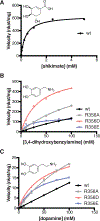
Pseudo first-order Michaelis-Menten kinetics of wild-type, R356A, R356D, and R356E CbHCTs against the native acyl acceptor substrate, shikimate (A), and two non-native acyl acceptor substrates, 3,4-dihydroxybenzylamine (B) and dopamine (C). p-coumaroyl-CoA was used as the acyl donor substrate in all assays at constant and excess concentration. Structures of acyl acceptor substrates are shown above the curves.
Figure 4.
Figure 4.
2D scatter plots of substrate position and orientation in CbHCT simulations. “Distance” measures the distance between the substrates and Arg356 (or Glu356). “Angle” measures the change in substrates’ orientations with reference to their initial structures (details in the Supplementary Experimental Procedures). Characterized simulations include (A) wild-type CbHCT with regard to shikimate, (B) CbHCT R356E with regard to shikimate, and (C) wild-type CbHCT with regard to 3-hydroxyacetophenone.
Figure 5.
Figure 5.
Structural and dynamic features of CbHCT in complex with p-coumaroyl-CoA and 3-hydroxyacetophenone. (A) Chemical structure of 3-hydroxyacetophenone (left) and the |2Fo-Fc| omit electron density map of 3-hydroxyacetophenone in the p-coumaroyl-CoA and 3-hydroxyacetophenone-bound CbHCT crystal structure contoured at 0.8 σ (right). (B) The conformation of 3-hydroxyacetophenone relative to the catalytic His153 in CbHCT active site. The AtHCT-p-coumaroylshikimate structure is overlaid as a comparison. (C) The conformational states of L1, L2, and L3 and Arg356 in various HCT structures. (D) Distribution of the distance change (Δd) between Arg356 and p-coumaroylshikimate from simulations of apo CbHCT (black), CbHCT in complex with p-coumaroyl CoA and shikimate (red), and CbHCT in complex with p-coumaroyl CoA and 3-hydroxyacetophenone (blue). Details can be found in the Supplementary Experimental Procedures. (E, F) The first cluster of Arg356 conformation from clustering analysis of shikimate- (E) and 3-hydroxyacetophenone-bound (F) CbHCT simulations. As an active site reference, p-coumaroylshikimate from the AtHCT crystal structure is shown in (E) and (F), and 3-hydroxyacetophenone from the CbHCT crystal structure is shown in (F). The conformation of Arg356 from the apo CbHCT crystal structure (E) and the 3-hydroxyacetophenone-bound CbHCT crystal structure (F) is shown in black.

References

    1. Weng JK, and Noel JP (2012) The remarkable pliability and promiscuity of specialized metabolism, Cold Spring Harb. Symp. Quant. Biol. 77, 309–320. - PubMed
    1. Jensen RA (1976) Enzyme Recruitment in Evolution of New Function, Annu Rev Microbiol 30, 409–425. - PubMed
    1. Peisajovich SG, and Tawfik DS (2007) Protein engineers turned evolutionists, Nat Methods 4, 991–994. - PubMed
    1. Campbell JH, Lengyel JA, and Langridge J (1973) Evolution of a second gene for beta-galactosidase in Escherichia coli, Proc Natl Acad Sci U S A 70, 1841–1845. - PMC - PubMed
    1. Aharoni A, Gaidukov L, Khersonsky O, Gould SM, Roodveldt C, and Tawfik DS (2005) The ‘evolvability’ of promiscuous protein functions, Nat Genet 37, 73–76. - PubMed

MeSH terms

LinkOut - more resources