

Review
doi: 10.1056/NEJMra1514296.
Molecular and Biochemical Aspects of the PD-1 Checkpoint Pathway
Affiliations
- PMID: 27806234
- PMCID: PMC5575761
- DOI: 10.1056/NEJMra1514296
Item in Clipboard
Review
Molecular and Biochemical Aspects of the PD-1 Checkpoint Pathway
N Engl J Med.
.
No abstract available
Figures
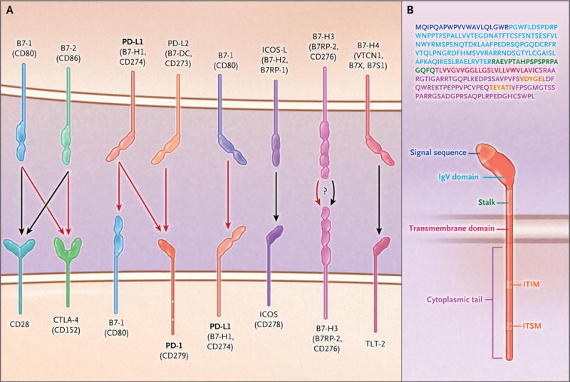
Panel A shows members of the B7–CD28–CTLA-4 family of costimulatory molecules. Black arrows indicate stimulatory signals, and red arrows indicate inhibitory signals. The receptors for B7-H3 and B7-H4 and their effects remain unclear. PD-L2 that is expressed on lung dendritic cells interacts with repulsive guidance molecule B, a coreceptor for bone morphogenetic proteins, expressed on alveolar macrophages, interstitial macrophages, and alveolar epithelial cells (not shown). Panel B shows the amino acid sequence and structure of human PD-1. The signal sequence is shown in purple, the immunoglobulin variable region (IgV)–like domain in light blue, the transmembrane segment in red, and the immunoreceptor tyrosine-based inhibition motif (ITIM) and immunoreceptor tyrosine-based switch motif (ITSM) in orange; the tyrosines within the ITIM and ITSM are highlighted in yellow. See the Supplementary Appendix, available with the full text of this article at NEJM.org , for a list of the protein abbreviations used in this review.

During T-cell receptor (TCR) crosslinking with antigen presented by major histocompatibility complex class II (MHCII) molecules (in CD4+ T cells) or class I molecules (in CD8+ T cells), the tyrosines of the cytoplasmic tail of PD-1 become phosphorylated (P). SHP-2 (and possibly SHP-1) is recruited to the ITSM, and an as-yet-unidentified partner is recruited to the ITIM. As a consequence, phosphorylation of TCR proximal signaling molecules, including Lck and ZAP-70, is impaired (Panel A). Activation of the PI3K–Akt–mTOR pathway (Panel B) and activation of the Ras–MEK–ERK pathway (Panel C) are inhibited. In contrast, other signaling events, such as the activation of the p38 pathway, remain unaffected or enhanced (e.g., BATF is up-regulated) (Panel D). The imbalanced activation of signaling pathways alters cell-cycle progression, gene transcription, metabolism, and epigenetic programs in T cells. Black arrows indicate activation signals, and red blocked arrows indicate inhibited signals.

Ligation of PD-1 that is expressed in activated T cells by PD-L1 expressed on antigen-presenting cells, nonhematopoietic parenchymal cells, or tumors alters T-cell metabolic reprogramming by inhibiting glycolysis, amino acid metabolism, and mitochondrial metabolism and promoting the accumulation of polyunsaturated fatty acids (PUFA) and activation of fatty acid oxidation (Panel A). By restraining T cells from remodeling their metabolism properly, PD-1 may alter T-cell differentiation, leading to impaired differentiation of T effector cells (TEFF) and T memory cells (TM) and enhanced differentiation of T regulatory cells (Treg) and T exhausted cells (TEX). PD-L1 functions as an inhibitory receptor to transmit antiapoptotic signals to cancer cells (Panel B). Because cancer cells are highly glycolytic and have enhanced activation of the PI3K–Akt pathway, expression of PD-L1 might result in increased levels of PI3K–Akt–mTOR activation and an elevated rate of tumor-intrinsic glycolysis as a consequence of improved survival.

In the tumor microenvironment, T cells that are capable of recognizing tumor neoantigens produce interferon-γ, which can induce the expression of PD-1 ligands on cancer cells and immune cell types, including macrophages, stromal cells, and dendritic cells, which become myeloid suppressor cells. The expression of PD-1 ligands on cancer cells is also mediated by cell-intrinsic mechanisms that are activated by oncogenic events, which result in altered activation of signaling pathways and altered gene-expression programs through transcriptional and epigenetic mechanisms. The term pMHC denotes peptide plus MHC (i.e., MHC of either class I or class II).
References
-
- Tivol EA, Borriello F, Schweitzer AN, Lynch WP, Bluestone JA, Sharpe AH. Loss of CTLA-4 leads to massive lymphoproliferation and fatal multiorgan tissue destruction, revealing a critical negative regulatory role of CTLA-4. Immunity. 1995;3:541–7. - PubMed
-
- Waterhouse P, Penninger JM, Timms E, et al. Lymphoproliferative disorders with early lethality in mice deficient in Ctla-4. Science. 1995;270:985–8. - PubMed
-
- Nishimura H, Nose M, Hiai H, Minato N, Honjo T. Development of lupus-like autoimmune diseases by disruption of the PD-1 gene encoding an ITIM motif-carrying immunoreceptor. Immunity. 1999;11:141–51. - PubMed
-
- Nishimura H, Okazaki T, Tanaka Y, et al. Autoimmune dilated cardiomyopathy in PD-1 receptor-deficient mice. Science. 2001;291:319–22. - PubMed
Publication types
MeSH terms
Substances
Grants and funding
LinkOut - more resources
Full Text Sources
Other Literature Sources
Medical