

In Vivo Interrogation of Spinal Mechanosensory Circuits
- PMID: 27806306
- PMCID: PMC5507199
- DOI: 10.1016/j.celrep.2016.10.010
In Vivo Interrogation of Spinal Mechanosensory Circuits
Abstract
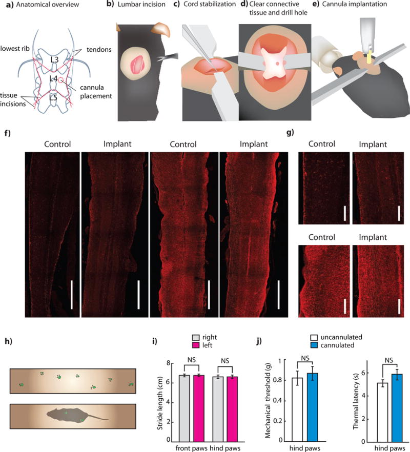
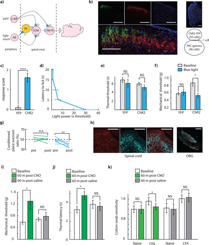
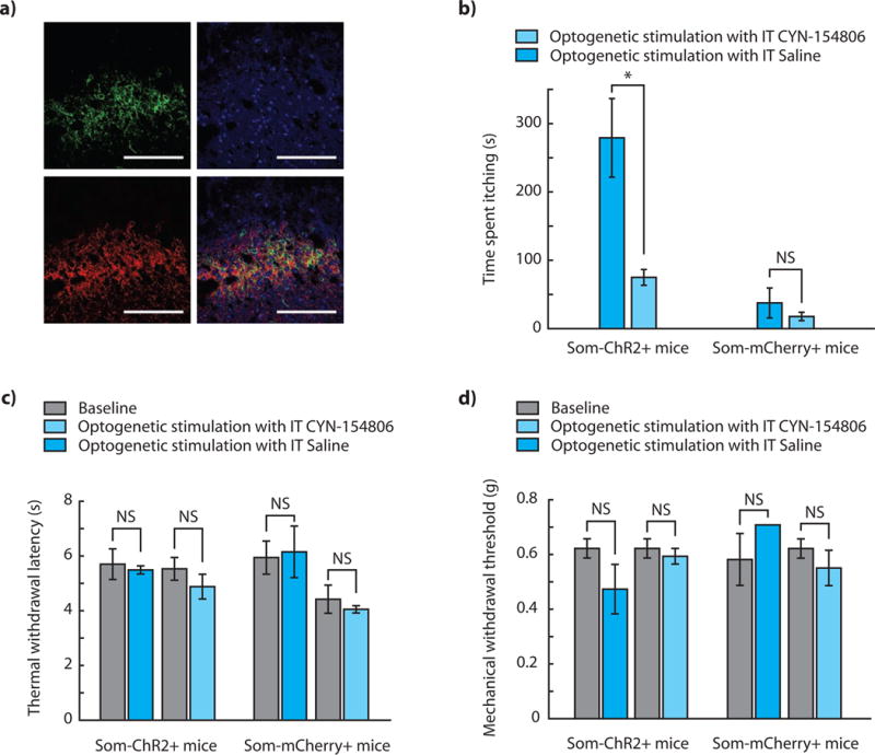
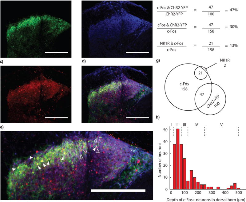
Spinal dorsal horn circuits receive, process, and transmit somatosensory information. To understand how specific components of these circuits contribute to behavior, it is critical to be able to directly modulate their activity in unanesthetized in vivo conditions. Here, we develop experimental tools that enable optogenetic control of spinal circuitry in freely moving mice using commonly available materials. We use these tools to examine mechanosensory processing in the spinal cord and observe that optogenetic activation of somatostatin-positive interneurons facilitates both mechanosensory and itch-related behavior, while reversible chemogenetic inhibition of these neurons suppresses mechanosensation. These results extend recent findings regarding the processing of mechanosensory information in the spinal cord and indicate the potential for activity-induced release of the somatostatin neuropeptide to affect processing of itch. The spinal implant approach we describe here is likely to enable a wide range of studies to elucidate spinal circuits underlying pain, touch, itch, and movement.
Keywords: itch; nociception; optogenetics; somatostatin; spinal cord; touch.
Copyright © 2016 The Author(s). Published by Elsevier Inc. All rights reserved.
Figures
Similar articles
-
Spinal somatostatin-positive interneurons transmit chemical itch.Pain. 2019 May;160(5):1166-1174. doi: 10.1097/j.pain.0000000000001499. Pain. 2019. PMID: 30913166
-
The Neurokinin-1 Receptor is Expressed with Gastrin-Releasing Peptide Receptor in Spinal Interneurons and Modulates Itch.J Neurosci. 2020 Nov 11;40(46):8816-8830. doi: 10.1523/JNEUROSCI.1832-20.2020. Epub 2020 Oct 13. J Neurosci. 2020. PMID: 33051347 Free PMC article.
-
Dorsal Horn Gastrin-Releasing Peptide Expressing Neurons Transmit Spinal Itch But Not Pain Signals.J Neurosci. 2019 Mar 20;39(12):2238-2250. doi: 10.1523/JNEUROSCI.2559-18.2019. Epub 2019 Jan 17. J Neurosci. 2019. PMID: 30655357 Free PMC article.
-
Spinal Circuits for Touch, Pain, and Itch.Annu Rev Physiol. 2018 Feb 10;80:189-217. doi: 10.1146/annurev-physiol-022516-034303. Epub 2017 Sep 27. Annu Rev Physiol. 2018. PMID: 28961064 Free PMC article. Review.
-
Insights Into Spinal Dorsal Horn Circuit Function and Dysfunction Using Optical Approaches.Front Neural Circuits. 2020 Jun 12;14:31. doi: 10.3389/fncir.2020.00031. eCollection 2020. Front Neural Circuits. 2020. PMID: 32595458 Free PMC article. Review.
Cited by
-
Surgical preparations, labeling strategies, and optical techniques for cell-resolved, in vivo imaging in the mouse spinal cord.Exp Neurol. 2019 Aug;318:192-204. doi: 10.1016/j.expneurol.2019.05.010. Epub 2019 May 13. Exp Neurol. 2019. PMID: 31095935 Free PMC article. Review.
-
Functional Divergence of Delta and Mu Opioid Receptor Organization in CNS Pain Circuits.Neuron. 2018 Apr 4;98(1):90-108.e5. doi: 10.1016/j.neuron.2018.03.002. Epub 2018 Mar 22. Neuron. 2018. PMID: 29576387 Free PMC article.
-
TRPA1 Agonist-Responsive Afferents Contribute to Central Sensitization by Suppressing Spinal GABAergic Interneurons Through Somatostatin 2A Receptors.J Pain. 2024 Dec;25(12):104686. doi: 10.1016/j.jpain.2024.104686. Epub 2024 Sep 23. J Pain. 2024. PMID: 39321909
-
Physiology and Pathophysiology of Itch.Physiol Rev. 2020 Jul 1;100(3):945-982. doi: 10.1152/physrev.00017.2019. Epub 2019 Dec 23. Physiol Rev. 2020. PMID: 31869278 Free PMC article. Review.
-
Optical Activation of the Dorsal Horn of the Thoracic Spinal Cord Prevents Ventricular Arrhythmias in Acute Myocardial Ischemia-Reperfusion Rats.Front Cardiovasc Med. 2022 Feb 7;9:753959. doi: 10.3389/fcvm.2022.753959. eCollection 2022. Front Cardiovasc Med. 2022. PMID: 35198610 Free PMC article.
References
-
- Adamantidis A, Arber S, Bains JS, Bamberg E, Bonci A, Buzsáki G, Cardin JA, Costa RM, Dan Y, Goda Y, Graybiel AM, Häusser M, Hegemann P, Huguenard JR, Insel TR, Janak PH, Johnston D, Josselyn SA, Koch C, Kreitzer AC, Lüscher C, Malenka RC, Miesenböck G, Nagel G, Roska B, Schnitzer MJ, Shenoy KV, Soltesz I, Sternson SM, Tsien RW, Tsien RY, Turrigiano GG, Tye KM, Wilson RI. Optogenetics: 10 years after ChR2 in neurons-views from the community. Nat Neurosci. 2015;18:1202–12. doi: 10.1038/nn.4106. - DOI - PubMed
-
- Bardoni R, Tawfik VL, Wang D, François A, Solorzano C, Shuster SA, Choudhury P, Betelli C, Cassidy C, Smith K, de Nooij JC, Mennicken F, O’Donnell D, Kieffer BL, Woodbury CJ, Basbaum AI, MacDermott AB, Scherrer G. Delta Opioid Receptors Presynaptically Regulate Cutaneous Mechanosensory Neuron Input to the Spinal Cord Dorsal Horn. Neuron. 2014;81:1443. doi: 10.1016/j.neuron.2014.03.006. - DOI - PubMed
Publication types
MeSH terms
Substances
Grants and funding
LinkOut - more resources
Full Text Sources
Other Literature Sources
Molecular Biology Databases
Research Materials
