Meta-Analysis
. 2016 Nov 29;7(48):78994-79007.
doi: 10.18632/oncotarget.12982.

Overexpression of stathmin promotes metastasis and growth of malignant solid tumors: a systemic review and meta-analysis

Affiliations
Meta-Analysis

Overexpression of stathmin promotes metastasis and growth of malignant solid tumors: a systemic review and meta-analysis

Rong Biaoxue et al. Oncotarget. .

Abstract

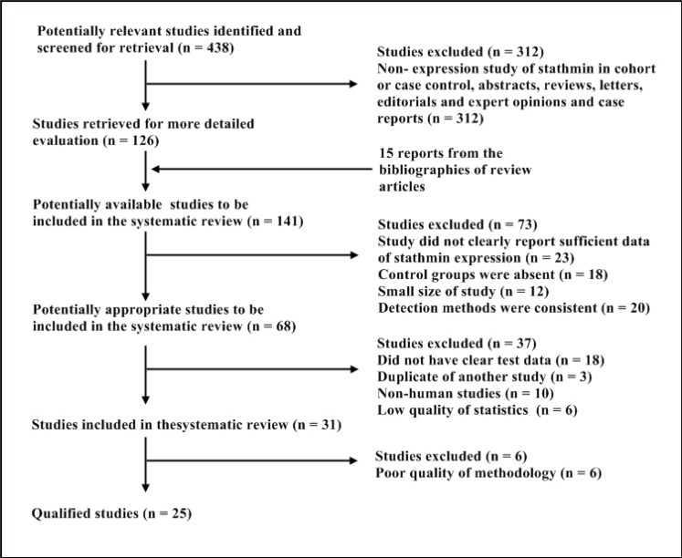
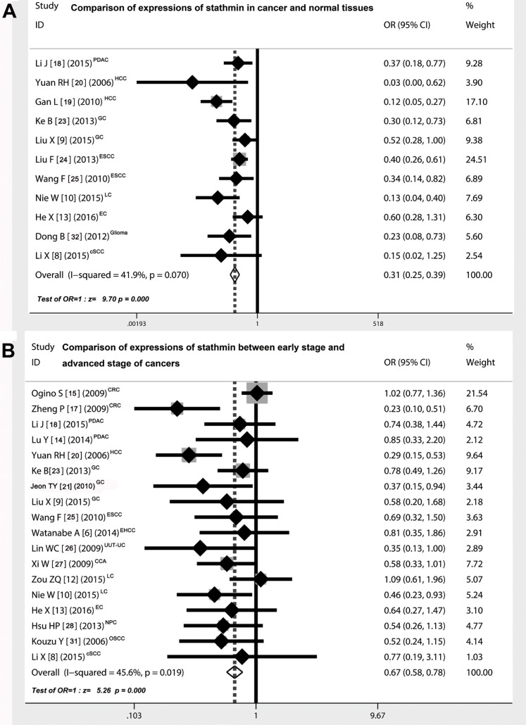
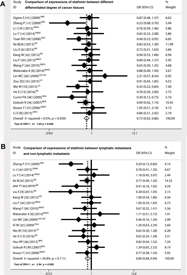
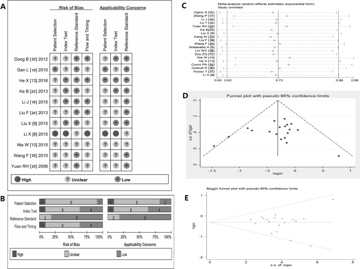
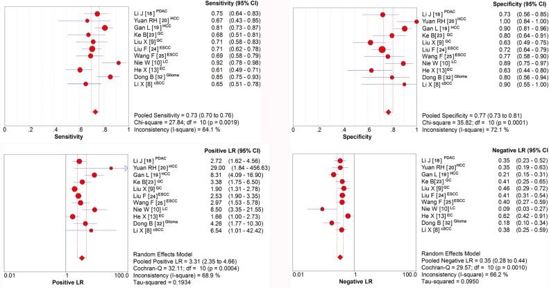
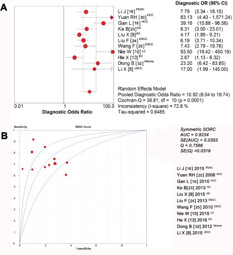
Stathmin has been investigated to be involved in development and progress of malignant tumors. This study was to clarify the relationship between expression of stathmin and tumors and assess its clinical significance. We identified 25 studies with a total of 3,571 individuals from the electronic bibliographic databases and strictly evaluated the quality and heterogeneity of included studies. We analysed the relationship between expression of stathmin and clinical characteristics by the fixed-effects and random-effects of meta-analysis and constructed a summary receiver-operator characteristic curve to estimate the test characteristics. The results showed that patients with cancer displayed a higher stathmin expression than those of non-cancer individuals (OR, 0.31), and overexpression of stathmin correlated with tumor cell differentiation (OR, 0.73), lymph node invasion (OR, 0.80) and high TNM stage (OR, 0.67). The pooled sensitivity of stathmin for distinguishing malignant tumors was 0.73 and the specificity was 0.77. The maximum balance joint for sensitivity and specificity (the Q-value) was 0.7566 and the area under the curve (AUC) was 0.8234. In conclusion, these results showed that overexpression of stathmin intimately correlated with malignant behavior of tumors, suggesting it could be a risk factor of malignant tumors. Stathmin had great sensitivity and specificity indicated it should be a significant molecular biomarker for malignant tumors.

Keywords: diagnosis; immunohistochemistry; meta-analysis; stathmin; tumors.


Conflict of interest statement

CONFLICTS OF INTEREST

The authors declare no conflicts of interest.

Figures

Figure 1
Figure 1. Selection of studies for review
Studies were retrieved from the electronic bibliographic databases such as PubMed, Embase, Cochrane Library and SCI database.
Figure 2
Figure 2. Comparison of expressions of stathmin in cancer and normal tissues
(A) the overall OR for the combined expressions of stathmin in cancer tissues versus normal tissues was 0.31 (95% CI = 0.25–0.39) in a fixed model (Z = 9.70, p < 0.001), suggesting that expression of stathmin was remarkably higher in cancer tissues than in normal tissues; (B) the overall OR for the combined expressions of stathmin in early stage versus advanced stage was 0.67 (95% CI = 0.58–0.78) in a random model (Z = 5.26, p < 0.001), suggesting that expression of stathmin was significantly higher in advanced stage of cancer patients than in those of early stage; OR, odds ratio; CRC, colorectal cancer; PDAC, pancreatic ductal adenocarcinoma; HCC, hepatocellular carcinoma; GC, gastric cancer; ESCC, esophageal squamous cell carcinoma; EHCC, extrahepatic cholangiocarcinoma; UUT-UC, upper urinary tract urothelial carcinoma; CCA, cervical carcinoma; LC, lung cancer EC, endometrial carcinoma; NPC, nasopharyngeal carcinoma; OSCC, oral squamous-cell carcinoma; cSCC, cutaneous squamous cell carcinoma.
Figure 3
Figure 3. Correlation of expressions of stathmin and different clinical characteristics
(A) the overall OR for the combined expressions of stathmin in early stage versus advanced stage was 0.73 (95% CI = 0.62–0.86) (Z = 3.83, p < 0.001), suggesting that expression of stathmin was remarkably higher in poor differentiated cancer tissues than in well and moderate differentiated cancer tissues; (B) the overall OR for the combined expressions of stathmin in lymphatic metastasis versus non-lymphatic metastasis was 0.80 (95% CI = 0.68–0.94) (Z = 2.64, p= 0.008), suggesting that expression of stathmin was remarkably higher in cancer tissues with lymphatic metastasis than those in without lymphatic metastasis; OR, odds ratio; CRC, colorectal cancer; PDAC, pancreatic ductal adenocarcinoma; HCC, hepatocellular carcinoma; GC, gastric cancer; ESCC, esophageal squamous cell carcinoma; EHCC, extrahepatic cholangiocarcinoma; UUT-UC, upper urinary tract urothelial carcinoma; CCA, cervical carcinoma; LC, lung cancer EC, endometrial carcinoma; NPC, nasopharyngeal carcinoma; BCA, breast carcinoma; OSCC, oral squamous-cell carcinoma; cSCC, cutaneous squamous cell carcinoma.
Figure 4
Figure 4. Summary of methodological quality, sensitivity analysis and assessment of publication bias
(AB) summary on basis of review authors' judgments on seven best differentiating items from QUADAS checklist for each study; (C) the exclusion of studies individually did not substantially modify the estimators, with OR values varying between 0.23 and 2.21; (D) the shape of the funnel in the funnel plot analysis of publication biases appeared to be approximately symmetrical; (E) Begg's test indicated publication biases did not have a significant influence on the results (SD = 28.58, and p = 0.421); OR, odds ratio; QUADAS, quality assessment of studies of diagnostic accuracy.
Figure 5
Figure 5. Sensitivity and specificity of stathmin in tissues for the diagnosis of cancer
(A) sensitivity of stathmin in tissues for the diagnosis of cancer was 0.73 (0.70 to 0.76); (B) sensitivity of stathmin in tissues for the diagnosis of cancer was 0.77 (0.73 to 0.81); (C) positive likelihood ratio (PLR) was 3.31 (2.35 to 4.66); (D) negative likelihood ratio (NLR) was 0.35 (0.28 to 0.44); PDAC, pancreatic ductal adenocarcinoma; HCC, hepatocellular carcinoma; GC, gastric cancer; ESCC, esophageal squamous cell carcinoma; LC, lung cancer EC, endometrial carcinoma; cSCC, cutaneous squamous cell carcinoma.
Figure 6
Figure 6. Diagnostic accuracy of stathmin in tissues for the diagnosis of cancer
(A) the mean DOR was 10.92, indicating that stathmin assay in cancer tissues could be helpful in the diagnosis of malignant tumors; (B) the maximum balance joint for sensitivity and specificity was 0.7566 and area under the curve (AUC) was 0.8234; DOR, diagnostic odds ratio; PDAC, pancreatic ductal adenocarcinoma; HCC, hepatocellular carcinoma; GC, gastric cancer; ESCC, esophageal squamous cell carcinoma; LC, lung cancer EC, endometrial carcinoma; cSCC, cutaneous squamous cell carcinoma.

References

    1. Siegel RL, Miller KD, Jemal A. Cancer statistics, 2015. CA Cancer J Clin. 2015;65:5–29. - PubMed
    1. Chen W, Zheng R, Baade PD, Zhang S, Zeng H, Bray F, Jemal A, Yu XQ, He J. Cancer statistics in China, 2015. CA Cancer J Clin. 2016;66:115–132. - PubMed
    1. Chen G, Jundong GU, Chen J, Liu Y, Song Z. Association between clinical pathology and multiple genes mRNA expression in Chinese patients with NSCLC. J Cancer Res Ther. 2013;9:S98–100. - PubMed
    1. Ng DCH, Byrne F. Cytoskeleton and Human Disease. Springer; 2012. Stathmin and Cancer; pp. 259–284.
    1. Rana S, Maples PB, Senzer N, Nemunaitis J. Stathmin 1: a novel therapeutic target for anticancer activity. Expert Rev Anticancer Ther. 2008;8:1461–1470. - PubMed

LinkOut - more resources