. 2017;9(1):94-108.
doi: 10.1159/000450576. Epub 2016 Nov 3.

Virus-Infected Human Mast Cells Enhance Natural Killer Cell Functions

Affiliations

Virus-Infected Human Mast Cells Enhance Natural Killer Cell Functions

Liliana Portales-Cervantes et al. J Innate Immun. 2017.

Abstract

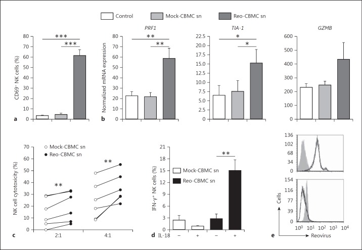
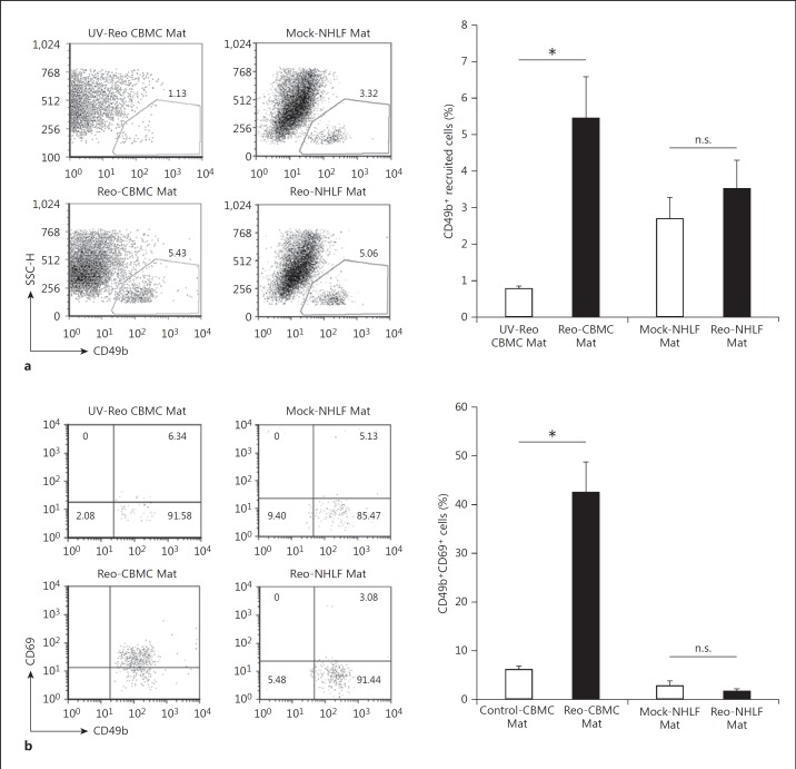
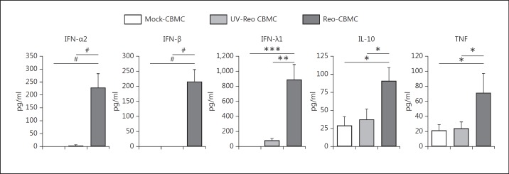
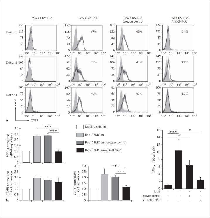
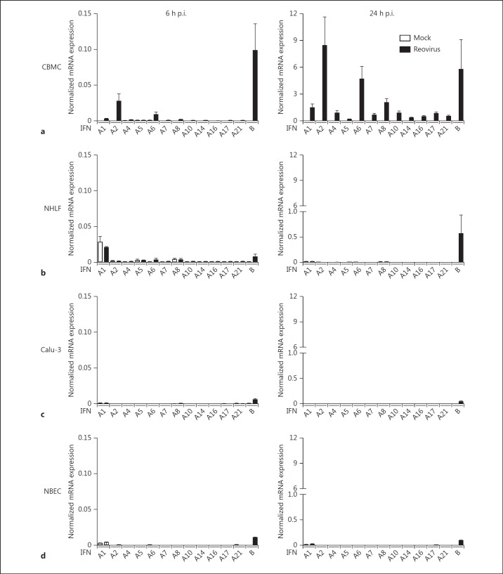
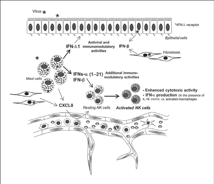
Mucosal surfaces are protected from infection by both structural and sentinel cells, such as mast cells. The mast cell's role in antiviral responses is poorly understood; however, they selectively recruit natural killer (NK) cells following infection. Here, the ability of virus-infected mast cells to enhance NK cell functions was examined. Cord blood-derived human mast cells infected with reovirus (Reo-CBMC) and subsequent mast cell products were used for the stimulation of human NK cells. NK cells upregulated the CD69 molecule and cytotoxicity-related genes, and demonstrated increased cytotoxic activity in response to Reo-CBMC soluble products. NK cell interferon (IFN)-γ production was also promoted in the presence of interleukin (IL)-18. In vivo, SCID mice injected with Reo-CBMC in a subcutaneous Matrigel model, could recruit and activate murine NK cells, a property not shared by normal human fibroblasts. Soluble products of Reo-CBMC included IL-10, TNF, type I and type III IFNs. Blockade of the type I IFN receptor abrogated NK cell activation. Furthermore, reovirus-infected mast cells expressed multiple IFN-α subtypes not observed in reovirus-infected fibroblasts or epithelial cells. Our data define an important mast cell IFN response, not shared by structural cells, and a subsequent novel mast cell-NK cell immune axis in human antiviral host defense.


Figures

Fig. 1
Fig. 1
Soluble mediator(s) produced by reovirus-infected mast cells activate NK cells. Purified peripheral blood human NK cells were cultured in Mock-CBMC sn, Reo-CBMC sn or culture medium (Control) for 24 h. a A purified lymphocyte population (≥90% CD3CD56+ cells) was analyzed for CD69 expression by FACS (n = 7). bPRF1 (n = 6), TIA-1 (n = 5) and GZMB (n = 5) gene expression was analyzed by qPCR and shown as normalized to GAPDH. c NK cells were resuspended in either Mock-CBMC sn or Reo-CBMC sn and cytotoxic activity against the K562 cell line was analyzed by LDH release assay at ratios of 2:1 and 4:1 (E:T) after 4 h of coculture (n = 6). All conditions were carried out in triplicate. d IFN-γ production was determined following stimulation with either Mock-CBMC sn or Reo-CBMC sn in the presence (+) or absence (-) of 100 ng/ml IL-18 (n = 7). Samples were analyzed by FACS. Data were compared using repeated-measures ANOVA with the Tukey post hoc test (a, b) or paired t test (c, d). * p < 0.05, ** p < 0.01, *** p < 0.001. Results are presented as mean ± SEM from at least 3 independent experiments performed on at least 5 different donors. e Reovirus infection was analyzed by FACS in CBMC (upper panel, n = 6) and NK cells (lower panel, n = 2) after culture with 20 MOI reovirus or Reo-CBMC sn, respectively. Isotype control (filled histograms) and anti-reovirus (empty histograms and black line) are shown.
Fig. 2
Fig. 2
NK cell recruitment and activation by reovirus-infected mast cells in vivo. Individual NOD SCID mice were each subcutaneously injected with Matrigel (Mat) containing cells under 2 different conditions, Reo-CBMC and either UV-Reo CBMC or Mock-CBMC. Similarly, NOD SCID mice received injections containing Reo-NHLF Mat and Mock-NHLF Mat. Matrigel plugs were harvested 24 h after injection. a Recruitment of NK cells was analyzed by FACS, first gating the leukocyte population by CD45 expression. CD49b+ cells were analyzed within this gate. b NK cell activation was determined by CD69 expression within the gate of CD49b+ cells. Control-CBMC Mat includes pooled results from both UV-Reo CBMC Mat (n = 1) and Mock-CBMC Mat (n = 4), which represent those control sites where sufficient NK cell recruitment occurred for analysis of cell activation. Results from the 5 animals where both control and Reo-CBMC Mat data were available were compared using the paired t test. * p < 0.05; all the available data from 9 (CBMC) or 6 (NHLF) NOD SCID are represented graphically as mean ± SEM from at least 5 independent experiments with at least 2 mice. n.s. = Not significant.
Fig. 3
Fig. 3
Cytokines produced by reovirus-infected mast cells. CBMC were infected with 20 MOI reovirus, cultured with 20 MOI UV-Reo or left uninfected for 24 h. Mast cell culture sn were analyzed by ELISA or multiplex to determine the production of IFN-α2 (n = 6), IFN-β (n = 5), IFN-λ1 (n = 8), IL-10 (n = 6), and TNF (n = 6). Data were compared using repeated measures with the Tukey (*) or Dunn (#) post hoc test. *, # p < 0.05, ** p < 0.01, *** p < 0.001. Results are presented as the mean ± SEM.
Fig. 4
Fig. 4
Response of NK cells to cytokines produced by reovirus-infected mast cells. NK cells were stimulated in parallel with Reo-CBMC sn, 10 ng/ml IFN-α2a, 100 ng/ml IFN-λ1 or 30 ng/ml IL-10 for 24 h. Mock-CBMC sn and 10% RPMI (control) were used as negative controls of stimulation. a CD69 upregulation (n = 5) was analyzed by FACS. bPRF1 gene expression (n = 6) was analyzed by qPCR. Results are shown as normalized to GAPDH. c For IFN-γ production (n = 5), IL-18 was added when indicated. Intracellular IFN-γ was analyzed by FACS. Data were compared using repeated-measures ANOVA with the Tukey post hoc test. * p < 0.05, ** p < 0.01, *** p < 0.001. Results are presented as the mean ± SEM from at least 3 independent experiments performed on at least 5 different donors. n.s. = Not significant.
Fig. 5
Fig. 5
Type I IFNs produced by reovirus-infected mast cells modulate NK cell activation. Purified NK cells were treated with 5 μg/ml anti-IFNAR, 5 μg/ml mouse IgG2a (isotype control) or left untreated for 1.5 h in 10% RPMI, followed by stimulation with Reo-CBMC sn for 24 h. a The number of CD69+ NK cells (black line, empty histograms) and control staining (filled histograms) are shown. b The expression of the genes PRF1, GZMB and TIA-1 was normalized to GAPDH, which was carried out as the value of 2 to the power of the difference between the threshold cycles for the amplification of GAPDH and either PRF1, TIA-1 or GZMB, and then presented graphically relative to the mock-treated cells for each condition (n = 6). c IFN-γ production in response to Reo-CBMC sn plus IL-18 was analyzed by FACS (n = 8). Data were compared using repeated-measures ANOVA with the Dunn post hoc test. * p < 0.05, *** p < 0.001. Results are presented as the mean ± SEM from at least 3 independent experiments performed on at least 5 different NK cell donors.
Fig. 6
Fig. 6
Reovirus-infected human mast cells are an important source of type I IFNs. CBMC (n = 7 and 9, a), NHLF (n = 4 and 5, b), Calu-3 (n = 3 and 4, c) and NBEC (n = 1, d) were infected with reovirus or left uninfected (Mock) for 6 or 24 h, respectively. Type I IFN data are presented as normalized mRNA expression relative to HPRT. Data are shown as mean ± SEM and are representative of at least 3 independent experiments. Where no bar is visible, no significant mRNA was detected.
Fig. 7
Fig. 7
Mast cells as sentinels in virus infections. Resident mature mast cells located close to blood vessels and epithelial barriers can recognize invading viruses and initiate innate immunity by recruitment and activation of NK cells through the production of CXCL8 and type I IFNs, respectively. Additional resident cells, such as fibroblasts, can also contribute to NK cell recruitment. At mucosal surfaces, the ability of mast cells, compared to structural cells, to express a wide range of IFNs-α (1–21) in addition to chemokines, may contribute to a more diverse cell infiltrate and more sustained IFN response, and therefore influence overall antiviral immunity. In addition to these mediators, mast cells can be considered as an important source of type III IFNs (IFN-λ), which have been reported to specifically protect epithelial surfaces more effectively than type I IFNs.

Similar articles

Cited by

References

    1. Bergelson JM. Virus interactions with mucosal surfaces: alternative receptors, alternative pathways. Curr Opin Microbiol. 2003;6:386–391. - PubMed
    1. Lodoen MB, Lanier LL. Natural killer cells as an initial defense against pathogens. Curr Opin Immunol. 2006;18:391–398. - PMC - PubMed
    1. Lanier LL. Up on the tightrope: natural killer cell activation and inhibition. Nat Immunol. 2008;9:495–502. - PMC - PubMed
    1. Zwirner NW, Domaica CI. Cytokine regulation of natural killer cell effector functions. BioFactors (Oxford) 2010;36:274–288. - PubMed
    1. Abraham SN, St John AL. Mast cell-orchestrated immunity to pathogens. Nat Rev Immunol. 2010;10:440–452. - PMC - PubMed

Publication types

Grants and funding