. 2016 Dec 9;291(50):26226-26238.
doi: 10.1074/jbc.M116.754853. Epub 2016 Nov 2.

Cellular Nuclear Export Factors TAP and Aly Are Required for HDAg-L-mediated Assembly of Hepatitis Delta Virus

Affiliations

Cellular Nuclear Export Factors TAP and Aly Are Required for HDAg-L-mediated Assembly of Hepatitis Delta Virus

Hsiu-Chen Huang et al. J Biol Chem. .

Abstract

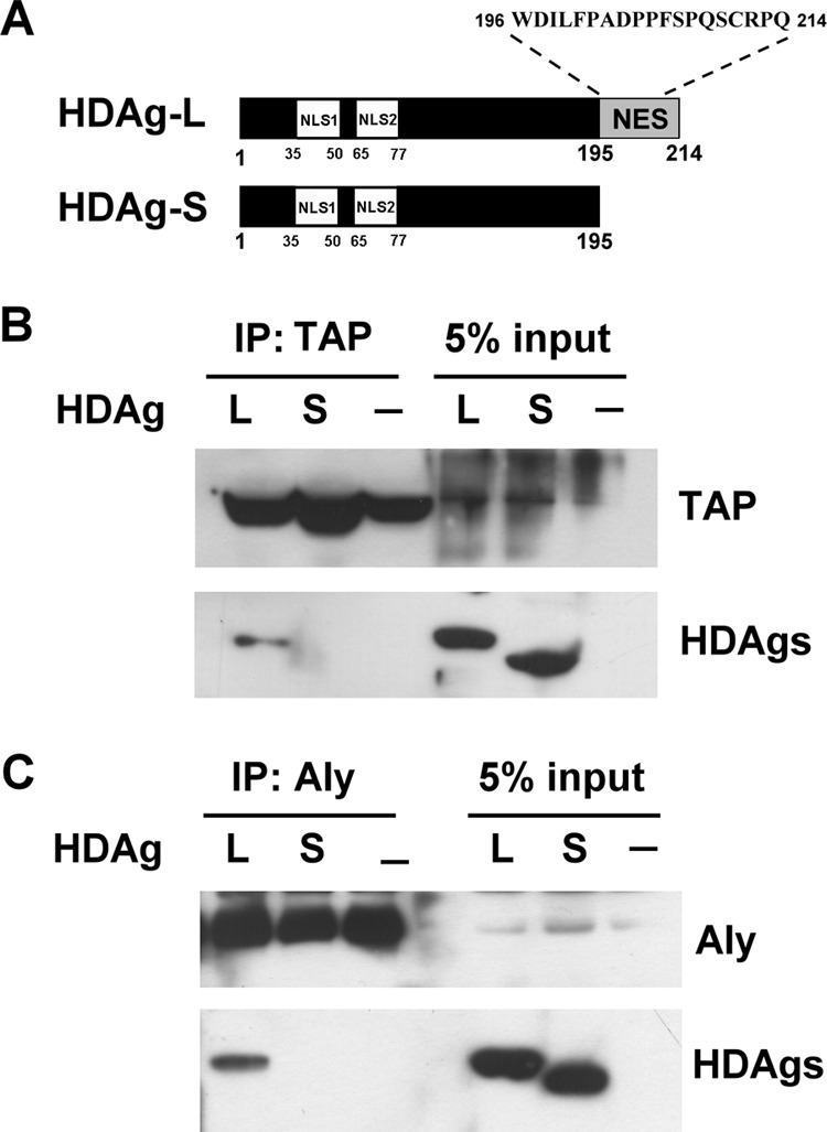
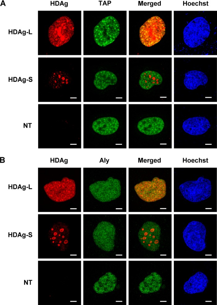
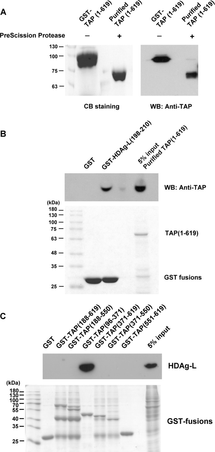
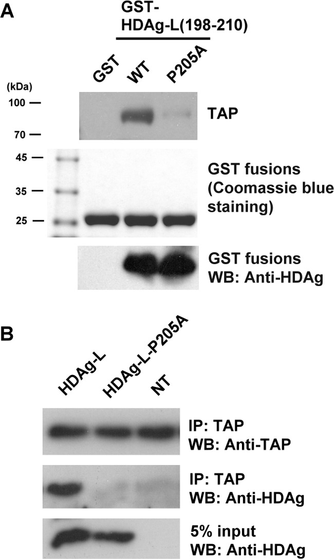
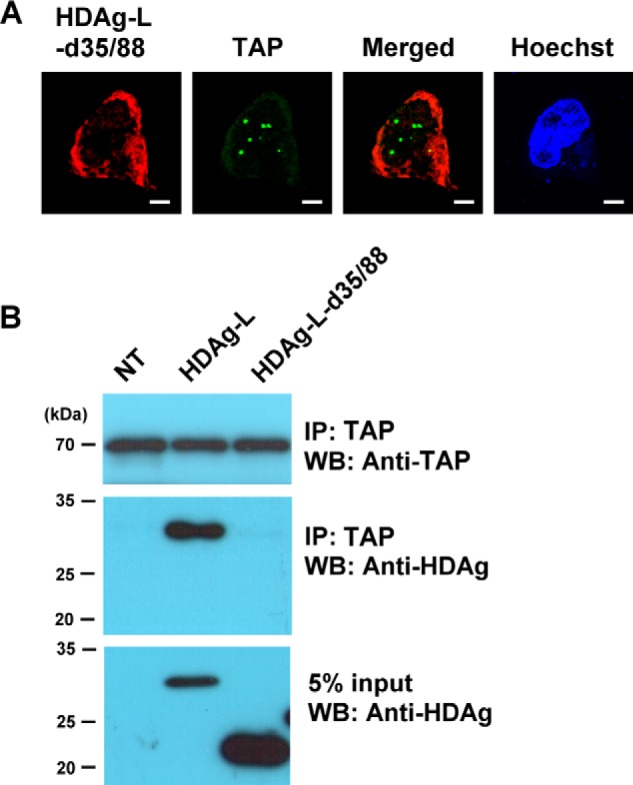
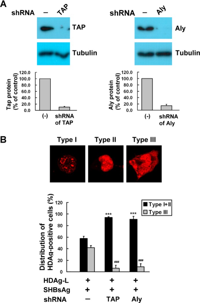
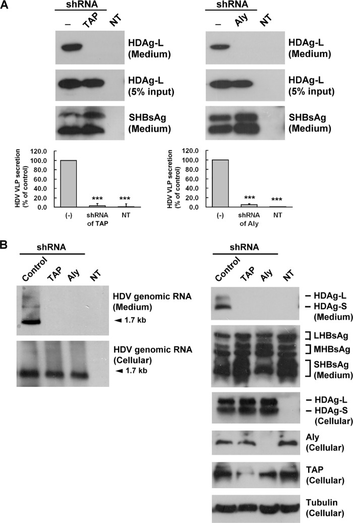
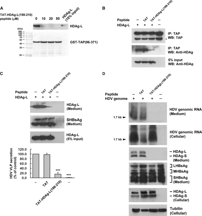
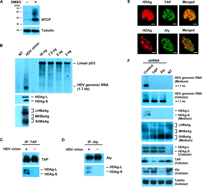
Hepatitis delta virus (HDV) is a satellite virus of hepatitis B virus (HBV). HDV genome encodes two forms of hepatitis delta antigen (HDAg), small HDAg (HDAg-S), which is required for viral replication, and large HDAg (HDAg-L), which is essential for viral assembly. HDAg-L is identical to HDAg-S except that it bears a 19-amino acid extension at the C terminus. Both HDAgs contain a nuclear localization signal (NLS), but only HDAg-L contains a CRM1-independent nuclear export signal at its C terminus. The nuclear export activity of HDAg-L is important for HDV particle formation. However, the mechanisms of HDAg-L-mediated nuclear export of HDV ribonucleoprotein are not clear. In this study, the host cellular RNA export complex TAP-Aly was found to form a complex with HDAg-L, but not with an export-defective HDAg-L mutant, in which Pro205 was replaced by Ala. HDAg-L was found to colocalize with TAP and Aly in the nucleus. The C-terminal domain of HDAg-L was shown to directly interact with the N terminus of TAP, whereas an HDAg-L mutant lacking the NLS failed to interact with full-length TAP. In addition, small hairpin RNA-mediated down-regulation of TAP or Aly reduced nuclear export of HDAg-L and assembly of HDV virions. Furthermore, a peptide, TAT-HDAg-L(198-210), containing the 10-amino acid TAT peptide and HDAg-L(198-210), inhibited the interaction between HDAg-L and TAP and blocked HDV virion assembly and secretion. These data demonstrate that formation and release of HDV particles are mediated by TAP and Aly.

Keywords: Aly; TAP; hepatitis delta antigen; hepatitis virus; nuclear export factor; nuclear transport; protein translocation; viral replication; virus assembly.


Figures

FIGURE 1.
FIGURE 1.
HDAg-L forms complexes with the cellular proteins TAP and Aly. A, schematic representation of HDAg-L and HDAg-S and the amino acid sequence of the C terminus of HDAg-L. The NLS and NES are shown. B and C, co-immunoprecipitation (IP) assay. Huh7 cells were left untreated or were transfected with plasmid pECE-d-BE or pECE-d-SM containing cDNAs encoding, respectively, HDAg-L and HDAg-S. At 2 days posttransfection, the cells were harvested and subjected to immunoprecipitation with anti-TAP antibodies (B), followed by Western blotting analysis with antibodies against TAP or HDAgs or with anti-Aly antibodies (C) followed by Western blotting analysis with antibodies against Aly and HDAgs.
FIGURE 2.
FIGURE 2.
Subcellular localization of HDAg-L, HDAg-S, TAP, and Aly in Huh7 cells. Huh7 cells were left untreated or were transfected with plasmid pECE-d-BE or pECE-d-SM containing cDNA encoding, respectively, HDAg-L or HDAg-S, then the subcellular localization of HDAg-L or HDAg-S and either TAP (A) or Aly (B) was examined by immunofluorescence staining with antibodies against HDAg, TAP, or Aly and confocal microscopy. NT, non-transfected. The bars on the images represent 20 μm.
FIGURE 3.
FIGURE 3.
Identification of TAP as an HDAg-L-interacting protein and mapping of the HDAg-L-binding domain in TAP. A, purification of TAP(1–619). GST-TAP(1–619) was incubated with PreScission Protease and then the glutathione-Sepharose beads were added to removed the cleaved GST, uncleaved GST-TAP(1–619), and the PreScission Protease (also a GST fusion protein). Following cleavage reactions, the purified proteins in the supernatant were detected by Coomassie Blue staining (left panel) or the Western blotting analysis was performed using antibodies against TAP (right panel). The positions of the molecular weight markers are shown on the left. B, GST pulldown assay with purified TAP(1–619) and GST-HDAg-L(198–210). The GST pulldown assay was performed with 100 μg of GST-HDAg-L(198–210) fusion protein precoupled to glutathione-Sepharose beads and 100 μg of purified TAP(1–619). 5% of input purified TAP(1–619) was used as control. Following GST pulldown, Western blotting analysis was performed using antibodies against TAP (top panel) or the pulled down proteins were detected by Coomassie Blue staining (bottom panel). The positions of the molecular weight markers are shown on the left. C, mapping of the HDAg-L-binding domain in TAP. The GST pulldown assay was performed with various GST-TAP fusion proteins precoupled to glutathione-Sepharose beads and lysates prepared from Huh7 cells transiently expressing HDAg-L. Following GST pulldown, Western blotting analysis was performed using antibodies against HDAg or Aly (top panel) or the pulled down proteins were detected by Coomassie Blue staining (bottom panel). The positions of the molecular weight markers are shown on the left.
FIGURE 4.
FIGURE 4.
The nuclear export mutant HDAg-L-P205A cannot bind to TAP. A, GST pulldown assay with TAP and GST fusion proteins containing either HDAg-L(198–210) or the P205A mutant. The GST pulldown assay was performed with either GST-HDAg-L(198–210) or the mutant precoupled to glutathione-Sepharose beads and lysates were prepared from Huh7 cells. Following GST pulldown, Western blotting analysis was performed using antibodies against TAP and HDAg (top and bottom panels) or the pulled down proteins were detected by Coomassie Blue staining (middle panel). The positions of the molecular weight markers are shown on the left. B, Huh7 cells were transfected with plasmid pECE-d-BE or pECE-d-BE(P205A) containing cDNA encoding, respectively, HDAg-L and HDAg-L-P205A, then, at 2 days posttransfection, were harvested and the cell lysates were subjected to immunoprecipitation (IP) with anti-TAP antibodies, followed by Western blotting analysis with antibodies against TAP or HDAgs. NT, non-transfected.
FIGURE 5.
FIGURE 5.
The HDAg-L NLS deletant mutant does not bind to TAP. A, Huh7 cells were transfected with plasmid pECEL-d35/88, then, at 2 days posttransfection, the subcellular localization of HDAg-L-d35/88 and TAP was examined by immunofluorescence staining with antibodies against HDAg and TAP using confocal microscopy. The bars on the images represent 20 μm. B, Huh7 cells were transfected with plasmid pECE-d-BE, coding for full-length HDAg-L, or pECEL-d35/88 lacking residues 35–88, then, at 2 days posttransfection, were harvested and the cell lysate was subjected to immunoprecipitation (IP) with anti-TAP antibodies, followed by Western blotting (WB) analysis with antibodies against TAP or HDAgs. The positions of the molecular weight markers are shown on the left. NT, non-transfected.
FIGURE 6.
FIGURE 6.
Knockdown of TAP and Aly inhibits nuclear export of HDAg-L. A, Huh7 cells were left untreated or were transfected with plasmid expressing TAP- or Aly-targeted shRNAs, then were harvested at 2 days posttransfection and analyzed by Western blotting analysis with antibodies against TAP and Aly. The top panel shows a typical result and the bottom panel the quantification of TAP and Aly expressed as the mean ± S.D. for three independent experiments. B, cellular distribution of HDAgs in the presence of TAP- or Aly-targeted shRNAs. Huh7 cells were transfected with HDAgs (L) alone or together with small HBsAg in the absence of presence of TAP or Aly shRNA, then, at 72 h posttransfection, immunofluorescence staining was performed using anti-HDAg antibodies. The top panels show the three HDAg staining patterns: type I, nucleolus; type II, both nucleolus and nucleoplasm; type III, nucleolus, nucleoplasm, and cytoplasm. The bottom panel shows the statistical analysis in which fields each containing at least 100 HDAg-positive cells were randomly selected and the number of cells with each type of HDAg staining pattern counted, and calculated as a percentage of the total number of the HDAg-positive cells in the same field. The results shown are the mean ± S.D. for three independent experiments. ***, p < 0.001 versus type I + type II control. ###, p < 0.001 versus type III control.
FIGURE 7.
FIGURE 7.
Interference with assembly and release of HDV virions in cells expressing TAP- or Aly-targeted shRNAs. A, Huh7 cells were cotransfected with plasmid pECE-C-ES encoding HBsAg and pECE-d-BE encoding HDAg-L in the presence or absence of TAP- or Aly-targeted shRNAs. Four days posttransfection, protein lysates were prepared from the transfected cells and HDV VLPs were collected from the culture medium. The HDV VLPs were subjected to Western blotting analysis with antibodies against HDAg or HBsAg (top panel) and the packaging activity calculated and normalized to HDAg-L in the absence of TAP- and Aly-targeted shRNAs (bottom panel). The results are the mean ± S.D. for three independent experiments. ***, p < 0.001 versus control. B, HepG2.2.15 cells were cotransfected with plasmid pSVD2 expressing a dimeric HDV RNA in the presence or absence of TAP- or Aly-targeted shRNAs or pLKO.1 control plasmid. Seven days posttransfection, protein lysates and RNA were prepared from the transfected cells and HDV virions were collected from the culture medium. A DIG-labeled HDV antigenomic RNA transcribed in vitro from plasmid pD3 was used as a probe to perform Northern blot analysis (left panel). Rabbit antiserum specific for HDAgs, Aly, TAP, and tubulin and goat polyclonal antibodies specific to HBsAg were used to perform Western blotting analysis (right panel) as indicated. NT, non-transfection.
FIGURE 8.
FIGURE 8.
Effect of the cell-permeable peptide TAT-HDAg-L(198–210) on the interaction between HDAg-L and TAP and on the release of HDV virions. A, competition between HDAg-L(198–210) and GST-TAP(96–371) for binding to HDAg-L. The GST pulldown competition assay was performed using GST-TAP(96–371) generated in bacteria, HDAg-L was prepared from transfected Huh7 cells and various amounts of HDAg-L(198–210). The top panel shows Western blotting (WB) analysis with antibodies against HDAgs and the bottom panel shows Coomassie Blue staining. B, effect of TAT-HDAg-L(198–210) on the binding of HDAg-L to TAP. Huh7 cells cotransfected with plasmids pSVD2 expressing a dimeric HDV RNA, pECE-C-ES expressing HBsAg, and pECE-d-BE expressing HDAg-L were cultured for 2 days in the presence or absence of 10 μm TAT-HDAg-L(198–210) or TAT peptide as control. They were then harvested and cell lysates were subjected to immunoprecipitation (IP) with anti-TAP antibodies followed by Western blotting analysis with antibodies against TAP or HDAgs. C, effects of TAT-HDAg-L(198–210) on the assembly of HDV virions. Huh7 cells were cotransfected with plasmid pSVD2 expressing a dimeric HDV RNA, pECE-C-ES encoding HBsAg, and pECE-d-BE encoding HDAg-L and incubated for 4 days with or without 10 μm TAT-HDAg-L(198–210) with TAT peptide as control. RNA and protein lysates were then prepared from the transfected cells and HDV virions were collected from the culture medium. Top panel, HDV virions subjected to Western blotting analysis with antibodies against HDAg or HBsAg. Bottom panel, HDV packaging activity calculated and normalized to HDAg-L in the absence of peptide. The results are the mean ± S.D. for three independent experiments. ***, p < 0.001 versus control. D, HepG2.2.15 cells were cotransfected with plasmid pSVD2 expressing a dimeric HDV RNA in the presence or absence of TAT-HDAg-L(198–210). Seven days posttransfection, protein lysates and RNA were prepared from the transfected cells and HDV virions were collected from the culture medium. Northern blot analysis was performed to detect cellular and secreted HDV genomic RNA. Western blotting analysis was performed to detect cellular and secreted HDAgs and HBsAg as indicated.
FIGURE 9.
FIGURE 9.
The role of TAP and Aly in viral assembly and release under conditions of HDV infection. A, Western blotting analysis of NTCP after DMSO treatment in HuS-E/2 cells. DMSO-differentiated HuS-E/2 cells were harvested and the cell lysate was subjected to Western blotting analysis with antibodies against NTCP. B, HDV virions were prepared and subjected to Northern blot analysis with a DIG-labeled probe (top panel) and Western blotting analysis with antibodies against HDAg or HBsAg (bottom panel). C and D, co-immunoprecipitation assay. DMSO-differentiated HuS-E/2 cells were left untreated or were infected with HDV. At 7 days post-infection, the cells were harvested and subjected to immunoprecipitation (IP) with anti-TAP antibodies (C), followed by Western blotting analysis with antibodies against TAP or HDAgs or with anti-Aly antibodies (D) followed by Western blotting analysis with antibodies against Aly and HDAgs. E, DMSO-differentiated HuS-E/2 cells were infected with HDV. At 7 days post-infection, cells were examined by immunofluorescence staining with antibodies against HDAg, TAP, or Aly and confocal microscopy. The bars on the images represent 20 μm. F, DMSO-differentiated HuS-E/2 cells were co-transfected with plasmid encoding TAP shRNA or Aly shRNA and plasmid p1.3HBcl encoding HBsAgs for 2 days. Then, cells were infected with HDV. Seven days post-infection, protein lysates and RNA were prepared from the infected cells and HDV virions were collected from the culture medium. Northern blot analysis was performed to detect secreted and cellular HDV genomic RNA. Western blotting analysis was performed to detect cellular TAP, Aly, HDAgs, and tubulin and secreted HDAgs and HBsAgs as indicated. NT, non-infection.

Similar articles

Cited by

References

    1. Rizzetto M. (1983) The delta agent. Hepatology 3, 729–737 - PubMed
    1. Bonino F., Heermann K. H., Rizzetto M., and Gerlich W. H. (1986) Hepatitis delta virus: protein composition of delta antigen and its hepatitis B virus-derived envelope. J. Virol. 58, 945–950 - PMC - PubMed
    1. Lai M. M. (1995) The molecular biology of hepatitis delta virus. Annu. Rev. Biochem. 64, 259–286 - PubMed
    1. Chang M. F., Baker S. C., Soe L. H., Kamahora T., Keck J. G., Makino S., Govindarajan S., and Lai M. M. (1988) Human hepatitis delta antigen is a nuclear phosphoprotein with RNA-binding activity. J. Virol. 62, 2403–2410 - PMC - PubMed
    1. Weiner A. J., Choo Q. L., Wang K. S., Govindarajan S., Redeker A. G., Gerin J. L., and Houghton M. (1988) A single antigenomic open reading frame of the hepatitis delta virus encodes the epitope(s) of both hepatitis delta antigen polypeptides p24 delta and p27 delta. J. Virol. 62, 594–599 - PMC - PubMed

MeSH terms

LinkOut - more resources