. 2016 Nov 15;113(46):E7297-E7306.
doi: 10.1073/pnas.1610784113. Epub 2016 Nov 2.

FEF inactivation with improved optogenetic methods

Affiliations

FEF inactivation with improved optogenetic methods

Leah Acker et al. Proc Natl Acad Sci U S A. .

Abstract

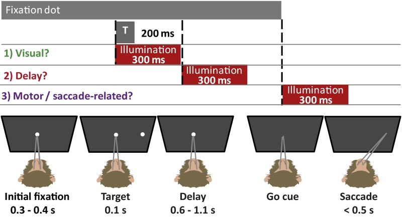
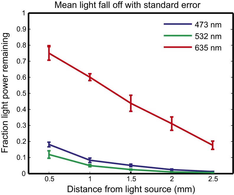
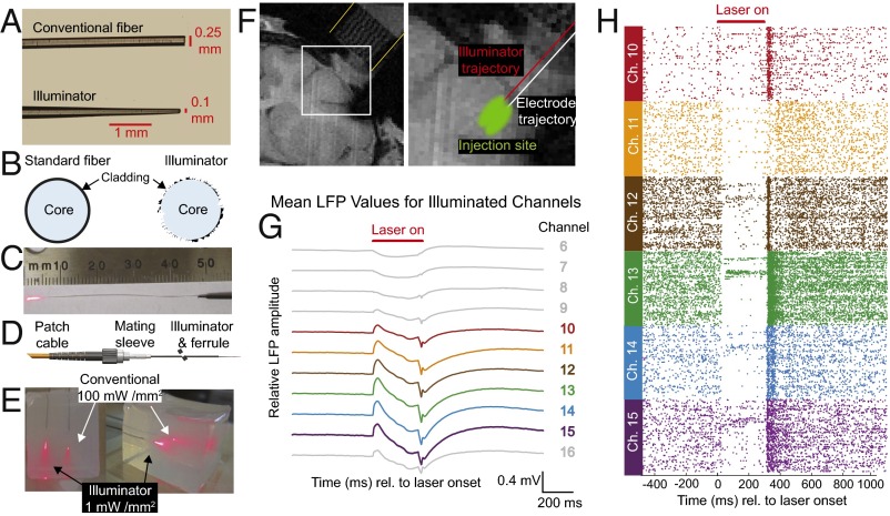
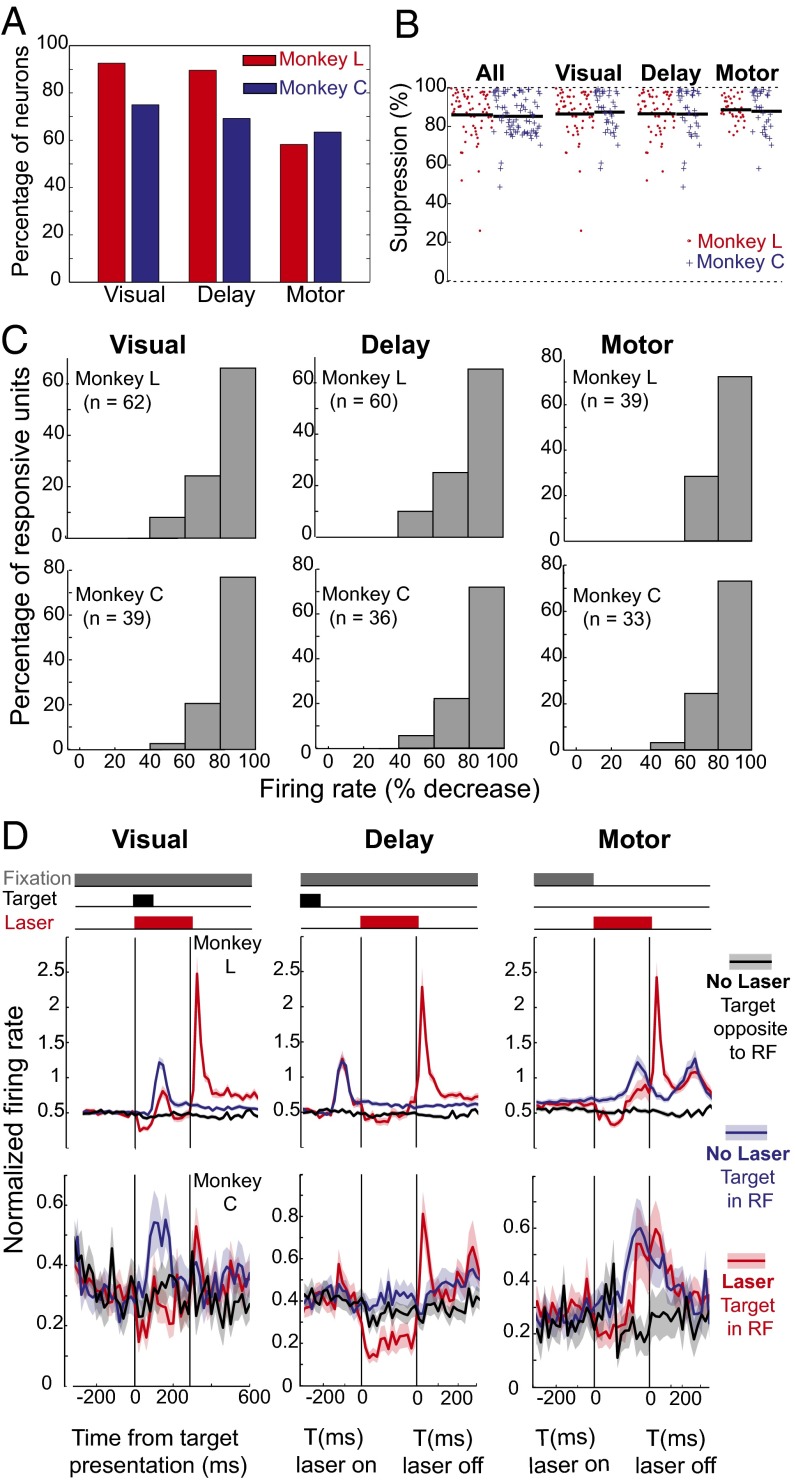
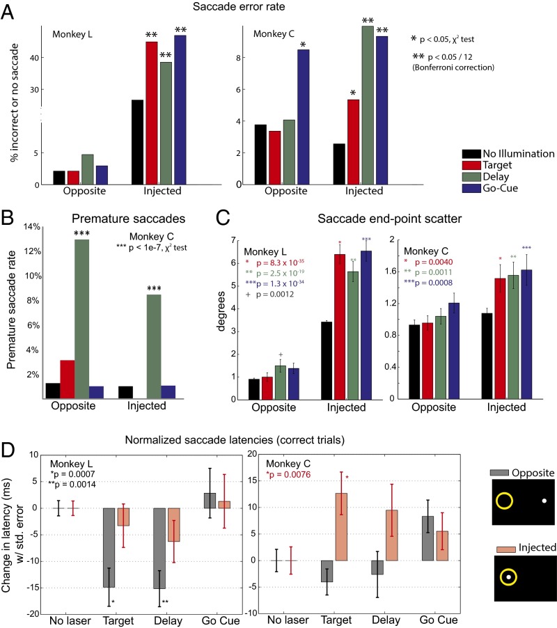
Optogenetic methods have been highly effective for suppressing neural activity and modulating behavior in rodents, but effects have been much smaller in primates, which have much larger brains. Here, we present a suite of technologies to use optogenetics effectively in primates and apply these tools to a classic question in oculomotor control. First, we measured light absorption and heat propagation in vivo, optimized the conditions for using the red-light-shifted halorhodopsin Jaws in primates, and developed a large-volume illuminator to maximize light delivery with minimal heating and tissue displacement. Together, these advances allowed for nearly universal neuronal inactivation across more than 10 mm3 of the cortex. Using these tools, we demonstrated large behavioral changes (i.e., up to several fold increases in error rate) with relatively low light power densities (≤100 mW/mm2) in the frontal eye field (FEF). Pharmacological inactivation studies have shown that the FEF is critical for executing saccades to remembered locations. FEF neurons increase their firing rate during the three epochs of the memory-guided saccade task: visual stimulus presentation, the delay interval, and motor preparation. It is unclear from earlier work, however, whether FEF activity during each epoch is necessary for memory-guided saccade execution. By harnessing the temporal specificity of optogenetics, we found that FEF contributes to memory-guided eye movements during every epoch of the memory-guided saccade task (the visual, delay, and motor periods).

Keywords: FEF; Jaws; memory-guided saccade; optogenetics; primate.


Conflict of interest statement

The authors declare no conflict of interest.

Figures

Fig. 1.
Fig. 1.
Memory-guided saccade task with illumination (or sham) at one of three task times (T). After an initial fixation of 300–400 ms (randomly assigned to eliminate timing cues), a target appears for 100 ms in one of the possible target locations (SI Appendix, Fig. S13). In a third of trials, a shutter (either the laser shutter or a sham shutter) opens with target presentation and closes 300 ms later to encapsulate all visually related activity in the FEF. In another third of trials, a shutter opens during the delay period, which is randomly distributed from 600 to 1,100 ms to prevent the monkey from using timing cues. In a final third of trials, a shutter opens with the go-cue (i.e., the disappearance of the fixation dot). The monkey has 500 ms to initiate a saccade.
Fig. 2.
Fig. 2.
In vivo measurement of visible light propagation. Average light decrease with distance from the illuminator: mean normalized fluence rates representing the fraction of applied light power reaching a given depth for blue (473 nm), green (532 nm), and red (635 nm) light as a function of distance from the illuminator with SE bars. (For blue, green, and red: n = 5, n = 6, and n = 5 mice, respectively).
Fig. 3.
Fig. 3.
Illuminator broadly distributes light to inhibit a large volume of primate cortex. (A) Illuminator and conventional fiber with the same diameter and material. (B) Etched core and cladding spread light broadly. (C) Light-emitting, 5-mm-long etched tip. (D) Optical fiber/mating sleeve/illuminator interface. (E) Illuminator and conventional optical fiber with equal total input light powers in 1-in. cubic brain phantom (1.75% agar). (F, Left) Coronal MRI from monkey L with a grid in the recording chamber (yellow lines). (F, Right) Enlarged view with an illuminator and electrode trajectories (1 mm apart), estimated virus injection region, and recording locations. (F, Right) The width of the chamber outlined with yellow lines is 1 in (25 mm) in diameter. (F, Left) The electrode and illuminator trajectories shown with red and white lines, respectively, are 1 mm apart. (G) LFP light artifact at 0.5-mm–spaced contacts. (H) Raster plots of corresponding neurons. For G and H, n = 462 trials.
Fig. 4.
Fig. 4.
Nearly universal inactivation of FEF neurons during the memory-guide saccade task dramatically increases error rates to targets in the inhibited receptive field. (A) Percentage of recorded FEF neurons with each of the three types of activity in each monkey. (B) Percentage of suppression with illumination by subtype with n values. (C) Distribution of firing rate decreases for each neuronal subtype in both monkeys. (D) Firing profile by subtype. Some neurons shown in B for monkey C were not recorded in enough trials for analysis in C because to sample more units, single-contact recordings had shorter durations. In monkey C, n = 19 (visual), n = 36 (delay), and n = 13 (motor). In monkey L, n is unchanged. RF, receptive field.
Fig. 5.
Fig. 5.
Optogenetic inactivation significantly increases error rates and alters saccade metrics. (A) Error rates increased significantly during delay and motor illumination. During target illumination, behavioral disruptions along the error/latency continuum included increased error rates (monkey L) and increased saccade latency. (B) Monkey C made many premature saccades. Data from monkey L are not shown because monkey L did not make any premature saccades. All n values are given as the number of premature saccade trials divided by total trials for that condition [no illumination: n = 9/611 (Injected), n = 14/592 (Opposite); Target: n = 0/272, n = 13/315; Delay: n = 29/284, n = 58/317; Go-Cue: n = 4/287, n = 8/290]. Premature saccades were not rewarded despite reaching the proper location. They likely reflect similar neuronal mechanisms to the decreased latency observed for targets to the opposite side in monkey C. (C) End-point scatter significantly increased for targets in the injected receptive field for all illumination conditions in both monkeys, a finding reflecting overall disruption of saccades and consistent with early primate FEF inactivation studies [monkey C: control trials: n = 409 (Injected), n = 403 (Opposite); Target: n = 207, n = 219; Delay: n = 176, n = 206; Go-Cue: n = 201, n = 208; monkey L: control trials: n = 1,470 (Injected), n = 1,500 (Opposite); Target: n = 187, n = 174; Delay: n = 167, n = 153; Go-Cue: n = 153, n = 160]. Mean end-point scatter is plotted with SE bars, and uncorrected P values (two-tailed Student’s t test) are shown. (D) Average latency for correct nonlaser trials was subtracted from the latencies for correct trials with illumination during the target, delay, or go-cue period for both monkeys to yield the average change in latency, plotted with SE bars [monkey C: control trials: n = 401 (Injected), n = 393 (Opposite); Target: n = 175, n = 215; Delay: n = 193, n = 198; Go-Cue: n = 214, n = 198; monkey L: control trials: n = 1,348 (Injected), n = 1,466 (Opposite); Target: n = 167, n = 169; Delay: n = 156, n = 142; Go-Cue: n = 137, n = 153]. Latency significantly increased during the target period for monkey C, which is consistent with the latency/error continuum. The optogenetic behavioral disruption during target presentation, which resulted in an increased error rate in monkey L, manifested itself differently in monkey C, as an increase in the latency of correct trials. As expected, we do not see an increase in the latency of correct trials for conditions where the error rate significantly increased. This figure also shows a significant decrease in saccade latency to the opposite hemifield with illumination for monkey L, a finding manifested as an increased premature saccade rate in monkey C.

References

    1. Chafee MV, Goldman-Rakic PS. Inactivation of parietal and prefrontal cortex reveals interdependence of neural activity during memory-guided saccades. J Neurophysiol. 2000;83(3):1550–1566. - PubMed
    1. Sommer MA, Tehovnik EJ. Reversible inactivation of macaque frontal eye field. Exp Brain Res. 1997;116(2):229–249. - PubMed
    1. Tehovnik EJ, Sommer MA. Effective spread and timecourse of neural inactivation caused by lidocaine injection in monkey cerebral cortex. J Neurosci Methods. 1997;74(1):17–26. - PubMed
    1. Dias EC, Kiesau M, Segraves MA. Acute activation and inactivation of macaque frontal eye field with GABA-related drugs. J Neurophysiol. 1995;74(6):2744–2748. - PubMed
    1. Dias EC, Segraves MA. Muscimol-induced inactivation of monkey frontal eye field: Effects on visually and memory-guided saccades. J Neurophysiol. 1999;81(5):2191–2214. - PubMed

Publication types

LinkOut - more resources