. 2016 Nov 15;113(46):E7212-E7221.
doi: 10.1073/pnas.1612626113. Epub 2016 Nov 2.

HIF-KDM3A-MMP12 regulatory circuit ensures trophoblast plasticity and placental adaptations to hypoxia

Affiliations

HIF-KDM3A-MMP12 regulatory circuit ensures trophoblast plasticity and placental adaptations to hypoxia

Damayanti Chakraborty et al. Proc Natl Acad Sci U S A. .

Abstract

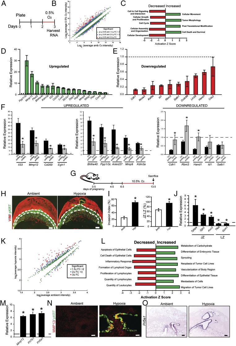
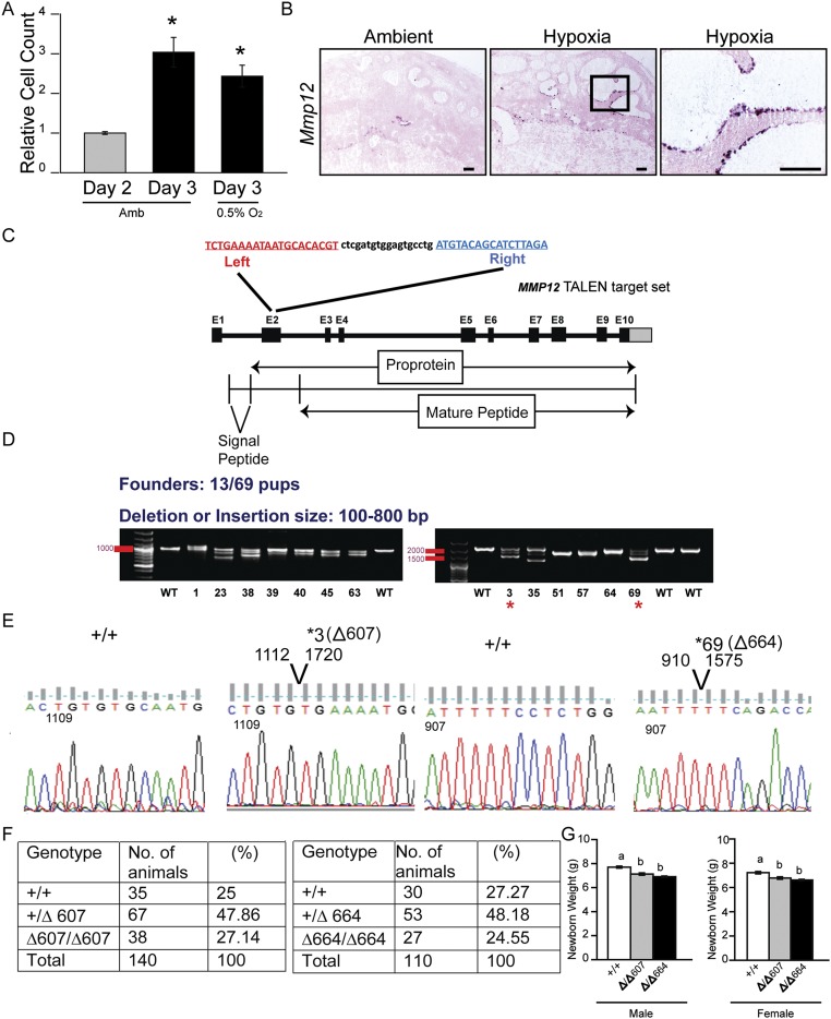
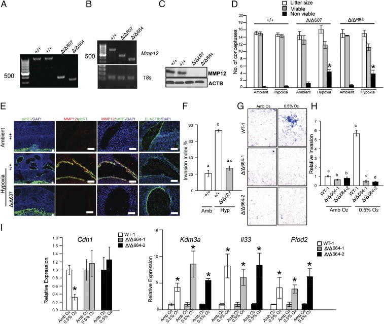
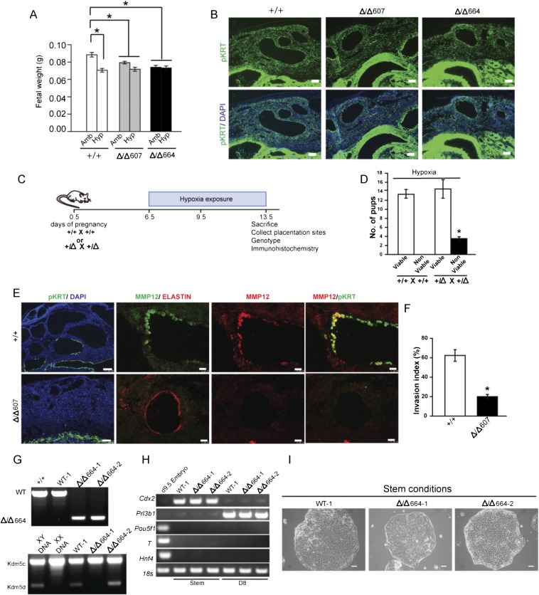
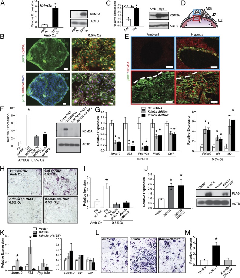
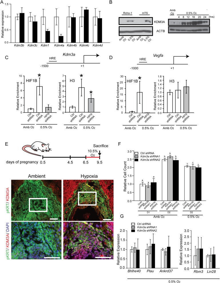
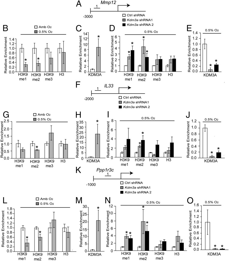
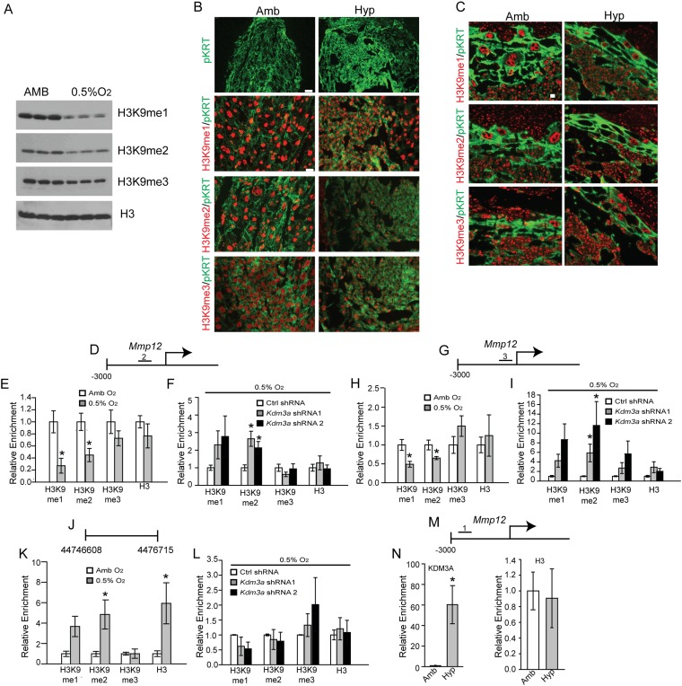
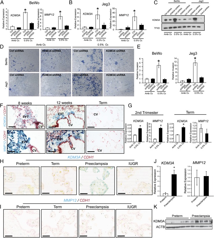
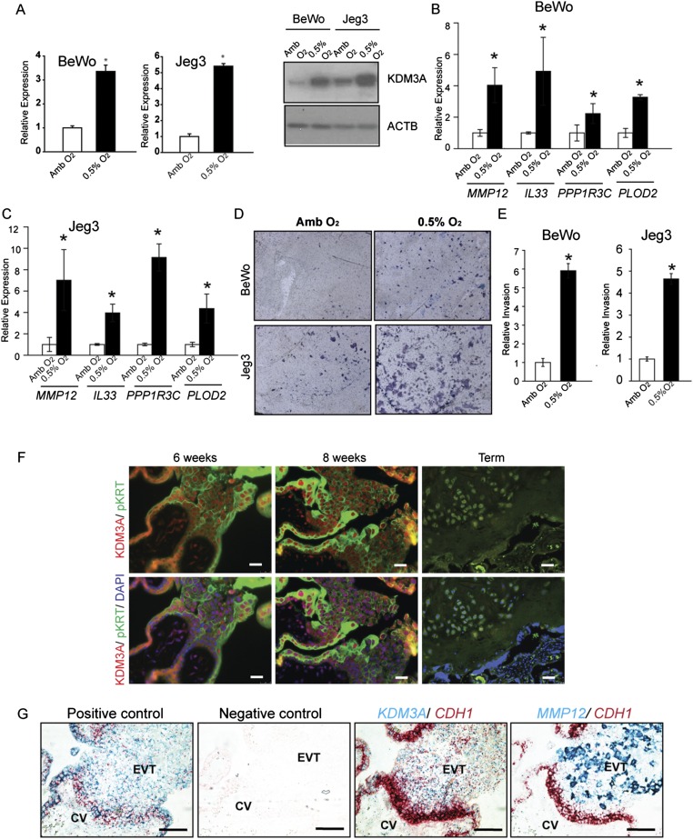
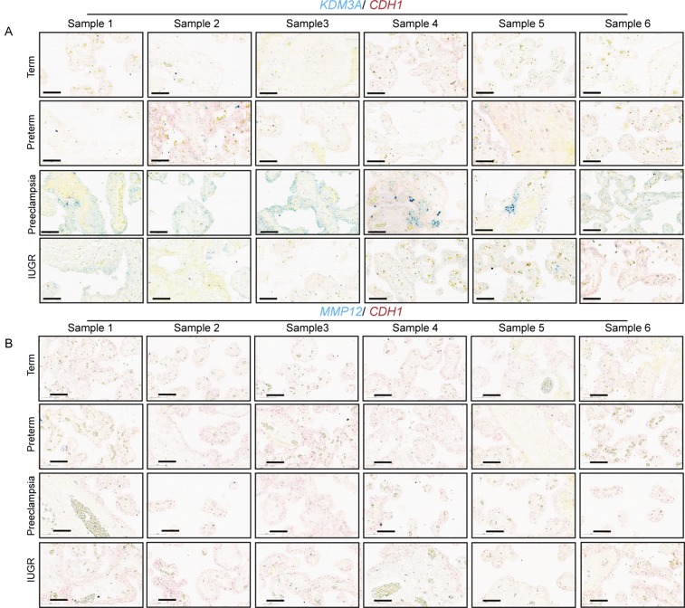
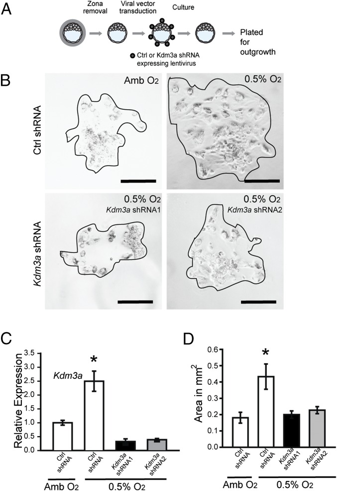
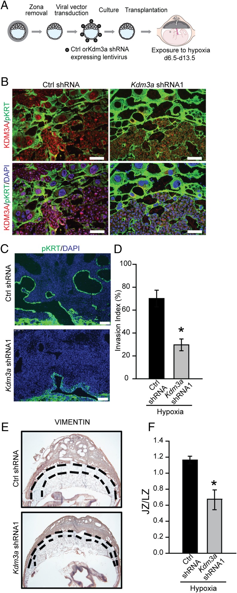
The hemochorial placenta develops from the coordinated multilineage differentiation of trophoblast stem (TS) cells. An invasive trophoblast cell lineage remodels uterine spiral arteries, facilitating nutrient flow, failure of which is associated with pathological conditions such as preeclampsia, intrauterine growth restriction, and preterm birth. Hypoxia plays an instructive role in influencing trophoblast cell differentiation and regulating placental organization. Key downstream hypoxia-activated events were delineated using rat TS cells and tested in vivo, using trophoblast-specific lentiviral gene delivery and genome editing. DNA microarray analyses performed on rat TS cells exposed to ambient or low oxygen and pregnant rats exposed to ambient or hypoxic conditions showed up-regulation of genes characteristic of an invasive/vascular remodeling/inflammatory phenotype. Among the shared up-regulated genes was matrix metallopeptidase 12 (MMP12). To explore the functional importance of MMP12 in trophoblast cell-directed spiral artery remodeling, we generated an Mmp12 mutant rat model using transcription activator-like nucleases-mediated genome editing. Homozygous mutant placentation sites showed decreased hypoxia-dependent endovascular trophoblast invasion and impaired trophoblast-directed spiral artery remodeling. A link was established between hypoxia/HIF and MMP12; however, evidence did not support Mmp12 as a direct target of HIF action. Lysine demethylase 3A (KDM3A) was identified as mediator of hypoxia/HIF regulation of Mmp12 Knockdown of KDM3A in rat TS cells inhibited the expression of a subset of the hypoxia-hypoxia inducible factor (HIF)-dependent transcripts, including Mmp12, altered H3K9 methylation status, and decreased hypoxia-induced trophoblast cell invasion in vitro and in vivo. The hypoxia-HIF-KDM3A-MMP12 regulatory circuit is conserved and facilitates placental adaptations to environmental challenges.

Keywords: epigenetics; hypoxia; placenta; plasticity; trophoblast invasion.


Conflict of interest statement

The authors declare no conflict of interest.

Figures

Fig. 1.
Fig. 1.
Hypoxia-dependent responses of TS cells and the placentation site. (A) Schematic representation of TS cell exposure to low oxygen tension (0.5% O2 for 24 h) before harvesting RNA for DNA microarray analysis. (B) Scatter plot presentation of in vitro hypoxia-responsive transcripts. (C) Presentation of pathway analysis of transcripts differentially expressed following TS cell exposure to 0.5% O2. (D and E) Validation of select differentially expressed transcripts by qRT-PCR. All hypoxia responses are significantly different from ambient control (n = 5/group; P < 0.05). (F) Examination of the dependence of hypoxia-dependent transcript changes on HIF signaling. TS cells were exposed to 0.5% O2 in the presence of control (Ctrl) or Hif1b shRNAs. RNA was harvested and transcript levels assessed by qRT-PCR (n = 4/group; ANOVA with Student–Newman–Keuls test, *P < 0.05). Dashed lines represent the ambient control values. (G) Schematic representation of in vivo maternal exposure to hypoxia (10.5% O2). (H) Representative cross sections of gd 13.5 placentation sites immunostained for vimentin and cytokeratin from pregnant rats exposed to ambient (Amb) or hypoxia (Hyp, 10.5% O2). The junctional zone (devoid of vimentin staining) is demarcated by the dashed white lines. (Scale bar, 1 mm.) (I) Quantification of invasion of trophoblast cells into the uterine mesometrial compartment and ratio of junctional and labyrinth zones (ambient, n = 10; hypoxia, n = 12; *P < 0.05). (J) Relative expression of transcripts associated with the junctional zone (JZ) and labyrinth zone (LZ) (n = 8/group, *P < 0.05). Dashed lines represent the ambient control values. (K) Scatter plot presentation of maternal hypoxia-responsive transcripts in the metrial gland. (L) Presentation of pathway analysis of differentially expressed transcripts in the metrial glands following hypoxia exposure. (M) Validation of selected differentially expressed transcripts by qRT-PCR (n = 10/group, *P < 0.05). Dashed lines represent the ambient control values. (N) Immunohistochemical analysis of MMP12 and pan cytokeratin (pKRT) staining in tissue sections from pregnant rats exposed to ambient or hypoxia conditions. (Scale bar, 50 μm.) (O) In situ hybridization analysis of Prl5a1 transcripts in placentation sites from pregnant rats exposed to ambient or hypoxia conditions. (Scale bar, 250 μm.) Data presented in DF, I, J, and M were analyzed with Mann–Whitney test.
Fig. S1.
Fig. S1.
Effects of low oxygen culture conditions on TS cell numbers and TALEN targeting of exon 2 within the rat Mmp12 locus. (A) Low oxygen effects on TS cell numbers. Note that 24-h exposure to ambient (Amb) or low oxygen (0.5% O2) conditions showed similar growth responses (n = 4/group, Mann–Whitney test, *P < 0.05). (B) In situ hybridization analysis of Mmp12 transcripts in gd 13.5 placentation sites from pregnant rats exposed to ambient or hypoxia conditions. (Scale bar, 250 μm.) (C) Schematic representation of the rat Mmp12 gene and the TALEN target site within exon 2 (NC_005107.4). Diagrammatic organization of the MMP12 protein. (D) PCR-based identification of Mmp12 mutant founders (13 founders identified from 69 offspring). Founder numbers 3 and 69 were used for expansion and characterization. (E) DNA sequence analysis showing two founder strains possessing deletions of 607 bp (Δ607) or 664 bp (Δ664). (F) Mendelian ratios generated from breeding +/Δ607 males x +/Δ607 females (Left) and +/Δ664 males x +/Δ664 females (Right). (G) Assessment of postpartum day 1 neonatal weights of WT (+/+), Δ/Δ 607, and Δ/Δ664 pups, sexed at birth (WT: males, n = 39, females, n = 33; Δ/Δ 607: males, n = 44, females n = 23; Δ/Δ664: males, n = 44, females, n = 43 females). Different letters above bars signify differences among means (ANOVA with Dunnett’s test, P < 0.05).
Fig. 2.
Fig. 2.
MMP12 and hypoxia-activated trophoblast-directed uterine spiral artery remodeling. (A) Genotyping of WT (+/+) and Mmp12 homozygous mutant rat strains generated by genome editing. (B) RT-PCR analysis for Mmp12 and 18s RNAs from spleens of WT (+/+) and Mmp12 mutant (Δ/Δ607 and Δ/Δ664) rats. (C) Western blotting for MMP12 and ACTB from spleens of WT (+/+) and Mmp12 mutant (Δ/Δ607 and Δ/Δ664) rats. (D) Effects of ambient and hypoxia (10.5% O2) conditions on litter size and the numbers of viable and nonviable conceptuses (+/+, n = 7; Δ/Δ 607, n = 5; Δ/Δ 664 n = 5; *P < 0.05). (E) Immunohistochemical analyses (pan cytokeratin, pKRT; MMP12; elastin) of the mesometrial placentation sites from WT (+/+) and Mmp12 mutant (Δ/Δ607) rats exposed to ambient or hypoxia conditions. (F) Quantification of trophoblast cell invasion into the uterine mesometrial compartment. Different letters above bars signify differences among means (n = 5/group, *P < 0.05). (G) Effects of low oxygen (0.5% O2) on invasive behavior of WT (WT-1) and two Mmp12-null (Δ/Δ664–1 and Δ/Δ664–2) TS cell populations. Images are representative filters. (H) Quantification of invasion through Matrigel. Different letters above bars signify differences among means (n = 5/group, P < 0.05). (I) qRT-PCR analysis of hypoxia responsive transcripts in WT (WT-1; white bars) and two Mmp12-null (Δ/Δ664–1; gray bars and Δ/Δ664–2; black bars) TS cell populations. Comparisons were between ambient (Amb) and 0.5% O2 conditions for each genotype (n = 3/group, Mann–Whitney test, *P < 0.05). Data presented in D, F, and H were analyzed with ANOVA and Holm–Sidak (D) or Newman–Keuls tests (F and H).
Fig. S2.
Fig. S2.
Role of MMP12 in placental and fetal adaptations to hypoxia. (A) Fetal weights at gd 13.5 from WT and Mmp12 mutant (Δ/Δ607 and Δ/Δ664) rat pregnancies exposed to ambient (Amb) or hypoxia (Hyp) conditions (Ambient, WT: n = 36; Hypoxia, WT: n = 44; Ambient, Δ/Δ607: n = 49; Hypoxia, Δ/Δ607: n = 41; Ambient, Δ/Δ664: n = 35; Hypoxia, Δ/Δ664: n = 41; *P < 0.05). (B) Immunohistochemical analyses (pKRT) of gd 18.5 WT (+/+), Δ/Δ607, and Δ/Δ664 placentation sites. (C) Schematic representation of in vivo maternal exposure to hypoxia (10.5% O2) and analyses. WT and Mmp12 mutant conceptuses were generated by +/Δ607 male × +/Δ607 female breeding. (D) Effects of hypoxia (10.5% O2) on the numbers of viable and nonviable conceptuses from WT × WT and heterozygous × heterozygous breeding. Heterozygous × heterozygous pregnancies exposed to hypoxia exhibited a higher number of nonviable conceptuses than did WT × WT (+/+) pregnancies (n = 5/group; *P < 0.05). (E) Immunohistochemical analyses (pan cytokeratin, pKRT; MMP12; elastin) of placentation sites from WT (+/+) and Mmp12 mutant (Δ/Δ607) conceptuses exposed to hypoxic conditions. (F) Quantification of trophoblast cell invasion into the uterine mesometrial compartment (n = 5/group, Mann–Whitney test, *P < 0.05). (G) Genotyping of WT (WT-1) and Mmp12-null (Δ/Δ664–1 and Δ/Δ664–2) TS cells for WT and Δ/Δ664 alleles (Top) and sex chromosome determination of WT and Mmp12-null TS cells (Bottom). WT-1 and Δ/Δ664–2 TS cells are X,Y and Δ/Δ664–1 TS cells are X,X. (H) Conventional RT-PCR analysis of embryonic and trophoblast-specific markers in stem and differentiated WT-1, Δ/Δ664–1, and Δ/Δ664–2 TS cells. (I) Representative images of stem state colonies for WT-1, Δ/Δ664–1, and Δ/Δ664–2 TS cells. Data presented in A and D were analyzed with ANOVA and Student–Newman–Keuls test.
Fig. 3.
Fig. 3.
KDM3A and hypoxia signaling in trophoblast cells. (A) Kdm3a transcript (qRT-PCR; Left) and protein (Western blot; Right) responses in TS cells cultured under ambient (Amb) or low oxygen (0.5% O2). Statistical analysis: n = 5/group, Mann–Whitney test, *P < 0.05. (B) Immunocytochemical staining for pan-cytokeratin (pKRT) and KDM3A in TS cells cultured under Amb or 0.5% O2. (Scale bar, 50 µm.) (C) In vivo placental Kdm3a transcript (qRT-PCR; Left) and protein (Western blot; Right) responses to maternal hypoxia (10.5% O2 from gd 6.5 to 13.5). (D) Schematic representation of a midgestation placentation site, consisting of the metrial gland (MG), junctional zone (JZ), and labyrinth zone (LZ). The blue box corresponds to the metrial gland region (Upper) of E and the red box corresponds to the junctional zone (Lower) of E. (E) Immunohistochemistry analysis (pan cytokeratin, pKRT; KDM3A) of gd 13.5 placentation site sections from pregnant rats exposed to ambient and hypoxic conditions. The dashed line in the bottom panels represents the border between the decidua and chorioallantoic placenta. (Scale bar, 100 µm.) (F) qRT-PCR and Western blot validation of Kdm3a shRNAs. TS cells expressing control (Ctrl) or Kdm3a shRNAs were examined in Amb or 0.5% O2 culture conditions (n = 4, *P < 0.05). (G) Effects of Kdm3a knockdown on hypoxia responsive transcripts in TS cells (n = 4, *P < 0.05). (H) Effects of Kdm3a knockdown on 0.5% O2 activated invasive behavior of TS cells. Images are representative filters. (I) Quantification of invasion through Matrigel (n = 3, Ctrl shRNA + 0.5% O2 vs. all other treatments, *P < 0.05). (J) Ectopic transcript (qRT-PCR; Left) and protein (Western blot, Right) expression of control (vector) and WT (Kdm3a) and mutant (Kdm3a-H1135Y) Kdm3a constructs stably transfected into TS cells. (K) Effects of ectopic expression of control (vector), WT (Kdm3a), and mutant Kdm3a (Kdm3a-H1135Y) constructs stably transfected into TS cells on hypoxia responsive transcripts (n = 4, Kdm3a vs. vector or Kdm3a-H1135Y, *P < 0.05). (L) Effects of ectopic expression of control (vector), WT (Kdm3a), and mutant Kdm3a (Kdm3a-H1135Y) constructs stably transfected into TS cells on invasive behavior. Images are representative filters. (M) Quantification of invasion through Matrigel (n = 4, Kdm3a vs. vector or Kdm3a-H1135Y, *P < 0.05). Data presented in F, G, I–K, and M were analyzed with ANOVA and Student–Keuls test. ACTB was used as a loading control for the Western blots shown in A, C, F, and J.
Fig. S3.
Fig. S3.
KDM3A, hypoxia, and placental expression. (A) qRT-PCR analysis of known histone H3K9 demethylases in rat TS cells maintained in ambient conditions (white bars) or following 24-h exposure to 0.5% O2 (black bars). Statistical analysis: n = 3/group, not significant. (B) KDM3A expression in Rcho-1, mouse TS (mTS), and rat TS cells. (Left) Western blot analysis of KDM3A expression after Rcho-1 TS cells and mouse TS cells exposed to Amb or 0.5% O2 for 24 h. (Right) Western blot analysis for KDM3A in rat TS cell exposed to 0.5% O2 for various time intervals. ACTB was used as a loading control for Western blots. (C) ChIP analysis for HIF1B and histone H3 at a conserved hypoxia response element (HRE) within the Kdm3a promoter. Rat TS cells were infected with lentiviral vectors containing control (Ctrl) or Hif1b shRNAs and exposed to ambient (Amb) or 0.5% O2 culture conditions (n = 4, *P < 0.05). (D) ChIP analysis for HIF1B and histone H3 at a conserved HRE within the Vegfa promoter. Rat TS cells were infected with lentiviral vectors containing Ctrl or Hif1b shRNAs and exposed to Amb or 0.5% O2 culture conditions (n = 4, *P < 0.05). (E) In vivo trophoblast KDM3A expression in response to maternal hypoxia. Rats were exposed to ambient or hypoxic (10.5% O2) conditions from gd 6.5 to 9.5 and euthanized, and placentation sites were dissected. Immunohistochemistry analysis (pan cytokeratin, pKRT; KDM3A) was performed on gd 9.5 placentation site tissue sections. White boxed areas shown in the upper images are presented in the lower panel. (Scale bars, 100 µm.) (F) Effects of Amb or 0.5% O2 culture conditions on cell numbers for control shRNA (Ctrl shRNA)-, Kdm3a shRNA1-, and Kdm3a shRNA2-expressing TS cells (n = 4/group, *P < 0.05). (G) Effects of Kdm3a knockdown on hypoxia responsive transcripts in TS cells (n = 4, not significant). Data presented in B was analyzed with Mann–Whitney test and C, D, F, and G were analyzed with ANOVA and Student–Newman–Keuls test.
Fig. 4.
Fig. 4.
Histone H3K9 methylation landscape associated with KDM3A targets in hypoxia-exposed TS cells. (A) Schematic layout of the rat Mmp12 gene and the location of one of the regions [No. 1: −2681 to −2568 bp upstream transcription start site (TSS)] surveyed by ChIP analysis in B–E. (B) ChIP analyses for histone H3K9 methylation (monomethylation, me1; dimethylation, me2; trimethylation, me3) and histone 3 (H3) at the Mmp12 locus in ambient (Amb) and low oxygen (0.5% O2) exposed TS cells. (C) ChIP analysis for KDM3A at the Mmp12 locus in Amb and 0.5% O2 exposed TS cells. (D) ChIP analyses for histone H3K9 methylation and H3 at the Mmp12 locus in 0.5% O2 exposed TS cells treated with control (Ctrl) or Kdm3a shRNAs. (E) ChIP analysis for KDM3A at the Mmp12 locus in 0.5% O2 exposed TS cells treated with Ctrl or Kdm3a shRNAs. (F) Schematic layout of the rat Il33 gene and the location of one of the regions (No. 1: −1372 to −1272 bp upstream of TSS) surveyed by ChIP analysis in G–J. (G) ChIP analysis for histone H3K9 methylation and H3 at the Il33 locus in Amb and 0.5% O2 exposed TS cells. (H) ChIP analysis for KDM3A at the Il33 locus in Amb and 0.5% O2 exposed TS cells. (I) ChIP analyses for histone H3K9 methylation and H3 at the Il33 locus in 0.5% O2 exposed TS cells treated with Ctrl or Kdm3a shRNAs. (J) ChIP analysis for KDM3A at the Il33 locus in 0.5% O2 exposed TS cells treated with Ctrl or Kdm3a shRNAs. (K) Schematic layout of the rat Ppp1r3c gene and the location of one of the regions (No. 1: −417 to −316 bp upstream of TSS) surveyed by ChIP analysis in L–O. (L) ChIP analyses for histone H3K9 methylation and H3 at the Ppp1r3c locus in Amb and 0.5% O2 exposed TS cells. (M) ChIP analysis for KDM3A at the Ppp1r3c locus in Amb and 0.5% O2 exposed TS cells. (N) ChIP analyses for histone H3K9 methylation and H3 at the Ppp1r3c locus in 0.5% O2 exposed TS cells treated with Ctrl or Kdm3a shRNAs. (O) ChIP analysis for KDM3A at the Ppp1r3c locus in 0.5% O2 exposed TS cells treated with Ctrl or Kdm3a shRNAs. Statistical analyses: n = 4; control vs. hypoxia exposed TS cell experiments: Mann–Whitney test, *P < 0.05; shRNA experiments: ANOVA with Dunnett’s test vs. the control, *P < 0.05).
Fig. S4.
Fig. S4.
Regulatory landscape associated with KDM3A target genes. (A) Western blot analysis for histone H3K9 methylation (monomethylation, me1; dimethylation, me2; trimethylation, me3) in lysates from rat TS cells exposed to ambient (Amb) or low oxygen (0.5% O2). (B) Immunolocalization of H3K9me1, H3K9me2, and H3K9me3 on gd 9.5 placentation sites from rats exposed to Amb or hypoxic (10.5% O2) conditions from gd 6.5 to 9.5. (C) Immunolocalization of H3K9me1, H3K9me2, and H3K9me3 on gd 13.5 placentation sites from rats exposed to Amb or 10.5% O2 from gd 6.5 to 13.5. (D) Schematic layout of the rat Mmp12 gene and location No. 2 (−920 to −765 bp upstream of TSS) within the 5′ flanking region surveyed by ChIP analysis in E and F. (E) ChIP analyses for histone H3K9 methylation and histone 3 (H3) at the Mmp12 locus in Amb and 0.5% O2 exposed TS cells. (F) ChIP analyses for histone H3K9 methylation and H3 at the Mmp12 locus in 0.5% O2 exposed TS cells treated with control (Ctrl) or Kdm3a shRNAs. (G) Schematic layout of the rat Mmp12 gene and location No. 3 (−414 to −276 bp upstream of TSS) within the 5′ flanking region surveyed by ChIP analysis in H and I. (H) ChIP analyses for histone H3K9 methylation and H3 at the Mmp12 locus in Amb and 0.5% O2 exposed TS cells. (I) ChIP analyses for histone H3K9 methylation and H3 at the Mmp12 locus in 0.5% O2 exposed TS cells treated with Ctrl or Kdm3a shRNAs. (J) Schematic layout of the gene desert location (Rnor_6.0, 44,746,608–44,746,715) surveyed by ChIP analysis in K and L. (K) ChIP analyses for histone H3K9 methylation and H3 at a gene desert location in Amb and 0.5% O2 exposed TS cells. (L) ChIP analyses for histone H3K9 methylation and H3 at the gene desert locus in 0.5% O2 exposed TS cells treated with Ctrl or Kdm3a shRNAs. (M) Schematic layout of the rat Mmp12 gene and location No. 1 (−2,681 to −2,568 bp upstream of the TSS) within the 5′ flanking region surveyed by ChIP analysis in N. (N) In vivo ChIP analyses for KDM3A and H3 in junctional zone tissue from gd 13.5 pregnant rats exposed to Amb or hypoxic (10.5% O2; Hyp) conditions. Statistical analyses: n = 4; control vs. hypoxia exposed TS cell experiments: Mann–Whitney test, *P < 0.05; shRNA experiments: ANOVA with Dunnett’s test vs. the control, *P < 0.05.
Fig. 5.
Fig. 5.
Conservation of hypoxia-dependent responses in human trophoblast cells. (A) Effects of KDM3A knockdown on low oxygen (0.5% O2) activated KDM3A and MMP12 transcript levels in BeWo human trophoblast cells (qRT-PCR; n = 3, *P < 0.05). (B) Effects of KDM3A knockdown on 0.5% O2 activated KDM3A and MMP12 transcript levels in Jeg3 human trophoblast cells (qRT-PCR; n = 3, *P < 0.05). (C) Effects of KDM3A knockdown on 0.5% O2 activated KDM3A protein in BeWo and Jeg3 human trophoblast cells. (D) Effects of KDM3A knockdown on 0.5% O2 induced invasive behavior of BeWo and Jeg3 human trophoblast cells. Images are representative Insets. (E) Quantification of invasion through Matrigel (n = 3, *P < 0.05). (F) In situ hybridization localization of CDH1 (red), KDM3A (blue, Top), and MMP12 (blue, Bottom) transcripts in first trimester (8 and 12 wk) and term human placental tissues. (Scale bar, 100 μm.) CV, chorionic villus; EVT, extravillous trophoblast. (G) Second trimester and term primary human trophoblast cell responses to 0.5% O2. KDM3A and MMP12 transcripts were measured by qRT-PCR (n = 3/group, Student t test, *P < 0.001). (H) In situ hybridization localization of CDH1 (red) and KDM3A (blue), transcripts in replicate representative placenta sections from preterm, term, preeclampsia, and intrauterine growth restriction (IUGR) pregnancies (low magnification: scale bar, 300 µm; high magnification: scale bar, 60 µm). (I) In situ hybridization localization of CDH1 (red) and MMP12 (blue), transcripts in replicate representative placenta sections from preterm, term, preeclampsia, and IUGR pregnancies. (Scale bar, 300 µm.) Data presented in A, B, and E were analyzed with ANOVA and Student–Newman–Keuls test. (J) qRT-PCR for Kdm3a and Mmp12 transcripts in placentas from preterm control and preeclamptic pregnancies (n = 6/group, Student t test, *P < 0.002). (K) Western blotting for KDM3A protein in placentas from preterm control and preeclamptic pregnancies (n = 5/group).
Fig. S5.
Fig. S5.
Hypoxia responses of human trophoblast cells and protein and transcript localization in human placental tissues. (A) Effects of low oxygen (0.5% O2) on KDM3A transcript (qRT-PCR) and protein (Western blot) expression in BeWo and Jeg3 human trophoblast cells. Amb, ambient. (B and C) Effects of 0.5% O2 on MMP12, IL33, PPP1R3C, and PLOD2 expression in BeWo (B) and Jeg3 (C) human trophoblast cells. Statistical analysis for A–C: n = 3/group; Mann–Whitney test, *P < 0.0.5. (D) Effects of 0.5% O2 on invasive behavior of BeWo and Jeg3 human trophoblast cells. Images are representative Insets. (E) Quantification of invasion through Matrigel (Student t test, *P < 0.05). (F) First trimester (6 and 8 wk) and term human placental tissues were processed for immunohistochemistry using KDM3A and pan cytokeratin (pKRT) antibodies. (Scale bar, 25 μm.) (G) In situ hybridization analysis of first trimester human placental tissue, including positive [POLR2A (red) and PPIB (blue)] and negative (DapB) controls, and colocalization of KDM3A (blue, third image) and MMP12 (blue, fourth image) with CDH1 (red). (Scale bar, 100 μm.)
Fig. S6.
Fig. S6.
KDM3A and MMP12 expression in diseased human placental tissues. (A) In situ hybridization localization of CDH1 (red) and KDM3A (blue), transcripts in replicate placenta sections from preterm, term, preeclampsia, and IUGR pregnancies (n = 6 for each). (Scale bar, 60 μm.) (B) In situ hybridization localization of CDH1 (red) and MMP12 (blue), transcripts in replicate placenta sections from preterm, term, preeclampsia, and IUGR pregnancies (n = 6 for each). (Scale bar, 60 μm.) The integrity and effectiveness of CDH1 and MMP12 probes were examined in first trimester placental tissue as a control.
Fig. 6.
Fig. 6.
Effects of oxygen tension and KDM3A expression on blastocyst outgrowth. (A) Schematic showing experimental plan for lentiviral transduction of blastocysts and outgrowth assay. Blastocysts were transduced with control (Ctrl) or Kdm3a shRNA and cultured for 72 h to allow hatching from the zona pellucida. The attached blastocysts were exposed to ambient (Amb) or low oxygen (0.5% O2) for 24 h and analyzed. (B) Representative images of blastocyst outgrowths from Ctrl shRNA and exposed to Amb, Ctrl shRNA and exposed to 0.5% O2, and Kdm3a shRNAs and exposed to 0.5% O2. (C) Measurement of Kdm3a transcripts in control and knockdown cultures was measured by qRT-PCR. Asterisks indicate significant differences among groups (n = 6/group; *P < 0.05). (D) The bar graph shows quantification of outgrowth area in square millimeters. The area of the outgrowth was measured using Image J software (Ctrl shRNA + Amb, n = 6; Ctrl shRNA + 0.5% O2, Kdm3a shRNA1 + 0.5% O2, n = 10; Kdm3a shRNA2 + 0.5% O2, n = 10; *P < 0.05). Data presented in C and D were analyzed with ANOVA and Student–Newman–Keuls test.
Fig. 7.
Fig. 7.
KDM3A and hypoxia-activated trophoblast-directed uterine spiral artery remodeling. (A) Schematic showing experimental plan for lentiviral transduction of blastocysts and in vivo transfer to pseudopregnant recipient animals. (B) Representative images of immunolocalization of KDM3A and pan cytokeratin (pKRT) on gd 13.5 placentation sites expressing control (Ctrl) shRNA or Kdm3a shRNA exposed to 10.5% O2 tension from gd 6.5 to 13.5. (C) Immunolocalization of pKRT on gd 13.5 placentation sites expressing Ctrl shRNA or Kdm3a shRNA exposed to 10.5% O2 tension from gd 6.5 to 13.5. (D) Quantification of the depth of cytokeratin-positive cell penetration into the uterine mesometrial vasculature (n = 6/group; *P < 0.05). (E) Localization of vimentin in placentation sites following Ctrl shRNA or Kdm3a shRNA transduction. (Scale bar, 1 mm.) Dashed black lines demarcate the location of the junctional zone (JZ) relative to the underlying labyrinth zone (LZ). (F) Ratio of cross-sectional areas of JZ vs. LZ from Ctrl shRNA and Kdm3a shRNA transduced placentation sites (n = 5/group; *P < 0.05). Data presented in D and F were analyzed with Student t test.

References

    1. Georgiades P, Ferguson-Smith AC, Burton GJ. Comparative developmental anatomy of the murine and human definitive placentae. Placenta. 2002;23(1):3–19. - PubMed
    1. Harris LK. Review: Trophoblast-vascular cell interactions in early pregnancy: How to remodel a vessel. Placenta. 2010;31(Suppl):S93–S98. - PubMed
    1. Maltepe E, Fisher SJ. Placenta: The forgotten organ. Annu Rev Cell Dev Biol. 2015;31:523–552. - PubMed
    1. Kaufmann P, Black S, Huppertz B. Endovascular trophoblast invasion: implications for the pathogenesis of intrauterine growth retardation and preeclampsia. Biol Reprod. 2003;69(1):1–7. - PubMed
    1. Pijnenborg R, Vercruysse L, Hanssens M. The uterine spiral arteries in human pregnancy: Facts and controversies. Placenta. 2006;27(9-10):939–958. - PubMed

Publication types

MeSH terms

Substances