. 2017 Jan 3;91(2):e01894-16.
doi: 10.1128/JVI.01894-16. Print 2017 Jan 15.

Molecular Architecture of the Cleavage-Dependent Mannose Patch on a Soluble HIV-1 Envelope Glycoprotein Trimer

Affiliations

Molecular Architecture of the Cleavage-Dependent Mannose Patch on a Soluble HIV-1 Envelope Glycoprotein Trimer

Anna-Janina Behrens et al. J Virol. .

Abstract

The formation of a correctly folded and natively glycosylated HIV-1 viral spike is dependent on protease cleavage of the gp160 precursor protein in the Golgi apparatus. Cleavage induces a compact structure which not only renders the spike capable of fusion but also limits further maturation of its extensive glycosylation. The redirection of the glycosylation pathway to preserve underprocessed oligomannose-type glycans is an important feature in immunogen design, as glycans contribute to or influence the epitopes of numerous broadly neutralizing antibodies. Here we present a quantitative site-specific analysis of a recombinant, trimeric mimic of the native HIV-1 viral spike (BG505 SOSIP.664) compared to the corresponding uncleaved pseudotrimer and the matched gp120 monomer. We present a detailed molecular map of a trimer-associated glycan remodeling that forms a localized subdomain of the native mannose patch. The formation of native trimers is a critical design feature in shaping the glycan epitopes presented on recombinant vaccine candidates.

Importance: The envelope spike of human immunodeficiency virus type 1 (HIV-1) is a target for antibody-based neutralization. For some patients infected with HIV-1, highly potent antibodies have been isolated that can neutralize a wide range of circulating viruses. It is a goal of HIV-1 vaccine research to elicit these antibodies by immunization with recombinant mimics of the viral spike. These antibodies have evolved to recognize the dense array of glycans that coat the surface of the viral molecule. We show how the structure of these glycans is shaped by steric constraints imposed upon them by the native folding of the viral spike. This information is important in guiding the development of vaccine candidates.

Keywords: furin; glycan; glycosylation; human immunodeficiency virus; neutralizing antibodies; oligosaccharides; structure; vaccines.


Figures

FIG 1
FIG 1
Design and glycosylation patterns of BG505 gp120, WT.SEKS, and SOSIP.664. (A) Schematic representation of the BG505 gp120, WT.SEKS, and SOSIP.664 constructs. Changes to the wild-type BG505 sequence are highlighted in blue. (B) HILIC-UPLC profiles of the enzymatically released N-linked glycans of the three constructs, transiently produced in HEK 293F cells and purified by 2G12 affinity chromatography followed by SEC. Oligomannose-type and hybrid glycans (green) were identified by their sensitivity to endo H digestion. Peaks corresponding to complex-type glycans are shown in pink. The peaks were integrated, and the pie charts summarize the quantification of the peak areas. Glycan symbols are as shown in Fig. 2.
FIG 2
FIG 2
Ion mobility mass spectrometry analysis of BG505 Env glycoproteins. Mobility-extracted singly charged negative-ion electrospray spectra are shown for N-linked glycans found on the following BG505 Env proteins: gp120 monomers (A), WT.SEKS pseudotrimers (B), and SOSIP.664 trimers (C). The inset in panel B shows an example of a DriftScope image derived from gp120 monomers, with singly charged ions encircled with a yellow oval. The peaks of the oligomannose series Man5–9GlcNAc2 are highlighted in green. A list of identified glycans is given in Table S1 in the supplemental material.
FIG 3
FIG 3
Quantitative site-specific N-glycosylation of BG505 Env glycoproteins. Relative quantification is shown for the N-glycosylation sites on the gp120 subunit (A) and the gp41 subunit (B) of gp120 monomers (no gp41 present), WT.SEKS pseudotrimers, and SOSIP.664 trimers. The proteins were digested with trypsin, chymotrypsin, pronase, GluC, or GluC plus trypsin and then analyzed by LC-ESI MS. Quantifications are based on the peak lists given in Tables S2, S3, and S4 in the supplemental material. The percentages corresponding to this figure are shown in Table S5. Glycans are categorized as oligomannose series (M5 to M9; Man5GlcNAc2 to Man9GlcNAc2), hybrids (H), and fucosylated hybrids (FH), and also by the number of branching antennae (A) of complex-type glycans. An, number (n) of antennae; Gn, number (n) of galactose residues; F, presence of a core fucose (50). The bar graphs represent the means for two analytical replicates, and the quantification of oligomannose-type (green) and complex/hybrid glycans (pink) on individual sites is summarized in the pie charts. The processing states of sites for which no quantitative analysis could be performed were classified by qualitative analysis of exoglycosidase-treated glycopeptides, as summarized by colored squares (Table S6).
FIG 4
FIG 4
Models of a fully glycosylated BG505 gp120 monomer and a SOSIP.664 trimer. Models of the glycosylated gp120 monomer (A) and the glycosylated SOSIP.664 trimer (B) were derived from one previously described elsewhere (50). The monomer is oriented as it would appear in situ as a subunit of the SOSIP.664 trimer. The glycans on the models are colored according to their oligomannose content, as derived in the present study. N-glycan sites for which quantitative results of sufficient quality could not be obtained were classified by qualitative analysis according to their susceptibility to endo H and PNGase F digestion (Table S6).
FIG 5
FIG 5
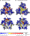
Heat maps on the surfaces of trimers, showing the increase in oligomannose glycans on BG505 SOSIP.664 trimers compared to gp120 monomers (A) and pseudotrimers (B). The increase in oligomannose content was calculated for sites for which quantitative data were available. To derive the heat map, a percentage point was calculated for each glycosylation site corresponding to an arithmetic difference of two percentage points; the percentage of oligomannose-type glycan for each of these sites on gp120 monomers or WT.SEKS pseudotrimers was subtracted from the corresponding percentage for SOSIP.664 trimers.
FIG 6
FIG 6
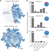
O-glycosylation of recombinant Env glycoproteins. (A) The location of the T499 O-glycosylation site is highlighted in red on the BG505 SOSIP.664 trimer model. (B) Quantification of the O-glycans identified on the gp120, WT.SEKS, and SOSIP.664 Env proteins, based on the peak list shown in Table S7.

References

    1. Lasky LA, Groopman JE, Fennie CW, Benz PM, Capon DJ, Dowbenko DJ, Nakamura GR, Nunes WM, Renz ME, Berman PW. 1986. Neutralization of the AIDS retrovirus by antibodies to a recombinant envelope glycoprotein. Science 233:209–212. doi:10.1126/science.3014647. - DOI - PubMed
    1. Wyatt R, Kwong PD, Desjardins E, Sweet RW, Robinson J, Hendrickson WA, Sodroski JG. 1998. The antigenic structure of the HIV gp120 envelope glycoprotein. Nature 393:705–711. doi:10.1038/31514. - DOI - PubMed
    1. Wei X, Decker JM, Wang S, Hui H, Kappes JC, Wu X, Salazar-Gonzalez JF, Salazar MG, Kilby JM, Saag MS, Komarova NL, Nowak MA, Hahn BH, Kwong PD, Shaw GM. 2003. Antibody neutralization and escape by HIV-1. Nature 422:307–312. doi:10.1038/nature01470. - DOI - PubMed
    1. Moore JP, Sodroski J. 1996. Antibody cross-competition analysis of the human immunodeficiency virus type 1 gp120 exterior envelope glycoprotein. J Virol 70:1863–1872. - PMC - PubMed
    1. Zhang M, Gaschen B, Blay W, Foley B, Haigwood N, Kuiken C, Korber B. 2004. Tracking global patterns of N-linked glycosylation site variation in highly variable viral glycoproteins: HIV, SIV, and HCV envelopes and influenza hemagglutinin. Glycobiology 14:1229–1246. doi:10.1093/glycob/cwh106. - DOI - PubMed

Substances

LinkOut - more resources