Review
. 2016 Nov;13(124):20160555.
doi: 10.1098/rsif.2016.0555.

Top-down models in biology: explanation and control of complex living systems above the molecular level

Affiliations
Review

Top-down models in biology: explanation and control of complex living systems above the molecular level

Giovanni Pezzulo et al. J R Soc Interface. 2016 Nov.

Abstract

It is widely assumed in developmental biology and bioengineering that optimal understanding and control of complex living systems follows from models of molecular events. The success of reductionism has overshadowed attempts at top-down models and control policies in biological systems. However, other fields, including physics, engineering and neuroscience, have successfully used the explanations and models at higher levels of organization, including least-action principles in physics and control-theoretic models in computational neuroscience. Exploiting the dynamic regulation of pattern formation in embryogenesis and regeneration requires new approaches to understand how cells cooperate towards large-scale anatomical goal states. Here, we argue that top-down models of pattern homeostasis serve as proof of principle for extending the current paradigm beyond emergence and molecule-level rules. We define top-down control in a biological context, discuss the examples of how cognitive neuroscience and physics exploit these strategies, and illustrate areas in which they may offer significant advantages as complements to the mainstream paradigm. By targeting system controls at multiple levels of organization and demystifying goal-directed (cybernetic) processes, top-down strategies represent a roadmap for using the deep insights of other fields for transformative advances in regenerative medicine and systems bioengineering.

Keywords: cognitive modelling; developmental biology; integrative; regeneration; remodelling; top-down.


Figures

Figure 1.
Figure 1.
Flexible, goal-directed shape homeostasis. (a) A tail grafted to the flank of a salamander slowly remodels to a limb, a structure more appropriate for its new location, illustrating shape homeostasis towards a normal amphibian body plan. Even the tail tip cells (in red) slowly become fingers, showing that the remodelling is not driven by only local information. Image taken with permission from [2]. (b) In some species of deer, the cell behaviour of bone growth during antler regeneration each year is modified by a memory of the three-dimensional location of damage made in prior years. Image taken with permission from [3]. (c) Kidney tubules in the newt are made with a constant size, whereas cell size can vary drastically under polyploidy (image taken with permission from: Fankhauser G. 1945 Maintenance of normal structure in heteroploid salamander larvae, through compensation of changes in cell size by adjustment of cell number and cell shape. J. Exper. Zool. 100, 445–455). Thus, the tubule pattern (a macroscopic goal state) can be implemented by diverse underlying molecular mechanisms such as cell : cell interactions (when there are many small cells) or cytoskeletal bending that curls one cell around itself, to make a tubule (when cells are very large). This illustrates the many-to-one relationship observed across scales of organization observed in statistical mechanics, computer science (implementation independence) and cognitive neuroscience (flexible pursuit of plans).
Figure 2.
Figure 2.
The functioning of the test, operate, test, exit (TOTE) model for action control and regeneration. (a) Generic functioning scheme of the TOTE model (which exemplifies a class of cybernetic models that operate using similar principles, see the main text). The ‘test’ operation corresponds to the comparison between desired and sensed state values. If a discrepancy is detected, then an action is activated (‘operate’) that tries to reduce it. When there is no discrepancy, the ‘exit’ operation is selected, corresponding to no action. (b) An example in the domain of action control. Here, the red box corresponds to a desired state value (handle grasped), which can trigger a series of (grasping) actions; see [69] for an example implementation. (c) The same mechanism at work in regeneration, where a discrepancy between the organism's target morphology and the current anatomical state (caused by injury) activates pattern homeostatic remodelling and growth. In highly regenerative animals, such as salamanders, cells proliferate, differentiate and migrate as needed to restore the correct pattern, and cease when the correct shape has been achieved.
Figure 3.
Figure 3.
An example of application of the free energy principle to morphogenesis. (a) Illustration of the target morphology that the cells have to achieve: a simple morphology with head, body and tail. The cells have to ‘find their place’ in this morphology, but are initially undifferentiated; this means that, in principle, each cell could become part of the head, body or tail (although constraints can be introduced on this process). (b) When cells occupy a given positions, they emit chemical signals that can be sensed by the surrounding cells. The figure illustrates the signals that each cell ‘expects’ to sense when it occupies a given spatial position. Of note, the cells can only sense these signals (and thus minimize their prediction error or free energy) if all the other cells are in the right place and emit the right signal. (c) In turn, this triggers a multi-agent migration (or differentiation) process, in which cells that ‘search’ their own place in the morphology influence and are influenced by the other cells. (d) There is only one solution to this multi-cell process—one in which each cell occupies its place and emits (and senses) the right signals. This corresponds to the target morphology. (e) It is only in this ‘solution’ state that the free energy of the system is minimized. Overall, it is a free energy minimization imperative that drives the pattern formation and morphogenetic process at the level of the whole system, with the cell–cell signalling mechanism playing a key role by permitting cells to influence one another. Images re-used according to the Creative Commons licence from ref. [52].
Figure 4.
Figure 4.
Bioelectric signalling in the brain and body. (a) The hardware of neural systems is composed of ion channel proteins which set the voltage level (node activation) of cells, and electrical synapses (gap junctions) that allow circuits to form via voltage propagation. These networks execute software—a diverse collection of modes and dynamics of bioelectrical states that evolve through time within cells (a true epigenetic system; transcriptional or translational changes are not required, as channels and gap junctions regulate and are regulated by voltage post-translationally). Efforts in computational neuroscience are ongoing to extract semantic content from readings of the electric activity (rightmost panel in row (a) is taken with permission from Naselaris T, Prenger RJ, Kay KN, Oliver M, Gallant JL. 2009 Bayesian reconstruction of natural images from human brain activity. Neuron 63, 902–915). (b) Precisely this system, albeit functioning at a slower timescale, occurs in non-excitable tissues, which use exactly those electrogenic proteins to form electrical networks in tissues during embryogenesis, regeneration and cancer. On the right-hand side is shown an image of an endogenous bioelectrical gradient (red = depolarized, blue = hyperpolarized) that serves as an instructive prepattern to regeneration in a planarian flatworm. The cracking of the bioelectric code to understand the anatomical outputs of bioelectric states is a key open problem in this field. Bioelectricity may be an important entry point to understand the integration of information across levels of organization during pattern regulation. Gene regulatory networks that regulate ion channel expression (c) constrain bioelectric circuits within tissue (d) which have their own dynamics that likewise regulate transcription of other genes. Together, these interplay to set up chemical and electrical gradients on an organism-wide scale which define anatomical regions and set organ size and identity (e). Events during remodelling and regeneration are controlled by high-level anatomical properties and topological interactions (f). Each of these levels have their own unique concepts, key variables, datasets and models. Top-down strategies may assist in the merging of these different levels of description towards an algorithmic (g) understanding of shape control, greatly facilitating rational control of outcomes. Images courtesy of Alexis Pietak, Jeremy Guay of Peregrine Creative, and Allegra Westfall.
Figure 5.
Figure 5.
Representation of multiple levels within bioelectric (neural-like) networks. (a) Neural networks of different kinds—e.g. deep nets, attractor nets—are able to learn internal representations of external entities directly from (unannotated) input data, at multiple levels, as they become tuned to the statistics of their inputs during learning [47]. For example, generative neural networks trained to reproduce in output the same figures (e.g. faces, objects or numbers) they receive in input often learn simpler-to-more-complex object or face features at increasingly higher hierarchical layers of the network, in ways that are (sometimes) biologically realistic [118,119]. Learning proceeds differently depending on the specific neural network architecture. For example, basins of attraction in the state space of attractor neural networks can represent specific memories [65]. If somatic tissues undergoing remodelling can be represented by similar frameworks, then it is possible that stable states in the network can represent specific patterning outcomes (like the one- or two-headed flatworms that result from editing of bioelectric networks in vivo [–122]). (b) One advantage of neural network architectures is that they show a proof of principle of how collections of cells can represent higher-level topological information; here are shown analogies between information representation in an ANN during a face recognition tasks and in flatworm regeneration. Neural network layers represent progressively increasingly abstract features of the input, in well-understood ways. The functioning of specific neural network architectures such as attractor, generative or deep nets (or other [–125]) may give clues as to how collections of somatic cells can learn and store memories about tissues, organs, and entire body plan layouts. The current state of the art in the field of developmental bioelectricity is that it is known, at the cellular level, how resting potentials are transduced into downstream gene cascades, as well as which transcriptional and epigenetic targets are sensitive to change in developmental bioelectrical signals [–128]. What is largely missing however (and may be provided by a network approach or other possible conceptualizations) is a quantitative understanding of how the global dynamics of bioelectric circuits make decisions that orchestrate large numbers of individual cells, spread out over considerable anatomical distances, towards specific pattern outcomes. The mechanisms by which bioelectrics and chromatin state interact at a single cell level will be increasingly clarified by straightforward reductive analysis. The more difficult, major advances in prediction and control will require a systems-level model of pattern memory and encoding implemented in somatic bioelectrical networks whose output is signals that control growth and form. Images in panels (a) and (b) drawn by Jeremy Guay of Peregrine Creative.

References

    1. Ross J, Arkin AP. 2009. Complex systems: from chemistry to systems biology. Proc. Natl Acad. Sci. USA 106, 6433–6434. (10.1073/pnas.0903406106) - DOI - PMC - PubMed
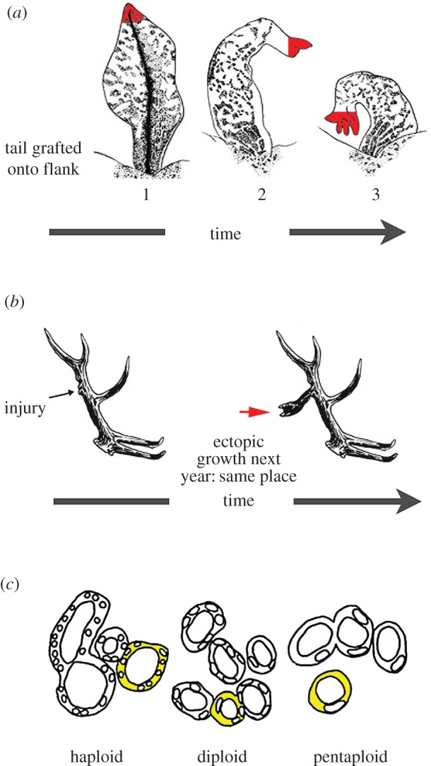
    1. Farinella-Ferruzza N. 1956. The transformation of a tail into limb after xenoplastic transplantation. Experientia 12, 304–305. (10.1007/BF02159624) - DOI
    1. Bubenik AB, Pavlansky R. 1965. Trophic responses to trauma in growing antlers. J. Exp. Zool. 159, 289–302. (10.1002/jez.1401590302) - DOI - PubMed
    1. Levin M. 2012. Morphogenetic fields in embryogenesis, regeneration, and cancer: non-local control of complex patterning. Biosystems 109, 243–261. (10.1016/j.biosystems.2012.04.005) - DOI - PMC - PubMed
    1. Mustard J, Levin M. 2014. Bioelectrical mechanisms for programming growth and form: taming physiological networks for soft body robotics. Soft Robot. 1, 169–191. (10.1089/soro.2014.0011) - DOI

Publication types

LinkOut - more resources