The effect of sling exercise on sagittal lumbosacral angle and intervertebral disc area of chronic low back pain patients
- PMID: 27807527
- PMCID: PMC5091064
- DOI: 10.12965/jer.1632676.338
The effect of sling exercise on sagittal lumbosacral angle and intervertebral disc area of chronic low back pain patients
Abstract
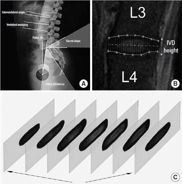
The purpose of this study was to observe the change of lumbosacral angle and intervertebral disc (IVD) area. The study was conducted on chronic low back pain (CLBP) female patients for 12 weeks by operating sling exercise and general physical therapy. The 57 CLBP were divided into 2 groups which, sling exercise group (SEG, n=34) and general physical therapy group (PTG, n=23). The experiment was conducted three times a week for 12 weeks. The lumbosacral angle, which means the angle between the L1-L2 lumbar was measured by plain radiography. The IVD area, which means the IVD height and volume was measured by magnetic resonance imaging. The pain was measured by visual analogue scale (VAS). As a result, after 12-week exercise, VAS had decreased in all groups. The angle of L3-4 and L4-5 and the height of IVD had increased in SEG. Also, IVD height and volume has more improved in SEG compare the PTG. Therefore, the sling exercise is proper treatment for CLBP patients' recovery because It improve the lumbosacral angle and IVD area.
Keywords: Chronic low back pain; Intervertebral disc area; Lumbosacral angle; Sling exercise; Visual analogue scale.
Figures
References
-
- Akuthota V, Nadler SF. Core strengthening. Arch Phys Med Rehabil. 2004;85(3 Suppl 1):S86–92. - PubMed
-
- Carpenter DM, Nelson BW. Low back strengthening for the prevention and treatment of low back pain. Med Sci Sports Exerc. 1999;31:18–24. - PubMed
-
- Derby R, Kim BJ, Lee SH, Chen Y, Seo KS, Aprill C. Comparison of discographic findings in asymptomatic subject discs and the negative discs of chronic LBP patients: can discography distinguish asymptomatic discs among morphologically abnormal discs? Spine J. 2005;5:389–394. - PubMed
LinkOut - more resources
Full Text Sources
Other Literature Sources
Research Materials