. 2017 Feb 15;595(4):1377-1392.
doi: 10.1113/JP273012. Epub 2016 Dec 11.

Cholinergic modulation of the parafacial respiratory group

Affiliations

Cholinergic modulation of the parafacial respiratory group

Rozlyn C T Boutin et al. J Physiol. .

Abstract

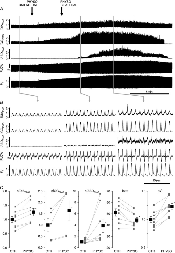
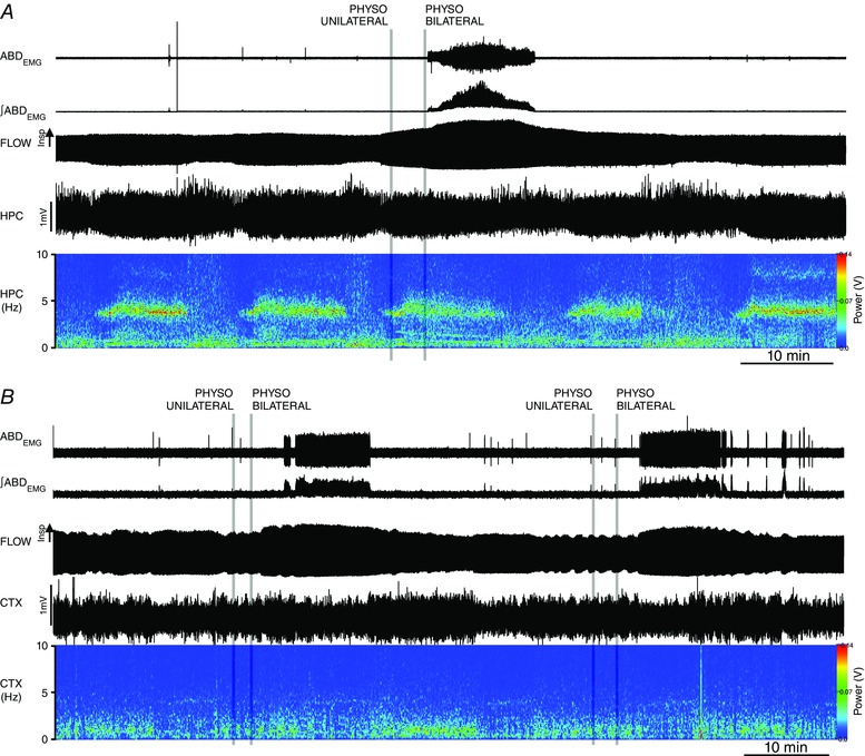
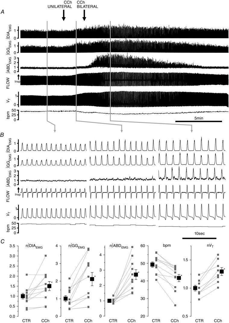
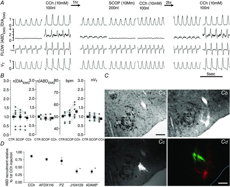
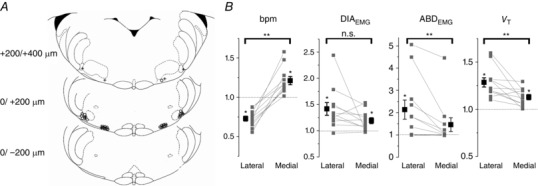
Key points: This study investigates the effects of cholinergic transmission on the expiratory oscillator, the parafacial respiratory group (pFRG) in urethane anaesthetized adult rats. Local inhibition of the acetyl cholinesterase enzyme induced activation of expiratory abdominal muscles and active expiration. Local application of the cholinomimetic carbachol elicited recruitment of late expiratory neurons, expiratory abdominal muscle activity and active expiration. This effect was antagonized by local application of the muscarinic antagonists scopolamine, J104129 and 4DAMP. We observed distinct physiological responses between the more medial chemosensitive region of the retrotrapezoid nucleus and the more lateral region of pFRG. These results support the hypothesis that pFRG is under cholinergic neuromodulation and the region surrounding the facial nucleus contains a group of neurons with distinct physiological roles.

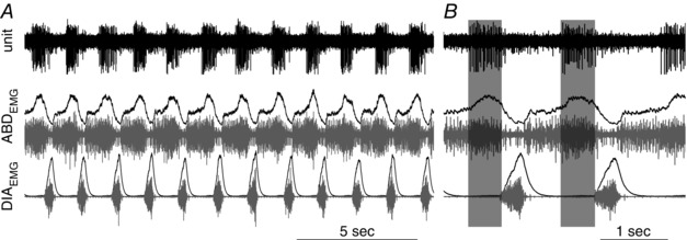
Abstract: Active inspiration and expiration are opposing respiratory phases generated by two separate oscillators in the brainstem: inspiration driven by a neuronal network located in the preBötzinger complex (preBötC) and expiration driven by a neuronal network located in the parafacial respiratory group (pFRG). While continuous activity of the preBötC is necessary for maintaining ventilation, the pFRG behaves as a conditional expiratory oscillator, being silent in resting conditions and becoming rhythmically active in the presence of increased respiratory drive (e.g. hypoxia, hypercapnia, exercise and through release of inhibition). Recent evidence from our laboratory suggests that expiratory activity in the principal expiratory pump muscles, the abdominals, is modulated in a state-dependent fashion, frequently occurring during periods of REM sleep. We hypothesized that acetylcholine, a neurotransmitter released in wakefulness and REM sleep by mesopontine structures, contributes to the activation of pFRG neurons and thus acts to promote the recruitment of expiratory abdominal muscle activity. We investigated the stimulatory effect of cholinergic neurotransmission on pFRG activity and recruitment of active expiration in vivo under anaesthesia. We demonstrate that local application of the acetylcholinesterase inhibitor physostigmine into the pFRG potentiated expiratory activity. Furthermore, local application of the cholinomimetic carbachol into the pFRG activated late expiratory neurons and induced long lasting rhythmic active expiration. This effect was completely abolished by pre-application of the muscarinic antagonist scopolamine, and more selective M3 antagonists 4DAMP and J104129. We conclude that cholinergic muscarinic transmission contributes to excitation of pFRG neurons and promotes both active recruitment of abdominal muscles and active expiratory flow.

Keywords: active expiration; carbachol; cholinergic modulation; parafacial respiratory group.


Figures

Figure 1
Figure 1. Distribution of cholinergic terminals in the parafacial area
A, low magnification image of the parafacial area immunoreacted for NeuN (blue) and VChaT (red). Boxes in A indicate the areas where images corresponding to the retrotrapezoid nucleus (B), facial nucles (C) and pFRG (D) have been acquired. Note the presence of fine VChaT positive puncta in both pFRG and RTN areas. Calibration bars: A: 500 μm, B–D: 50 μm.
Figure 2
Figure 2. Local application of physostigmine (PHYSO, 5 mm, 200 nl) in pFRG induces long lasting ABD recruitment and active expiration
A, long trace recordings of ∫DIAEMG, ∫GGEMG and ∫ABDEMG, respiratory airflow and tidal volume (V T) during local bilateral application of PHYSO (black arrows indicate time of injections on the two sides of the ventrolateral medulla). B, details of respiratory traces before (left) and after PHYSO application (centre and right). Note progressive activation of expiratory modulated ABDEMG activity. C, single experiment data (grey) and averaged data (black) on the effect of bilateral application of PHYSO on ∫DIAEMG, ∫GGEMG and ∫ABDEMG, breaths per minute (bpm) and tidal volume (V T). EMG and tidal volume data are normalized (n) to control values (CTR) and asterisks indicate statistical significance (< 0.05) relative to pre‐injection CTR values.
Figure 3
Figure 3. Local application of physostigmine (PHYSO, 5 mm, 200 nl) in pFRG does not affect spontaneous brain state alternations
A, long trace recordings of ABDEMG, ∫ABDEMG, respiratory airflow, hippocampal activity (HPC) and its power spectrogram during local bilateral application of PHYSO (grey lines indicate time of injections on the two sides of the ventrolateral medulla) during activated state (high power at 4 Hz frequency). Note lack of effect in spontaneous brain state alternation and the persistence of expiratory activity with brain state changes. B, long trace recordings of ABDEMG, ∫ABDEMG, respiratory airflow, cortical activity (CTX) and its power spectrogram during local bilateral application of PHYSO during deactivated state (high power at 1 Hz frequency). Note lack of effect in spontaneous brain state alternation and the onset of expiratory activity occurring with activated states (i.e., low power in the CTX 1 Hz frequency range).
Figure 4
Figure 4. Local application of carbachol (CCh, 10 mm, 100 nl) in pFRG induces long lasting ABD recruitment and active expiration
A, effect of bilateral application of CCh on ∫DIAEMG, ∫GGEMG and ∫ABDEMG, respiratory airflow, tidal volume (V T), and respiratory rate (bpm). Black arrows indicate time of injections. B, details of respiratory traces before (left), following the first (unilateral, centre) and second (bilateral, right) CCh injections into the pFRG of urethane anaesthetized rats. Note progressive activation of expiratory modulated ABDEMG activity following CCh injection. C, single experiment (grey) and averaged data results (black) on the effect of bilateral application of CCh on ∫DIAEMG, ∫GGEMG and ∫ABDEMG, breaths per minute (bpm) and tidal volume (V T). Respiratory muscle EMGs and tidal volume data are normalized to control and asterisks indicate statistical significance (< 0.05) relative to pre‐injection control values.
Figure 5
Figure 5. Effective CCh injection sites are localized in the pFRG area
Representative sections of adult rat brainstem indicating CCh injection sites in the pFRG of urethane anaesthetized rats that promoted recruitment of ABD activity and active expiration (modified from Paxinos and Watson, 1998); 0 μm corresponds to the caudal tip of facial nucleus (VII). Each symbol represents an injection site and lines connect the two injection sites performed in each experiment (n = 10). NA: nucleus ambiguus.
Figure 6
Figure 6. Late expiratory neurons are located at the site of CCh injection during recruitment of active expiration
Multiunit activity recorded at injection site, integrated (black) and raw (grey) DIAEMG and ABDEMG activity recorded during expression of active expiration induced by local microinjection of CCh into the pFRG. A, long trace recording; B, detail of multiunit activity across two respiratory events.
Figure 7
Figure 7. Scopolamine blocks CCh induced recruitment of ABDEMG and active expiration
A, details of traces from ∫DIAEMG, ∫ABDEMG, respiratory flow and tidal volume (V T) before and after first CCh injection (left), before and after SCOP and CCh (centre), and CCh injection after the recovery period (right). B, effect of bilateral application of SCOP followed by CCh injection on normalized ∫DIAEMG and ∫ABDEMG, bpm and normalized tidal volume (V T). Note lack of significant changes on respiratory parameters (> 0.05) after SCOP and CCh injection relative to pre‐injection control values (grey, individual experiments; black, averaged data). C, images taken from a transverse section at the caudal tip of the facial nucleus counterstained with thionine (a) and illustrating multiple injection sites for CCh (b) and injection site for SCOP (c); d illustrate proximity of injections of the two drugs (SCOP, green; CCh, red). D, effects of the muscarinic antagonists AF‐DX116, PZ, J104129 and 4DAMP on CCh‐induced ABDEMG activity. Data are reported as relative to initial (first injection) CCh‐induced contraction of ABD muscles calculated in the first 5 min post‐injection. Significant changes relative to second CCh injection are indicated with an asterisk. Calibration bar Ca–c: 200 μm; Cd: 100 μm. NA, nucleus ambiguus; VII, facial nucleus.
Figure 8
Figure 8. Local application of carbachol (CCh, 1 mm, 50–100 nl) in the region lateral or medial to the facial nucleus induces different respiratory responses
A, representative sections of adult rat brainstem (modified from Paxinos and Watson, 1998) indicating location of medial and lateral injection sites along the rostro‐caudal axis. 0 corresponds to the caudal tip of facial nucleus (VII). Each symbol represents an injection site. Open circles identify injections sites where strong abdominal muscle recruitment was observed. Crosses indicate injection sites that did not elicit ABD recruitment. B, summary plots indicating changes in respiratory rate (bpm), peak ∫DIAEMG and ∫ABDEMG activity, and tidal volume (V T) (relative to pre‐injection values (grey, individual experiments; black, averaged data). Connecting lines indicate differential responses across sites in the same rat. *Significant change upon CCh injection compared to pre‐injection values; **significant changes between lateral and medial injections (P < 0.05). n.s., not significant.

References

    1. Abbott SB, Stornetta RL, Coates MB & Guyenet PG (2011). Phox2b‐expressing neurons of the parafacial region regulate breathing rate, inspiration, and expiration in conscious rats. J Neurosci 31, 16410–16422. - PMC - PubMed
    1. Abbott SB, Stornetta RL, Fortuna MG, Depuy SD, West GH, Harris TE & Guyenet PG (2009). Photostimulation of retrotrapezoid nucleus phox2b‐expressing neurons in vivo produces long‐lasting activation of breathing in rats. J Neurosci 29, 5806–5819. - PMC - PubMed
    1. Andrews CG & Pagliardini S (2015). Expiratory activation of abdominal muscle is associated with improved respiratory stability and an increase in minute ventilation in REM epochs of adult rats. J Appl Physiol 119, 968–974. - PMC - PubMed
    1. Boers J, Kirkwood PA, de Weerd H & Holstege G (2006). Ultrastructural evidence for direct excitatory retroambiguus projections to cutaneous trunci and abdominal external oblique muscle motoneurons in the cat. Brain Res Bull 68, 249–256. - PubMed
    1. Boucetta S, Cisse Y, Mainville L, Morales M & Jones BE (2014). Discharge profiles across the sleep‐waking cycle of identified cholinergic, GABAergic, and glutamatergic neurons in the pontomesencephalic tegmentum of the rat. J Neurosci 34, 4708–4727. - PMC - PubMed

Publication types

LinkOut - more resources