. 2016 Oct 31;17(11):1815.
doi: 10.3390/ijms17111815.

In Vivo Follow-up of Brain Tumor Growth via Bioluminescence Imaging and Fluorescence Tomography

Affiliations

In Vivo Follow-up of Brain Tumor Growth via Bioluminescence Imaging and Fluorescence Tomography

Coralie Genevois et al. Int J Mol Sci. .

Abstract

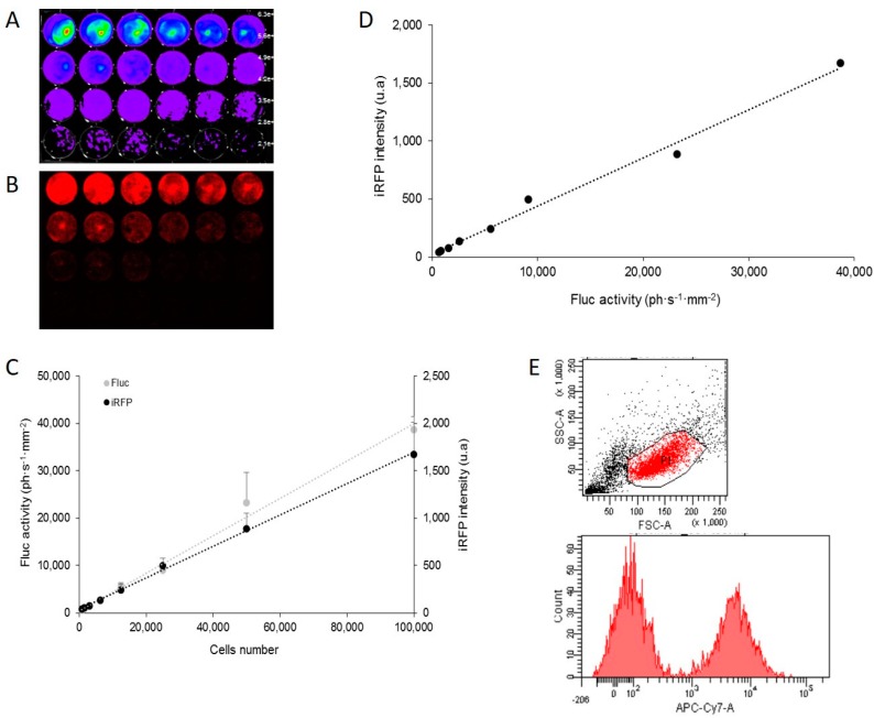
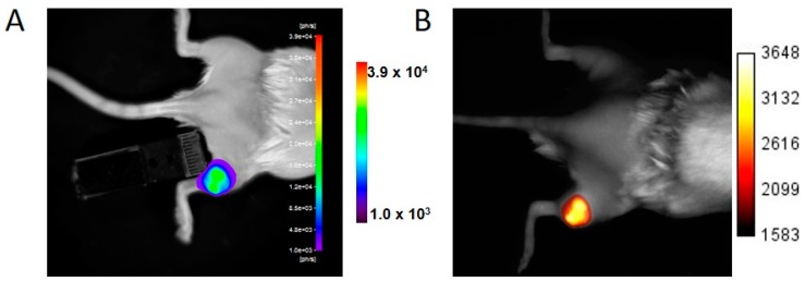
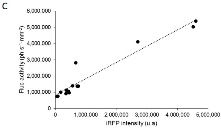
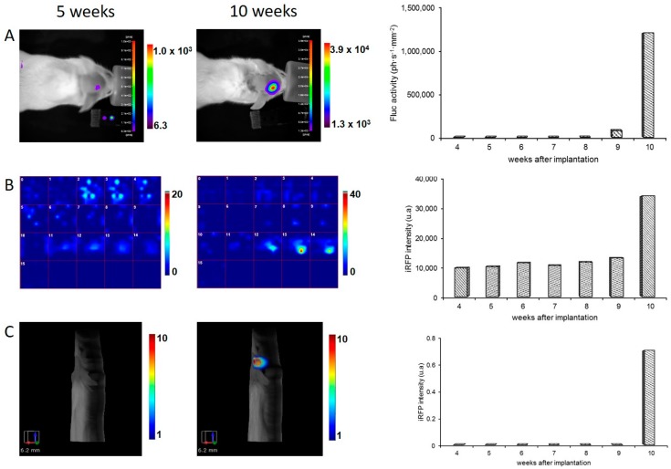
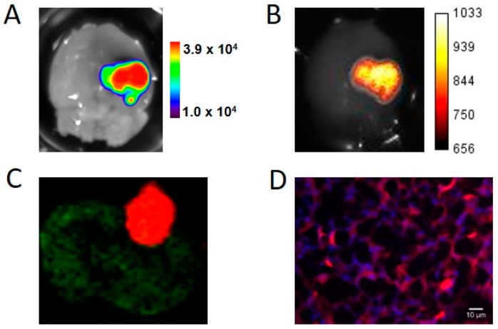
Reporter gene-based strategies are widely used in experimental oncology. Bioluminescence imaging (BLI) using the firefly luciferase (Fluc) as a reporter gene and d-luciferin as a substrate is currently the most widely employed technique. The present paper compares the performances of BLI imaging with fluorescence imaging using the near infrared fluorescent protein (iRFP) to monitor brain tumor growth in mice. Fluorescence imaging includes fluorescence reflectance imaging (FRI), fluorescence diffuse optical tomography (fDOT), and fluorescence molecular Imaging (FMT®). A U87 cell line was genetically modified for constitutive expression of both the encoding Fluc and iRFP reporter genes and assayed for cell, subcutaneous tumor and brain tumor imaging. On cultured cells, BLI was more sensitive than FRI; in vivo, tumors were first detected by BLI. Fluorescence of iRFP provided convenient tools such as flux cytometry, direct detection of the fluorescent protein on histological slices, and fluorescent tomography that allowed for 3D localization and absolute quantification of the fluorescent signal in brain tumors.

Keywords: bioluminescence; cancer; fluorescence tomography; glioblastoma; optical imaging; reporter gene.


Conflict of interest statement

The authors declare no conflict of interest. The founding sponsors had no role in the design of the study; in the collection, analyses, or interpretation of data; in the writing of the manuscript, or in the decision to publish the results.

Figures

Figure 1
Figure 1
In vitro correlation between firefly luciferase (Fluc) activity and infrared fluorescent protein (iRFP) expression in U87-iRFP+-Fluc+ cells. Bioluminescence imaging (BLI) (A) and fluorescence reflectance imaging (FRI) (B) of successive dilutions (1:2) of U87-iRFP+-Fluc+ cells (from 781 to 100,000 cells) two days after plating. Fluorescent signals of iRFP and Fluc activities were plotted versus cell number (C). Fluorescent signals of iRFP were plotted versus bioluminescence signals (D). Representative distribution of iRFP-expressing cells (E) was determined by flux cytometry (10,000 cells). u.a, units arbitrary.
Figure 2
Figure 2
In vivo detection of Fluc activity and iRFP fluorescence by subcutaneous tumors U87-iRFP+-Fluc+. BLI (A) and FRI (B) of a representative mouse. Bioluminescence signals were plotted versus iRFP fluorescence signals (C) (n = 8).
Figure 2
Figure 2
In vivo detection of Fluc activity and iRFP fluorescence by subcutaneous tumors U87-iRFP+-Fluc+. BLI (A) and FRI (B) of a representative mouse. Bioluminescence signals were plotted versus iRFP fluorescence signals (C) (n = 8).
Figure 3
Figure 3
In vivo detection of Fluc activity and iRFP fluorescence by brain tumors U87-iRFP+-Fluc+. (A) BLI of a mouse at 5 weeks and at 10 weeks after cell injection. The graph represents the bioluminescence quantification of the mouse; (B) detection of the iRFP-fluorescent signal by fluorescence diffuse optical tomography (fDOT) of the same mouse at 5 and 10 weeks after cell injection. Z cross sections (1 mm thickness) are presented in the same color scale from z = 0 (ventral) to z = 15 (dorsal). Quantification of the fluorescence signal recovered from fDOT imaging at different times are plotted on the graph; (C) 3D representation of the iRFP-fluorescent signal by FMT® of a mouse at 5 and 10 weeks after cell injection. Quantification of the fluorescence signal recovered from FMT® imaging at different times are plotted on the graph.
Figure 4
Figure 4
Ex vivo imaging and histology of U87-iRFP+-Fluc+ brain tumors. An excised brain was sequentially imaged by BLI (A) and FRI (B) to reveal tumors; (C) Cryosection of the brain tumor was scanned for fluorescence using the Odyssey scanner showing iRFP fluorescence at 700 nm (red) and brain autofluorescence at 800 nm (green); (D) Fluorescence signals of iRFP (red) and nucleus (DAPI, blue) were revealed using epifluorescence microscopy.

References

    1. Contag P.R., Olomu I.N., Stevenson D.K., Contag C.H. Bioluminescent indicators in living mammals. Nat. Med. 1998;4:245–247. doi: 10.1038/nm0298-245. - DOI - PubMed
    1. Weissleder R., Pittet M.J. Imaging in the era of molecular oncology. Nature. 2008;452:580–589. doi: 10.1038/nature06917. - DOI - PMC - PubMed
    1. Berger F., Paulmurugan R., Bhaumik S., Gambhir S.S. Uptake kinetics and biodistribution of 14C-d-luciferin—A radiolabeled substrate for the firefly luciferase catalyzed bioluminescence reaction: Impact on bioluminescence based reporter gene imaging. Eur. J. Nucl. Med. Mol. Imaging. 2008;35:2275–2285. doi: 10.1007/s00259-008-0870-6. - DOI - PMC - PubMed
    1. Weissleder R., Ntziachristos V. Shedding light onto live molecular targets. Nat. Med. 2003;9:123–128. doi: 10.1038/nm0103-123. - DOI - PubMed
    1. Garofalakis A., Dubois A., Kuhnast B., Dupont D.M., Janssens I., Mackiewicz N., Dollé F., Tavitian B., Ducongé F. In vivo validation of free-space fluorescence tomography using nuclear imaging. Opt. Lett. 2010;35:3024–3026. doi: 10.1364/OL.35.003024. - DOI - PubMed

Substances

LinkOut - more resources