. 2016 Oct 31;21(11):1452.
doi: 10.3390/molecules21111452.

Anti-Inflammatory Potential of Ethyl Acetate Fraction of Moringa oleifera in Downregulating the NF-κB Signaling Pathway in Lipopolysaccharide-Stimulated Macrophages

Affiliations

Anti-Inflammatory Potential of Ethyl Acetate Fraction of Moringa oleifera in Downregulating the NF-κB Signaling Pathway in Lipopolysaccharide-Stimulated Macrophages

Palanisamy Arulselvan et al. Molecules. .

Abstract

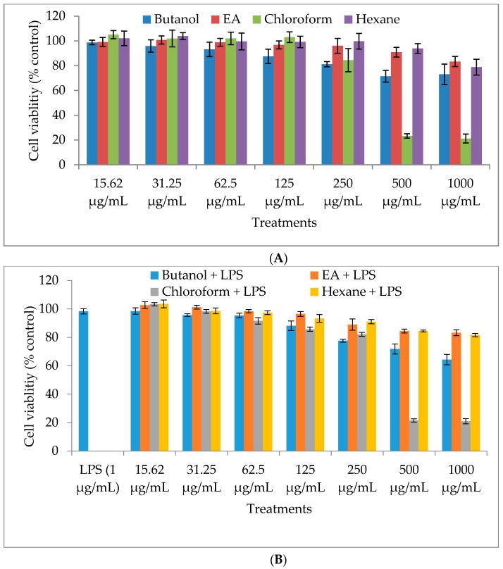
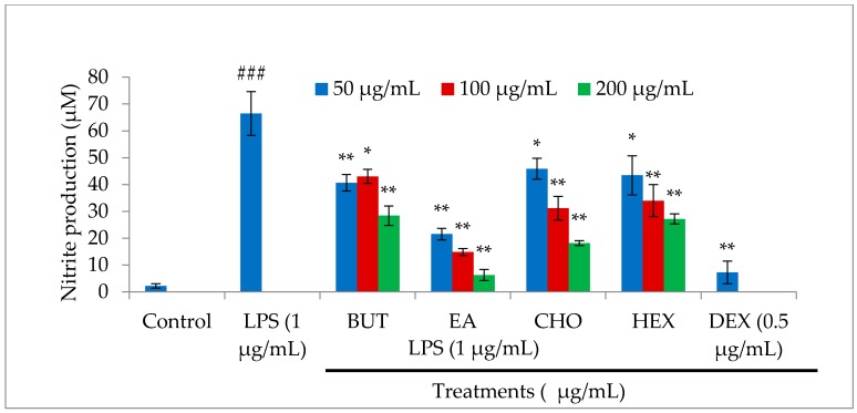
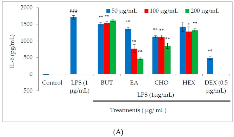
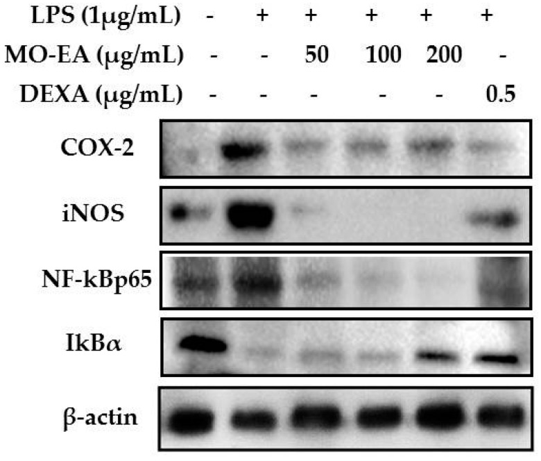
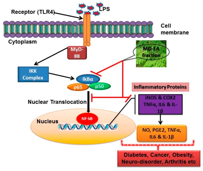
In the present investigation, we prepared four different solvent fractions (chloroform, hexane, butanol, and ethyl acetate) of Moringa oleifera extract to evaluate its anti-inflammatory potential and cellular mechanism of action in lipopolysaccharide (LPS)-induced RAW264.7 cells. Cell cytotoxicity assay suggested that the solvent fractions were not cytotoxic to macrophages at concentrations up to 200 µg/mL. The ethyl acetate fraction suppressed LPS-induced production of nitric oxide and proinflammatory cytokines in macrophages in a concentration-dependent manner and was more effective than the other fractions. Immunoblot observations revealed that the ethyl acetate fraction effectively inhibited the expression of inflammatory mediators including cyclooxygenase-2, inducible nitric oxide synthase, and nuclear factor (NF)-κB p65 through suppression of the NF-κB signaling pathway. Furthermore, it upregulated the expression of the inhibitor of κB (IκBα) and blocked the nuclear translocation of NF-κB. These findings indicated that the ethyl acetate fraction of M. oleifera exhibited potent anti-inflammatory activity in LPS-stimulated macrophages via suppression of the NF-κB signaling pathway.

Keywords: IκBα; RAW264.7 cells; inflammation; inflammatory mediators; proinflammatory cytokines.


Conflict of interest statement

The authors declare that there are no conflicts of interest.

Figures

Figure 1
Figure 1
Effect of different solvent fractions (butanol (BUT), ethyl acetate (EA), chloroform (CHO), and hexane (HEX)) of M. oleifera extract on cell viability of RAW264.7 macrophages. The macrophages were treated with various concentrations of the fractions without lipopolysaccharide (LPS) (A) and with LPS (B) incubated for 24 h. After the incubation period, cell viability was determined by MTT (3-(4,5-dimethylthiazol-2-yl)-2,5-diphenyltetrazolium bromide) assay. Values are expressed as the percentage viability of the control cells. All experiments were conducted in triplicate and results are expressed as mean ± SD.
Figure 2
Figure 2
Effect of different solvent fractions (butanol, ethyl acetate, chloroform, and hexane) of M. oleifera extract on nitric oxide (NO) production in LPS-stimulated macrophages. Macrophages were treated with LPS in the absence or presence of the fractions at various concentrations (50, 100, and 200 µg/mL) for 24 h. After the incubation period, cell culture supernatants were harvested and NO production was determined by Griess reagent. Values are expressed as described in the Methods section. All experiments were conducted in triplicate and results are expressed as mean ± SD. ### p < 0.001 vs. control; ** p < 0.01 and * p < 0.05 vs. LPS treatment.
Figure 3
Figure 3
Effect of different solvent fractions (butanol, ethyl acetate, chloroform, and hexane) of M. oleifera extract on interleukin (IL)-6 (A), tumor necrosis factor (TNF)-α (B), IL-1β (C), and prostaglandin E2 (PGE2) (D) production in LPS-stimulated macrophages. Macrophages were treated with LPS in the absence or presence of the fractions at various concentrations (50, 100, and 200 µg/mL) for 24 h. After the incubation period, cell culture supernatants were harvested, and IL-6, TNF-α, IL-1β, and PGE2 production was determined by ELISA. Values are expressed as described in the methods section. All experiments were conducted in triplicate and results are expressed as mean ± SD. ### p < 0.001 vs. control; *** p < 0.001, ** p < 0.01, and * p < 0.05 vs. LPS treatment.
Figure 3
Figure 3
Effect of different solvent fractions (butanol, ethyl acetate, chloroform, and hexane) of M. oleifera extract on interleukin (IL)-6 (A), tumor necrosis factor (TNF)-α (B), IL-1β (C), and prostaglandin E2 (PGE2) (D) production in LPS-stimulated macrophages. Macrophages were treated with LPS in the absence or presence of the fractions at various concentrations (50, 100, and 200 µg/mL) for 24 h. After the incubation period, cell culture supernatants were harvested, and IL-6, TNF-α, IL-1β, and PGE2 production was determined by ELISA. Values are expressed as described in the methods section. All experiments were conducted in triplicate and results are expressed as mean ± SD. ### p < 0.001 vs. control; *** p < 0.001, ** p < 0.01, and * p < 0.05 vs. LPS treatment.
Figure 4
Figure 4
Effect of the ethyl acetate fraction of M. oleifera extract on the expression of inflammatory mediators, cyclooxygenase (COX)-2, inducible nitric oxide synthase (iNOS), nuclear factor (NF)-κB p65, and inhibitor of κB (IκBα) in LPS-stimulated macrophages. Macrophages were treated with LPS in the absence or presence of the ethyl acetate fraction at various concentrations (50, 100, and 200 µg/mL) for 24 h. After the incubation period, proteins were extracted from treated cells and subjected to electrophoresis, and inflammatory mediator expression was detected by Western blots. All experiments were conducted in triplicate and images shown are representatives of triplicate experiments.
Figure 5
Figure 5
Effect of ethyl acetate fraction of M. oleifera extract on NF-κB signaling pathway in LPS-stimulated RAW264.7 macrophages. Cells were pretreated with ethyl acetate fraction or dexamethasone for 2 h and then stimulated with LPS to induce the inflammatory signaling cascade. Cells were fixed and processed for immunostaining with specific antibodies. Nuclei were counterstained with Hoechst stain (blue color) and observed with a fluorescent microscope (magnification of images, ×600).
Figure 6
Figure 6
Schematic illustration of the possible inhibitory mechanism of ethyl acetate fraction of M. oleifera in suppressing LPS-activated inflammatory pathway.

Similar articles

Cited by

References

    1. Ariel A., Serhan C.N. Resolvins and protectins in the termination program of acute inflammation. Trends Immunol. 2007;28:176–183. doi: 10.1016/j.it.2007.02.007. - DOI - PubMed
    1. Bhatelia K., Singh K., Singh R. Tlrs: Linking inflammation and breast cancer. Cell. Signal. 2014;26:2350–2357. doi: 10.1016/j.cellsig.2014.07.035. - DOI - PubMed
    1. Aggarwal B.B. Nuclear factor-κB: The enemy within. Cancer Cell. 2004;6:203–208. doi: 10.1016/j.ccr.2004.09.003. - DOI - PubMed
    1. Ueda F., Iizuka K., Tago K., Narukawa Y., Kiuchi F., Kasahara T., Tamura H., Funakoshi-Tago M. Nepetaefuran and leonotinin isolated from leonotis nepetaefolia R. Br. Potently inhibit the LPS signaling pathway by suppressing the transactivation of NF-κB. Int. Immunopharmacol. 2015;28:967–976. doi: 10.1016/j.intimp.2015.08.015. - DOI - PubMed
    1. Vo V.A., Lee J.-W., Shin S.-Y., Kwon J.-H., Lee H.J., Kim S.-S., Kwon Y.-S., Chun W. Methyl p-hydroxycinnamate suppresses lipopolysaccharide-induced inflammatory responses through akt phosphorylation in RAW264.7 cells. Biomol. Ther. 2014;22:10–16. doi: 10.4062/biomolther.2013.095. - DOI - PMC - PubMed

LinkOut - more resources